This website uses cookies to ensure you get the best experience on our website.


- Table of Contents
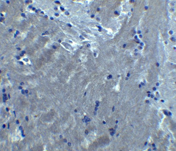
Plan chromogenic S1PR2 IHC in paraffin sections using high staining in duodenal glandular cells as a positive reference and absent staining in adipose tissue adipocytes as a negative reference (HPA tissue IHC). The catalog antibody lists 10 μg/ml for IHC, with an IHC image from mouse brain tissue (datasheet).

Expected localisation, validated protocols, controls and antibodies — the at-a-glance facts below, then the full design guide.
| Expected localisation | Cell membrane expected (UniProt); microvilli observed (HPA tissue IHC) | |
| Staining pattern | Distinct staining in erythrocytes and intestinal microvilli (HPA tissue IHC) | |
| Antigen retrieval | Citrate pH 6.0 HIER, 95–98 °C, 20 min (rule: cytoplasmic / membrane antigen) | |
| Positive control | Duodenum+2 more · see all | |
| Negative control | Adipose tissue+4 more · see all |
| Fixation | Keep fixation consistent across sections (standard IHC practice; not target-specific) | |
| Caveat | Erythrocyte staining may complicate scoring (HPA tissue IHC) | |
| Regulation | Expression regulation not annotated (UniProt) | |
| Isoform / epitope | No isoforms annotated; check extracellular vs cytoplasmic epitope location (UniProt) |
The catalog antibody protocol is paired with published S1PR2 IHC methods for paraffin sections and frozen tissue (PMC4587559; PMC10949307; PMC8972069).
| Sample | Tissue sections; selected-image fixative not specified (standard IHC workflow) |
| Fixation | Image fixative and duration unreported (datasheet A02904); verify before use. |
| Sectioning | 4–5 µm sections on charged slides (standard) |
| Deparaffinisation | Xylene, graded ethanol series to water (standard) |
| Antigen retrieval | Heat-induced epitope retrieval in citrate buffer, pH 6.0, 20 min at 95–98 °C (standard rule: cytoplasmic / membrane antigen) |
| Peroxidase block | 3% H2O2, 10 min, room temperature (standard) |
| Blocking | 10% normal serum of the secondary host, 30 min, room temperature (standard) |
| Primary antibody | Rabbit anti-S1PR2, 10 μg/ml (datasheet A02904) |
| Primary incubation | Overnight at 4 °C (standard) |
| Detection | HRP-polymer secondary, DAB chromogen 5–10 min (standard) |
| Counterstain | Hematoxylin, blue, dehydrate and mount (standard) |
| Expected result | S1PR2-positive staining in glandular cells of duodenum (HPA tissue IHC: High). HPA tissue profile: Distinct expression in erythrocytes and intestinal microvilli. No signal in the no-primary control. |
S1PR2 is a seven transmembrane cell membrane receptor (UniProt O95136 topology and subcellular location). In paraffin section IHC, look first for staining along intestinal glandular cell surfaces, especially the microvilli described by HPA; duodenal and small intestinal glandular cells are scored High, and kidney tubular cells Medium (HPA tissue IHC). HPA rates its tissue IHC profile Approved, with medium consistency between staining and RNA expression (HPA tissue IHC).
| Intestinal glandular cells show strong apical or microvillar staining. | This fits HPA's High staining in duodenal and small intestinal glandular cells and its distinct intestinal microvillar pattern (HPA tissue IHC). A membrane associated distribution is consistent with S1PR2's seven transmembrane topology (UniProt O95136 topology). Compare the stained edge with tissue morphology before scoring it. |
| Staining is predominantly nuclear, or fills cells without a discernible membrane associated pattern. | That pattern is discordant with the UniProt cell membrane annotation and should prompt an artefact check (UniProt O95136 subcellular location). HPA reports uncertain nuclear speckles in ICC/IF, but that observation does not establish a nuclear pattern for paraffin section IHC (HPA subcellular ICC/IF). |
| Strong staining appears in a cell population HPA lists as not detected. | For example, HPA scores adipocytes in adipose tissue and respiratory epithelial cells in bronchus as Not detected (HPA tissue IHC). Check morphology and controls before interpreting signal there as S1PR2; antibody cross reactivity or endogenous detection activity are possible explanations (general IHC practice). |
| Color covers stroma, empty spaces, and many unrelated cells with little structure. | A broadly diffuse deposit is difficult to reconcile with the reported intestinal microvillar pattern (HPA tissue IHC). It may reflect background from the antibody or detection system; compare a no primary control and assess blocking and washing before assigning a cellular score (general IHC practice). |
| Duodenal or small intestinal glandular cells have no detectable signal. | These are useful positive reference populations because HPA scores their staining High (HPA tissue IHC). First check that the expected cells and microvilli are present, then review antibody suitability, antigen retrieval, detection, and the control slide (general IHC practice). One negative section alone cannot establish absent S1PR2. |
| Membrane topology | S1PR2 has seven membrane spanning segments and extracellular and cytoplasmic regions (UniProt O95136 topology). Interpret a sharply outlined cell surface or microvillar pattern in that context; topology alone does not specify which epitope the IHC antibody recognizes. |
| Reference tissue and cell choice | HPA scores duodenal and small intestinal glandular cells High and kidney tubular cells Medium (HPA tissue IHC). Use the intestinal cells as the clearer positive reference, while keeping the tissue's cell level annotation in view when comparing sections. |
| Antibody evidence | HPA014307 is Approved for IHC; HPA014255 has no listed IHC status in the supplied antibody record (HPA antibodies). The tissue profile itself has medium staining to RNA consistency (HPA tissue IHC), so interpret a new compartment or cell type with controls rather than treating approval as universal specificity. |
| Erythrocyte signal | HPA describes distinct erythrocyte staining (HPA tissue IHC). Record it separately from glandular cell staining. If using an enzyme based chromogenic system, evaluate whether endogenous activity contributes to blood cell color with an appropriate control (general IHC practice). |
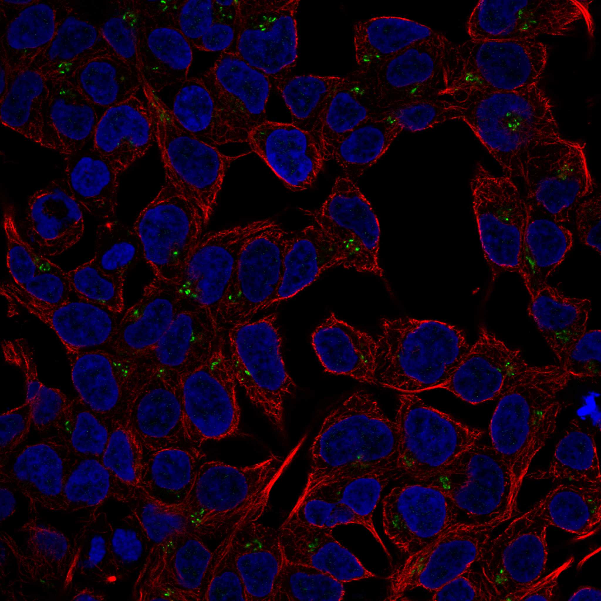
| IF/ICC Q: should the reported intracellular pattern guide this IHC score? | A: HPA places S1PR2 mainly at the Golgi apparatus and additionally at nuclear speckles in ICC/IF, marking both locations uncertain (HPA subcellular ICC/IF). These observations warrant caution when comparing modalities; they do not override UniProt's cell membrane annotation or HPA's tissue IHC pattern. |
| Situation | Likely cause | Next action |
|---|---|---|
| The intestinal positive reference is blank. | The expected glandular cells may be absent from the section, or staining conditions may have failed; HPA reports High staining in those cells (HPA tissue IHC). | Confirm glandular morphology on the section and inspect the positive control. Review antibody suitability, retrieval, and detection using the established IHC workflow (general IHC practice). |
| Only nuclei are strongly colored. | Dominant nuclear IHC staining conflicts with the cell membrane annotation (UniProt O95136 subcellular location). HPA's nuclear speckle assignment comes from uncertain ICC/IF data (HPA subcellular ICC/IF). | Compare the slide with a no primary control and recheck tissue morphology and antibody specificity before scoring nuclear color as S1PR2 (general IHC practice). |
| Color is widespread and obscures the intestinal brush border. | Diffuse background can obscure the distinct microvillar pattern reported by HPA (HPA tissue IHC; general IHC practice). | Review blocking, washing, antibody concentration, and detection background with matched controls; then reassess whether a structured microvillar edge remains (general IHC practice). |
| An HPA negative population stains as strongly as intestinal glands. | Cell identification may be mistaken, or the signal may reflect cross reactivity or endogenous detection activity (general IHC practice); HPA records several cell populations as Not detected (HPA tissue IHC). | Verify the cell type and compare its signal with a no primary control. Keep cell specific scores separate rather than assigning the whole tissue one result (general IHC practice). |
| Erythrocytes dominate the chromogenic signal. | Erythrocyte staining is part of HPA's reported profile (HPA tissue IHC), while endogenous activity can also produce color in enzyme based detection (general IHC practice). | Check a no primary control and the detection system's endogenous activity control; evaluate glandular cell staining independently (general IHC practice). |
| Kidney tubules stain more weakly than intestinal glands. | That relative pattern is compatible with HPA's Medium score in kidney tubular cells and High score in intestinal glandular cells (HPA tissue IHC). | Score each specified cell population against its own expected level and compare slides processed together before calling the kidney result a failure (general IHC practice). |
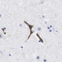


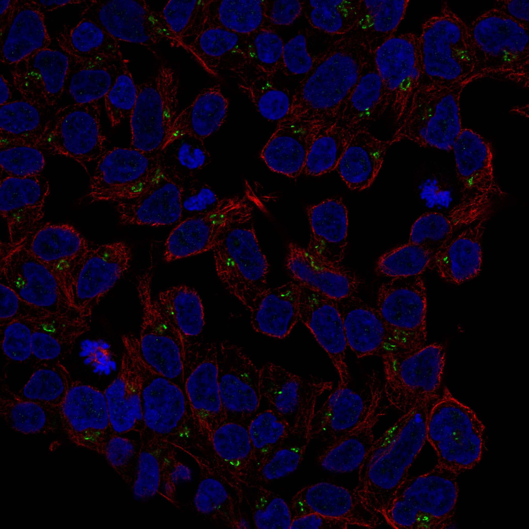
Comprehensive Human Protein Atlas IHC scoring per tissue (reliability: Approved — Medium consistency between antibody staining and RNA expression data. Antibody staining in cells/structures not annotated, view images.). Rows are taken directly from the HPA tissue chart — click any row's HPA link to view the source.
| Tissue | Cell type | Level | Evidence | Source |
|---|---|---|---|---|
| Duodenum | Glandular cells | High | Protein (IHC) | HPA → |
| Small intestine | Glandular cells | High | Protein (IHC) | HPA → |
| Kidney | Cells in tubules | Medium | Protein (IHC) | HPA → |
| Tissue | Cell type | Level | Evidence | Source |
|---|---|---|---|---|
| Adipose tissue | Adipocytes | Not detected | Protein (IHC) | HPA → |
| Adrenal gland | Glandular cells | Not detected | Protein (IHC) | HPA → |
| Appendix | Glandular cells | Not detected | Protein (IHC) | HPA → |
| Bone marrow | Hematopoietic cells | Not detected | Protein (IHC) | HPA → |
| Breast | Adipocytes | Not detected | Protein (IHC) | HPA → |
Use matched controls and compartment aware scoring to troubleshoot S1PR2 staining in paraffin sections; assess fluorescence separately.
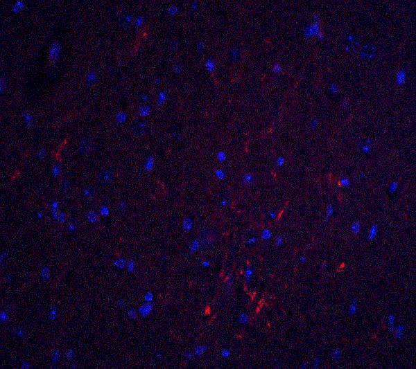
A02904 has mouse brain IHC and IF images (catalog image captions). Human, mouse, and rat reactivity is listed for both antibodies (catalog reactivity).
The rendered card is A02904, with IHC-P listed and a mouse brain IHC image at 10 μg/ml (catalog applications; IHC image caption). A02904 also has a mouse brain IF image at 20 μg/ml (IF image caption).
Which to pick: For tissue IHC, choose A02904: IHC-P is listed, and its own image shows mouse brain staining at 10 μg/ml (catalog applications; IHC image caption); the fixative is unreported (IHC image caption). For IF/ICC, A02904-2 lists both applications and is polyclonal, but has no supplied image; A02904 has a mouse brain IF image (catalog applications; dilution details; image captions). Both list human, mouse, and rat reactivity, while the supplied IHC image documents mouse tissue only (catalog reactivity; IHC image caption).