SERPINA5 / Plasma serine protease inhibitor · IHC design guide

Design Immunohistochemistry for SERPINA5

Plan SERPINA5 paraffin IHC with seminal vesicle glandular cells or kidney tubules as positive reference tissue (HPA tissue IHC). This guide covers expected staining and the catalog antibody’s 0.5–1 μg/ml IHC range (datasheet A01916-1).

Evidence assembled Oct 2026 · For research use; verify linked source records and product datasheet before use
Immunohistochemistry protocol sheet for SERPINA5 (IHC for SERPINA5): expected localisation Cytoplasmic staining (HPA tissue IHC); extracellular secretion (UniProt), antibody A01916-1, validated IHC image, and IHC protocol steps
Printable SERPINA5 IHC protocol sheet — expected localisation Cytoplasmic staining (HPA tissue IHC); extracellular secretion (UniProt), antibody A01916-1, controls and protocol steps. Open the full SERPINA5 IHC guide →

SERPINA5 Immunohistochemistry Experimental Design Guide

Expected localisation, validated protocols, controls and antibodies — the at-a-glance facts below, then the full design guide.

Must know before staining
Expected localisation Cytoplasmic staining (HPA tissue IHC); extracellular secretion (UniProt)
Staining pattern Prominent glandular epithelial cytoplasmic staining (HPA tissue IHC)
Antigen retrieval EDTA pH 8.0 HIER, heat-mediated (datasheet A01916-1)
Positive control ⓘ Adrenal gland+4 more · see all
Negative control ⓘ Breast+4 more · see all
Important caveats
Reasons your staining may differ from the expected pattern.
Fixation Keep fixation consistent across sections. (standard IHC practice; not target-specific)
Caveat Secretion can separate protein staining from RNA expression (HPA tissue IHC)
Regulation Staining regulators are not specified (UniProt)
Isoform / epitope No isoforms; mature chain lacks residues 1–25 (UniProt)
Section 1

Recommended SERPINA5 IHC & IF Protocols

The catalog antibody protocol uses EDTA pH 8.0 heat retrieval (datasheet A01916-1). The published SERPINA5 IHC protocols below cover brain and placenta (PMC10270469; PMC8516267; PMC8055900).

Recommended immunohistochemistry (IHC-P) protocol parameters
SampleParaffin-embedded mouse kidney tissue; fixative not specified (datasheet A01916-1)
FixationImage fixative and duration unreported (datasheet A01916-1); verify before use.
Sectioning4–5 µm sections on charged slides (standard)
DeparaffinisationXylene, graded ethanol series to water (standard)
Antigen retrievalHeat retrieval: EDTA pH 8.0 (datasheet A01916-1); 20 min, 95–100 °C (standard)
Peroxidase block3% H2O2, 10 min, room temperature (standard)
Blocking10% goat serum (datasheet A01916-1)
Primary antibodyRabbit anti-SERPINA5, 0.5-1μg/ml (datasheet A01916-1)
Primary incubationOvernight at 4 °C (datasheet A01916-1)
DetectionStreptavidin-biotin complex (SABC), DAB chromogen (datasheet A01916-1)
CounterstainHematoxylin, blue, dehydrate and mount (standard)
Expected resultSERPINA5-positive staining in glandular cells of adrenal gland (HPA tissue IHC: High). HPA tissue profile: Cytoplasmic and plasma expression in several tissues. Prominent in glandular epithelium. No signal in the no-primary control.
💡Decision noteStart with EDTA pH 8.0 heat retrieval for the catalog antibody (datasheet A01916-1). The placental protocol also used heated EDTA; its pH was unreported (PMC8516267).
Section 2

What Is the Expected SERPINA5 Staining Pattern?

SERPINA5 is secreted, with no transmembrane segment; tissue IHC shows cytoplasmic and plasma staining, especially in glandular epithelium (UniProt P05154 topology; HPA: tissue profile). Expect staining in glandular cells of seminal vesicle, adrenal gland, cervix and endometrium, and in kidney tubule cells (HPA: High in these cells). HPA rates tissue staining Enhanced, while reporting medium consistency between staining and RNA expression for this secreted protein (HPA: reliability).

What am I looking at on my slide?
Strong cytoplasmic staining in seminal-vesicle glandular cells or kidney tubule cells, possibly with extracellular staining.This fits the reported High staining in those cell types and the cytoplasmic and plasma tissue profile (HPA: tissue IHC). Extracellular signal is plausible because SERPINA5 is secreted, but the distribution alone does not identify which cells produced it (UniProt P05154: secreted; HPA: reliability description).
Predominantly nuclear staining in an otherwise appropriate glandular or tubular cell.A nuclear dominant pattern does not match HPA's cytoplasmic and plasma profile or UniProt's secreted location (HPA: tissue profile; UniProt P05154: subcellular location). Treat it as suspect until controls and the expected compartment agree; nuclear color alone is insufficient to assign SERPINA5.
Strong staining in breast adipocytes, lung alveolar cells or lymph-node germinal-center cells.HPA reports SERPINA5 as Not detected in those specific cells (HPA: tissue IHC). Investigate cross-reactivity or endogenous chromogenic activity before calling the staining positive (general IHC practice). The HPA result concerns those cell types, not every cell in each organ.
A diffuse chromogenic haze obscures cell borders and appears across positive and negative regions.This is not the discrete glandular or tubular pattern used to interpret this target (HPA: tissue IHC). Background from detection reagents, inadequate blocking or excessive antibody concentration can obscure localization; review the negative control and adjust one workflow variable at a time (general IHC practice).
No staining in seminal-vesicle glandular cells or kidney tubule cells on a slide expected to be positive.Both are reported High by HPA, so an absent signal warrants a technical check (HPA: tissue IHC). First confirm tissue identity and preservation, then review the IHC antibody, detection reagents and retrieval conditions (general IHC practice). One negative section cannot establish absent SERPINA5 expression.
💡Expected SERPINA5 appearanceCall a section positive when glandular or kidney tubule cells show convincing cytoplasmic staining, with possible plasma staining, at the reported High level; isolated nuclear color or strong signal in HPA-negative cell types is suspect (HPA: tissue profile and cell-level IHC; UniProt P05154: secreted).
How each factor affects the staining
Secretion and cell-of-origin inferenceSERPINA5 enters extracellular space and lacks a transmembrane segment (UniProt P05154: subcellular location and topology). Staining in plasma or secretions can therefore be spatially separate from its production site; HPA explicitly cautions that tissue RNA and protein locations may differ (HPA: reliability description).
Cell type and tissue selectionHPA reports High staining in seminal-vesicle glandular cells and kidney tubule cells, and Not detected in breast adipocytes and lung alveolar cells (HPA: tissue IHC). Compare named cell types within sections; an organ-wide positive or negative label loses that distinction.
Protein processing and epitopeUniProt lists a signal peptide at residues 1–19, a propeptide at 20–25 and a mature chain at 26–406 (UniProt P05154: processing). The payload does not locate the antibody epitope, so it cannot establish whether staining detects precursor, mature protein or both.
Antibody evidenceHPA lists antibody HPA078846 as IHC Enhanced, and its tissue summary cites external characterization while noting medium staining–RNA consistency (HPA: antibody validation and reliability). Use those observations to frame expected tissue staining; they do not establish a target-specific retrieval or fixation effect.
Why is my staining missing, weak or wrong?
SituationLikely causeNext action
A known-positive glandular or tubular region is blank.Possible tissue mismatch, failed detection or an unsuitable staining condition (general IHC practice); HPA reports High staining in seminal-vesicle glandular and kidney tubule cells (HPA: tissue IHC).Confirm the named cell population and run a known-positive section alongside the sample. Check reagent order, retrieval, antibody concentration and chromogen development using general IHC controls; no SERPINA5-specific retrieval setting is supplied.
The entire section has weak, uniform brown color.Nonspecific background or endogenous detection activity may mask the cell-level pattern (general IHC practice). HPA instead describes prominent glandular expression (HPA: tissue profile).Inspect a negative reagent control, check endogenous peroxidase blocking for chromogenic detection, and adjust blocking, washing or antibody concentration as indicated by the control (general IHC practice). Reassess named cells after background clears.
Nuclei dominate the staining.That compartment conflicts with the reported cytoplasmic and plasma tissue profile and secreted location (HPA: tissue profile; UniProt P05154: subcellular location).Compare with a matched control and check counterstain and chromogen interpretation (general IHC practice). Do not score nuclear-only staining as the expected SERPINA5 pattern without independent evidence.
A presumed negative cell population stains strongly.Cross-reactivity or endogenous activity is possible (general IHC practice). HPA reports breast adipocytes, lung alveolar cells and lymph-node germinal-center cells as Not detected (HPA: tissue IHC).Verify cell identity and compare a negative reagent control. If staining persists, seek independent antibody evidence before assigning SERPINA5; avoid extending a cell-specific HPA negative call to an entire tissue.
Extracellular material stains while nearby cells are faint.SERPINA5 is secreted and occurs in body fluids (UniProt P05154: location and function); HPA notes that protein and RNA tissue locations may differ (HPA: reliability description).Record extracellular and cellular staining separately. Use the HPA cell-level pattern for tissue interpretation, and avoid inferring the producing cell from extracellular staining alone.
Can this IHC pattern be assumed for IF/ICC?HPA lists SERPINA5 as secreted but provides no main ICC-IF location or cell-line images; the listed antibody has no ICC validation status (HPA: subcellular and antibody records).Use the separate IF/ICC guide for assay design. Treat this page's IHC tissue pattern as context only, and establish IF/ICC localization with assay-specific controls (general IF practice).

Sample controls for SERPINA5 IHC & IF

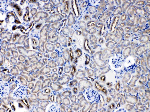
🧪Run kidney first and assess staining in tubular cells (HPA: High in kidney cells in tubules; selected A01916-1 IHC caption: mouse kidney staining). Use esophageal squamous epithelial cells as the negative comparator (HPA: Not detected in esophageal squamous epithelial cells); on the kidney slide, nontubular cells with background-level signal are candidate internal negatives, but their SERPINA5 status is unverified (HPA: kidney result specifies cells in tubules).
Positive control tissue: Adrenal gland (Glandular cells, HPA High)
Negative control tissue: Breast (HPA Not detected)
ICC-IF cell lines (HPA subcellular resource): HPA carries no ICC-IF cell line for SERPINA5; derive a cell-line control from the positive tissue's cell type (Glandular cells) and confirm it by RNA or western blot first.
Technical controls: Include no-primary (secondary-only) and concentration-matched rabbit IgG isotype controls, plus SERPINA5-knockout tissue or a validated antigen-peptide competition control (selected A01916-1 IHC caption: rabbit primary; standard IHC practice). Check endogenous peroxidase and biotin background in kidney before interpreting DAB staining (selected A01916-1 IHC caption: SABC/DAB detection; standard IHC practice).
⚠️Feasibility: A target-specific fixation window or fixation effect is unreported, and the selected paraffin-section caption does not state the fixative (selected A01916-1 IHC caption). The caption uses heat retrieval in EDTA at pH 8.0, but does not establish that retrieval is required (selected A01916-1 IHC caption). Neither frozen sections nor IF/ICC is established as easier by the supplied evidence (HPA subcellular: no ICC-IF images); kidney staining with SABC/DAB warrants an endogenous biotin check (selected A01916-1 IHC caption; standard IHC practice).

HPA tissue IHC evidence for SERPINA5

Comprehensive Human Protein Atlas IHC scoring per tissue (reliability: Enhanced — Medium consistency between antibody staining and RNA expression data. Secreted protein, tissue location of RNA and protein is expected to differ. External characterization data supports antibody staining.). Rows are taken directly from the HPA tissue chart — click any row's HPA link to view the source.

Positive expression · recommended positive controls

TissueCell typeLevelEvidenceSource
Adrenal gland Glandular cells High Protein (IHC) HPA →
Cervix Glandular cells High Protein (IHC) HPA →
Endometrium Glandular cells High Protein (IHC) HPA →
Kidney Cells in tubules High Protein (IHC) HPA →
Seminal vesicle Glandular cells High Protein (IHC) HPA →

Undetected expression · recommended negative controls

TissueCell typeLevelEvidenceSource
Breast Adipocytes Not detected Protein (IHC) HPA →
Caudate Glial cells Not detected Protein (IHC) HPA →
Cerebellum Cells in granular layer Not detected Protein (IHC) HPA →
Cerebral cortex Endothelial cells Not detected Protein (IHC) HPA →
Esophagus Squamous epithelial cells Not detected Protein (IHC) HPA →
Section 3

Advanced SERPINA5 IHC Tips

Troubleshoot SERPINA5 staining in paraffin sections by checking retrieval, tissue context, extracellular signal and detection controls before assigning biological meaning.

How should I optimize retrieval when SERPINA5 staining is weak in paraffin sections?
Start with heat mediated EDTA antigen retrieval at pH 8.0 for paraffin sections (datasheet A01916-1). Keep retrieval conditions consistent across the test section and a positive control so differences in staining can be interpreted (standard IHC practice). The catalog antibody stained mouse kidney after this retrieval, followed by 1 μg/ml primary antibody overnight at 4°C (datasheet A01916-1). If staining remains weak, adjust retrieval duration incrementally while checking tissue morphology and background on adjacent sections (standard IHC practice). Score tubular cells separately from luminal material because SERPINA5 is secreted and kidney tubular cells can stain strongly (UniProt P05154; HPA tissue IHC).
Could fixation explain absent or uneven SERPINA5 staining?
Target specific fixation sensitivity is unknown: the selected paraffin section caption does not state a fixative (datasheet A01916-1). Record fixation method and duration for each specimen, and compare sections processed together before attributing a signal difference to SERPINA5 expression (standard IHC practice). Check whether poorly stained regions also show altered morphology or uneven retrieval, using adjacent sections when available (standard IHC practice). A documented starting workflow uses heat mediated EDTA at pH 8.0 and primary antibody overnight at 4°C, but it establishes no fixation requirement (datasheet A01916-1).
Where should convincing SERPINA5 staining appear in tissue sections?
Assess cell associated and extracellular staining separately because SERPINA5 is secreted and lacks a transmembrane segment (UniProt P05154 topology). Kidney tubular cells and seminal vesicle glandular cells are useful anatomical reference populations with high reported IHC staining (HPA tissue IHC). Cytoplasmic signal can fit the reported tissue staining profile, while luminal or extracellular material may contain secreted protein (HPA tissue IHC; UniProt P05154 subcellular location). Do not treat every membrane associated signal as artefact: SERPINA5 has documented surface localisation in spermatozoa and activated platelets (UniProt P05154 subcellular location). Compare the stained structure with morphology and controls before assigning a cellular compartment (standard IHC practice).
Can processing or glycosylation change how I interpret SERPINA5 staining?
The record lists no isoforms, but it describes a 1–19 signal peptide, a 20–25 propeptide and a 26–406 mature chain (UniProt P05154 processing). It also lists four glycosylation sites at residues 39, 249, 262 and 338 (UniProt P05154 glycosylation). The selected caption does not identify the antibody epitope, so staining cannot establish which processed or glycosylated form was detected (datasheet A01916-1). Compare staining under the documented EDTA pH 8.0 retrieval conditions before changing preparation, and interpret any change as an assay effect until independently validated (datasheet A01916-1; standard IHC practice).
How can I assess SERPINA5 by multiplex IF alongside a cell type marker?
Treat IF as a separate assay requiring its own validation; the supplied catalog example demonstrates chromogenic staining in a paraffin section (datasheet A01916-1). Pair SERPINA5 with a marker for the expected cell population, such as tubular epithelium in kidney, and check each channel individually (HPA tissue IHC; standard IF practice). Choose spectrally separated fluorophores and favor a longer wavelength channel when tissue autofluorescence obscures the target signal (standard IF practice). For an intracellular epitope, test gentle permeabilisation; for an accessible extracellular or surface epitope, compare a nonpermeabilised condition (standard IF practice; UniProt P05154 subcellular location). Include single label and secondary only controls before interpreting overlap (standard IF practice).
What controls distinguish SERPINA5 staining from chromogenic background?
Run a no primary control and inspect diffuse staining, vessel contents and section edges before calling a weak signal positive (standard IHC practice). Quench endogenous peroxidase for DAB detection and assess endogenous biotin when using the documented streptavidin–biotin detection method (standard IHC practice; datasheet A01916-1). The illustrated workflow used 10% goat serum blocking, 1 μg/ml primary antibody overnight at 4°C, and secondary antibody for 30 minutes at 37°C (datasheet A01916-1). Compare cellular staining with a reference population such as kidney tubular cells and with a low signal population such as lung alveolar cells (HPA tissue IHC).
How should I score SERPINA5 when cellular and extracellular staining coexist? ⚠ ANSWER MARKED FOR VERIFICATION
Define the region and compartment before scoring because SERPINA5 is secreted and kidney tubular cells can show strong staining (UniProt P05154 subcellular location; HPA tissue IHC). For cellular staining, record the percentage of positive viable target cells and intensity, or calculate an H-score from those two measures (standard IHC practice). Normalise cellular results to the number of evaluable target cells rather than all cells in the section (standard IHC practice). Measure extracellular staining separately as positive area or signal density per mm² of viable tissue, using identical thresholds and imaging conditions (standard IHC practice). Exclude folds and necrotic areas consistently across specimens (standard IHC practice).
How do I decide whether an unexpected SERPINA5 signal is biological?
First compare the stained cells with expected populations: kidney tubular and seminal vesicle glandular cells show high IHC staining, whereas lung alveolar cells were not detected (HPA tissue IHC). Consider extracellular signal separately because SERPINA5 is secreted and occurs in body fluids (UniProt P05154 subcellular location and tissue specificity). Review unexpected nuclear or widespread uniform staining against morphology and no primary controls before assigning it to SERPINA5 (standard IHC practice; UniProt P05154 subcellular location). Edge accentuation, necrotic deposits and endogenous enzyme signal can mimic specific DAB staining, so repeat assessment on intact regions with detection controls (standard IHC practice).
Boster reagents

Best SERPINA5 / Plasma serine protease inhibitor IHC Antibodies

A01916-1 has real IHC images from paraffin-embedded mouse and rat kidney sections (image captions: A01916-1); the catalog lists human, mouse and rat reactivity (catalog: A01916-1).

Real IHC data IHC analysis of SERPINA5 using anti-SERPINA5 antibody (A01916-1). SERPINA5 was detected in a paraffin-embedded section of mouse kidney tissue. Heat mediated antigen retrieval was performed in EDTA buffer (pH 8.0, epitope retrieval solution). The tissue section was blocked with 10% goat serum. The tissue section was then incubated with 1 μg/ml rabbit anti-SERPINA5 Antibody (A01916-1) overnight at 4°C. Biotinylated goat anti-rabbit IgG was used as secondary antibody and incubated for 30 minutes at 37°C. The tissue section was developed using Strepavidin-Biotin-Complex (SABC) (Catalog # SA1022) with DAB as the chromogen.
Anti-Protein C inhibitor/SERPINA5 Antibody ®
Cat # A01916-1

The only card is A01916-1, listed for IHC and WB with human, mouse and rat reactivity (catalog: A01916-1). Its IHC images show paraffin-embedded mouse and rat kidney sections (image captions: A01916-1).

Which to pick: Choose A01916-1 for paraffin-section IHC; its listed IHC dilution is 0.5–1 μg/ml (datasheet: A01916-1), and its images show mouse and rat kidney sections (image captions: A01916-1). For cross-species work, A01916-1 lists human, mouse and rat reactivity, while its IHC images cover mouse and rat (catalog: A01916-1; image captions: A01916-1). No listed SKU has IF/ICC validation, and the IHC captions do not report the fixative (catalog: A01916-1 applications; image captions: A01916-1).

Each figure is that product's own IHC / IF validation image from its datasheet.

References

  1. UniProt Consortium. UniProt entry P05154 (IPSP_HUMAN, Plasma serine protease inhibitor).
  2. Human Protein Atlas. SERPINA5 tissue IHC expression (reliability: Enhanced).
  3. Human Protein Atlas. SERPINA5 subcellular location (ICC-IF): Secreted.
  4. Human Protein Atlas. SERPINA5 antibody validation summary (1 antibodies).
  5. The SERPINA5 coding variant E228Q does not contribute to clinicopathologic characteristics in Alzheimer's disease: A cross-sectional study. Medicine 2023 — PMC10270469.
  6. Plasma SerpinA5 in conjunction with uterine artery pulsatility index and clinical risk factor for the early prediction of preeclampsia. PloS one 2021 — PMC8516267.
  7. Bioinformatics analysis of the proteome in the pathway of complement and coagulation cascades in COVID-19: discovering potential biomarkers of FGB and SERPINA5. BMC infectious diseases 2025 — PMC12506357.
  8. Transcriptomic analysis to identify genes associated with selective hippocampal vulnerability in Alzheimer's disease. Nature communications 2021 — PMC8055900.
  9. PubMed PMID:3027058 — UniProt-cited evidence.
  10. PubMed PMID:2173165 — UniProt-cited evidence.
  11. PubMed PMID:1714450 — UniProt-cited evidence.