This website uses cookies to ensure you get the best experience on our website.


- Table of Contents
Plan chromogenic SERPINB2 IHC on paraffin sections with the human IHC-validated antibody at 0.5–1 μg/mL (datasheet). Use placental trophoblasts as a high-staining reference and interpret cytoplasmic signal with HPA's cross-gene caution in mind (HPA tissue IHC).

Expected localisation, validated protocols, controls and antibodies — the at-a-glance facts below, then the full design guide.
| Expected localisation | Cytoplasmic in tissue (HPA tissue IHC); also secreted (UniProt) | |
| Staining pattern | Cytoplasmic trophoblasts and squamous epithelial cells (HPA tissue IHC) | |
| Antigen retrieval | Citrate pH 6.0 HIER, 95–98 °C, 20 min (rule: cytoplasmic / membrane antigen) | |
| Positive control | Placenta+2 more · see all | |
| Negative control | Adipose tissue+4 more · see all |
| Fixation | Matched tissue-IHC evidence does not establish the fixation claim. Validate the specimen-specific method before use. (selected-SKU IHC image PB9355) | |
| Caveat | Signal may include protein from another gene (HPA tissue IHC) | |
| Regulation | Expression regulation is not annotated (UniProt) | |
| Isoform / epitope | No isoforms or propeptide annotated; epitope location unknown (UniProt) |
The catalog antibody protocol is followed by published SERPINB2 IHC methods for xenograft tumors and human lung adenocarcinoma (PMC4997607; PMC5710879).
| Sample | Tissue sections; selected-image fixative not specified (standard IHC workflow) |
| Fixation | Image fixative and duration unreported (datasheet PB9355); verify before use. |
| Sectioning | 4–5 µm sections on charged slides (standard) |
| Deparaffinisation | Xylene, graded ethanol series to water (standard) |
| Antigen retrieval | Heat-induced epitope retrieval in citrate buffer, pH 6.0, 20 min at 95–98 °C (standard rule: cytoplasmic / membrane antigen) |
| Peroxidase block | 3% H2O2, 10 min, room temperature (standard) |
| Blocking | 10% normal serum of the secondary host, 30 min, room temperature (standard) |
| Primary antibody | Rabbit anti-SERPINB2, 0.5-1μg/ml (datasheet PB9355) |
| Primary incubation | Overnight at 4 °C (standard) |
| Detection | HRP-polymer secondary, DAB chromogen 5–10 min (standard) |
| Counterstain | Hematoxylin, blue, dehydrate and mount (standard) |
| Expected result | SERPINB2-positive staining in trophoblastic cells of placenta (HPA tissue IHC: High). HPA tissue profile: Cytoplasmic expression in placenta and squamous epithelia. No signal in the no-primary control. |
Expect predominantly cytoplasmic SERPINB2 staining in placental trophoblastic cells, with weaker staining in esophageal squamous epithelial cells and skin basal-layer cells (HPA: tissue IHC). UniProt places SERPINB2 in the cytoplasm and extracellular space and lists no transmembrane segment (UniProt P05120: subcellular location and topology). HPA rates the tissue pattern Enhanced but notes only medium agreement with RNA data and possible staining of proteins from more than one gene (HPA: tissue IHC reliability).
| Strong cytoplasmic stain in placental trophoblastic cells, with medium stain in esophageal squamous epithelium. | This matches the clearest reported tissue pattern: placental trophoblastic cells are High and esophageal squamous epithelial cells are Medium (HPA: tissue IHC). Judge localisation within the named cells, alongside the counterstain, rather than calling a section positive from color alone (general IHC practice). |
| Signal is chiefly nuclear or forms a crisp plasma-membrane outline. | Neither pattern matches the reported cytoplasmic tissue profile or the absence of a transmembrane segment (HPA: tissue IHC; UniProt P05120: topology). Treat it as an unexpected result and check controls and detection background before assigning it to SERPINB2 (general IHC practice). |
| Strong stain appears in cells reported as undetected, such as adipocytes in adipose tissue. | HPA reports no detection in those adipocytes (HPA: tissue IHC). Unexpected cellular stain raises concern for antibody cross-reactivity or endogenous detection activity; HPA also cautions that the tissue stain may include proteins from more than one gene (HPA: reliability; general IHC practice). |
| Color covers stroma, empty spaces, and many unrelated cell types without clear cell boundaries. | A broad haze makes the reported cell-specific cytoplasmic pattern difficult to assess (HPA: tissue IHC). Check reagent-only controls for nonspecific detection or endogenous activity, then review blocking, washes, and chromogen development (general IHC practice). |
| No stain is visible in placental trophoblastic cells on a control section. | Placental trophoblastic cells are reported High, so a blank known-positive control makes a negative study section uninterpretable (HPA: tissue IHC; general IHC practice). Check section quality, antibody application, retrieval conditions, and detection reagents before interpreting absence of SERPINB2. |
| Tissue and cell choice | Placental trophoblastic cells provide the strongest reported positive reference; esophageal squamous cells are Medium, while skin basal-layer and tonsillar squamous cells are Low (HPA: tissue IHC). A weak result in the latter cells carries less weight than a blank placental control. |
| Compartment and extracellular protein | UniProt lists cytoplasmic and secreted, extracellular SERPINB2, with no transmembrane segment (UniProt P05120: location and topology). HPA's tissue readout is cytoplasmic, so extracellular color alone does not establish the expected cellular IHC pattern (HPA: tissue IHC). |
| Antibody validation | Two listed antibodies have Enhanced IHC status and one has Supported status; none has ICC status in the supplied list (HPA: antibody validation). HPA's tissue reliability note cautions that staining may include proteins from more than one gene (HPA: tissue IHC reliability). |
| IF/ICC localisation? | HPA supplies no main ICC-IF location or cell-line images here (HPA: subcellular record). Its IHC cytoplasmic pattern can guide an IF localisation check, but this record cannot establish an IF-positive cell line or an IF protocol (HPA: tissue IHC; HPA: subcellular record). |
| Situation | Likely cause | Next action |
|---|---|---|
| Placental positive control is blank. | An assay step may have failed; HPA reports High staining in trophoblastic cells (HPA: tissue IHC; general IHC practice). | Confirm the trophoblastic cells are present, then check antibody application, the chosen retrieval conditions, detection reagents, and counterstain on the control section (general IHC practice). |
| Study tissue is blank while placental control stains correctly. | Expression may be lower in the sampled cells; HPA reports Medium esophageal staining and Low skin basal-layer and tonsillar squamous staining (HPA: tissue IHC). | Identify the cell type before scoring and compare it with the reported level. Record an absent or below-detection result for that specimen without treating the valid placental control as a guarantee of local expression (HPA: tissue IHC; general IHC practice). |
| All cells show similar brown signal. | Diffuse background or endogenous detection activity may obscure the cell-specific pattern (general IHC practice; HPA: tissue IHC). | Inspect reagent-only controls, review the detection block, washes, and chromogen development, and score only interpretable cellular stain (general IHC practice). |
| Nuclear or membrane-edge staining dominates. | The compartment differs from HPA's cytoplasmic tissue profile and UniProt's topology (HPA: tissue IHC; UniProt P05120: topology). | Review the stained section beside a positive control and reagent-only control; report the localisation as unexpected unless a specific signal can be resolved (general IHC practice). |
| Strong stain appears in HPA-undetected adipocytes. | Cross-reactivity or endogenous activity is possible; HPA warns that its tissue stain may include proteins from more than one gene (HPA: tissue IHC reliability; general IHC practice). | Check detection controls and cell identity, then seek independent antibody or orthogonal evidence before attributing that stain to SERPINB2 (general IHC practice). |
| An IF/ICC image appears to contradict the IHC result. | This payload provides no HPA ICC-IF images or main subcellular location for comparison (HPA: subcellular record). | Describe the IF observation separately and assess it with its own controls; use the placental cytoplasmic IHC pattern only as a tissue reference (HPA: tissue IHC; general IHC practice). |

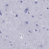


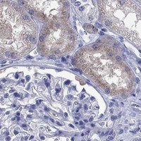
Comprehensive Human Protein Atlas IHC scoring per tissue (reliability: Enhanced — Medium consistency between antibody staining and RNA expression data. Caution, targets protein from more than one gene.). Rows are taken directly from the HPA tissue chart — click any row's HPA link to view the source.
| Tissue | Cell type | Level | Evidence | Source |
|---|---|---|---|---|
| Placenta | Trophoblastic cells | High | Protein (IHC) | HPA → |
| Esophagus | Squamous epithelial cells | Medium | Protein (IHC) | HPA → |
| Skin | Cells in basal layer | Low | Protein (IHC) | HPA → |
| Tissue | Cell type | Level | Evidence | Source |
|---|---|---|---|---|
| Adipose tissue | Adipocytes | Not detected | Protein (IHC) | HPA → |
| Adrenal gland | Glandular cells | Not detected | Protein (IHC) | HPA → |
| Appendix | Glandular cells | Not detected | Protein (IHC) | HPA → |
| Bone marrow | Hematopoietic cells | Not detected | Protein (IHC) | HPA → |
| Breast | Adipocytes | Not detected | Protein (IHC) | HPA → |
Troubleshoot SERPINB2 staining in chromogenic IHC of paraffin sections, using the placenta control and the reported cytoplasmic pattern to assess results (PB9355 tissue-IHC caption; HPA tissue IHC).
The catalog has one human-reactive anti-SERPINB2 antibody with a real paraffin-section IHC image from human placenta; no IF image is supplied (catalog: PB9355 reactivity, applications and image captions).
PB9355 is listed for human IHC and WB (catalog: PB9355 applications and reactivity). Its own IHC(P) image caption shows human placenta tissue (PB9355 IHC image caption).
Which to pick: For tissue IHC, choose PB9355: it is a rabbit antibody listed for human IHC, and its own caption supports paraffin sections of human placenta; the fixative is unreported (catalog: PB9355 host, applications and reactivity; PB9355 IHC image caption). PB9355 has no reported clone, IF/ICC application, IF image or IF dilution, so this catalog provides no IF/ICC option (catalog: PB9355 clone, applications and IF fields). It also provides no supported cross-species option because PB9355 lists Human reactivity only (catalog: PB9355 reactivity).