This website uses cookies to ensure you get the best experience on our website.


- Table of Contents
Plan SIRT6 chromogenic IHC in paraffin sections around the expected nuclear staining pattern (HPA tissue IHC). This guide covers fixation, antibody use, controls and scoring, with attention to the broad tissue distribution (HPA tissue IHC).

Expected localisation, validated protocols, controls and antibodies — the at-a-glance facts below, then the full design guide.
| Expected localisation | Nuclear in tissue (HPA tissue IHC); ER fraction (UniProt) | |
| Staining pattern | Ubiquitous nuclear staining across cell types (HPA tissue IHC) | |
| Antigen retrieval | Tris-EDTA pH 9.0 HIER, 95–98 °C, 20 min (rule: nuclear antigen) | |
| Positive control | Adrenal gland+4 more · see all | |
| Negative control | None in HPA (detected in all 45 tissues); use no-primary + isotype controls |
| Fixation | Keep fixation consistent across sections (standard IHC practice; not target-specific); Matched tissue-IHC evidence does not establish the fixation claim. Validate the specimen-specific method before use. (selected-SKU IHC image PB9357) | |
| Caveat | Widespread nuclear staining limits negative tissue controls (HPA tissue IHC) | |
| Regulation | Low tissue specificity (HPA tissue RNA) | |
| Isoform / epitope | 2 isoforms; epitope differences unspecified (UniProt) |
The catalog antibody’s IHC-P protocol is paired with a published SIRT6 staining protocol for cardiac paraffin sections (PMC11187832).
| Sample | Tissue sections; selected-image fixative not specified (standard IHC workflow) |
| Fixation | Image fixative and duration unreported (datasheet PB9357); verify before use. |
| Sectioning | 4–5 µm sections on charged slides (standard) |
| Deparaffinisation | Xylene, graded ethanol series to water (standard) |
| Antigen retrieval | Heat-induced epitope retrieval in Tris-EDTA buffer, pH 9.0, 20 min at 95–98 °C (standard rule: nuclear antigen) |
| Peroxidase block | 3% H2O2, 10 min, room temperature (standard) |
| Blocking | 10% normal serum of the secondary host, 30 min, room temperature (standard) |
| Primary antibody | Rabbit anti-SIRT6, 0.5-1μg/ml (datasheet PB9357) |
| Primary incubation | Overnight at 4 °C (standard) |
| Detection | HRP-polymer secondary, DAB chromogen 5–10 min (standard) |
| Counterstain | Hematoxylin, blue, dehydrate and mount (standard) |
| Expected result | SIRT6-positive staining in glandular cells of adrenal gland (HPA tissue IHC: High). HPA tissue profile: Ubiquitous nuclear expression. No signal in the no-primary control. |
SIRT6 is predominantly nuclear, with a smaller endoplasmic reticulum fraction; it has no transmembrane segment (UniProt Q8N6T7). Expect nuclear staining across many cell types, including glandular, hematopoietic, epithelial and neuronal cells reported as High by HPA (HPA: tissue IHC). HPA describes the tissue pattern as ubiquitous nuclear expression and rates its reliability Supported, with medium consistency between staining and RNA data (HPA: tissue IHC).
| Clear nuclear chromogen in an HPA-listed cell population, with nuclei distinguishable from surrounding tissue. | This fits the predominant nuclear location (UniProt Q8N6T7) and HPA's ubiquitous nuclear IHC profile (HPA: tissue IHC). Judge intensity against cells on the same slide and a known-positive section; HPA's High category does not specify a chromogen shade or require every cell to stain equally (HPA: tissue IHC; general IHC practice). |
| Cytoplasmic staining dominates while nuclei are faint or unstained. | A dominant cytoplasmic pattern conflicts with the expected predominant nuclear signal (UniProt Q8N6T7; HPA: tissue IHC). Review specificity and detection controls before accepting it as SIRT6. Do not treat every extranuclear signal as false: UniProt reports an endoplasmic reticulum fraction, and HPA reports uncertain vesicular localization in ICC-IF (UniProt Q8N6T7; HPA: subcellular). |
| Staining appears chiefly outside an HPA-listed High population in a tested tissue. | For example, HPA reports High staining in adrenal glandular cells and bone marrow hematopoietic cells (HPA: tissue IHC). If those populations are negative while other cells stain strongly, check tissue identification and antibody or detection specificity (general IHC practice). HPA also describes broad expression, so an unlisted stained cell type alone does not establish cross-reactivity (HPA: tissue IHC). |
| A diffuse chromogen haze obscures nuclear boundaries throughout the section. | The expected distribution is nuclear (HPA: tissue IHC; UniProt Q8N6T7). Haze that also appears in a primary-antibody omission control points toward background from the detection workflow; review blocking, washes and endogenous detection activity (general IHC practice). Haze by itself cannot establish a SIRT6-specific tissue pattern. |
| No nuclear signal appears in a section containing an HPA-listed High population. | An absent result in, for example, bronchial respiratory epithelial or cerebral cortical neuronal cells conflicts with their reported High IHC levels (HPA: tissue IHC). First verify the control tissue and staining run, then review antibody conditions and detection reagents (general IHC practice). HPA reports medium staining-to-RNA consistency, so one negative result does not resolve target absence (HPA: tissue IHC). |
| Compartment | Predominant nuclear localization sets the main scoring compartment (UniProt Q8N6T7; HPA: tissue IHC). UniProt also reports association with pericentric and telomeric heterochromatin and a smaller endoplasmic reticulum fraction; routine chromogenic IHC need not resolve those finer structures (UniProt Q8N6T7; general IHC practice). |
| Tissue reference | HPA calls the tissue pattern ubiquitous nuclear expression and RNA specificity Low tissue specificity (HPA: tissue IHC). Its listed High examples span glandular, hematopoietic, respiratory epithelial and neuronal populations (HPA: tissue IHC). The supplied HPA record lists no negative or low IHC tissues, so it offers no validated negative tissue comparator (HPA: tissue IHC). |
| Antibody evidence | HPA071776 has Supported IHC status; HPA049729 has no IHC status in the supplied antibody record (HPA: antibodies). The overall tissue reliability is Supported with medium staining-to-RNA consistency (HPA: tissue IHC). Interpret a discrepant section with controls rather than treating antibody status as proof that every stained cell is specific (general IHC practice). |
| Isoforms and epitope | UniProt lists two isoforms and a deacetylase sirtuin-type domain at residues 27–272 (UniProt Q8N6T7). The supplied evidence does not locate the IHC antibody's epitope or establish isoform-specific staining. Report what the assay shows without assigning a nuclear or extranuclear signal to one isoform. |
| Processing and modifications | UniProt lists no signal peptide, propeptide, transmembrane segment or glycosylation site, and records modified residues (UniProt Q8N6T7). These annotations do not show that a modification changes IHC staining. Target-specific fixation sensitivity and epitope retrieval requirements are unreported in the supplied evidence; optimize retrieval using assay controls (general IHC practice). |
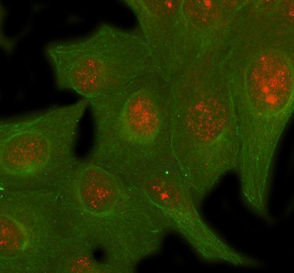
| IF/ICC: Where should signal appear? | Mainly in the nucleoplasm: HPA rates that ICC-IF location Supported (HPA: subcellular). HPA also reports vesicles with Uncertain status, while UniProt describes predominantly nuclear SIRT6 and an endoplasmic reticulum fraction (HPA: subcellular; UniProt Q8N6T7). Treat vesicular signal cautiously and use the separate IF/ICC guide for that assay. |
| Situation | Likely cause | Next action |
|---|---|---|
| An HPA-listed High control population has no nuclear chromogen. | The run, antibody conditions or detection may have failed; the supplied sources do not identify a SIRT6-specific fixation cause (HPA: tissue IHC; general IHC practice). | Confirm the section contains the intended HPA-listed cell population, inspect run controls, then check primary antibody, antigen retrieval and detection conditions within the assay's validated workflow (HPA: tissue IHC; general IHC practice). |
| Cytoplasmic staining overwhelms a weak or absent nuclear signal. | The compartment pattern disagrees with predominant nuclear localization, though a smaller extranuclear fraction is reported (UniProt Q8N6T7; HPA: tissue IHC). | Compare a known-positive section and primary-antibody omission control, and assess whether the signal follows cell boundaries or diffuse background (general IHC practice). Require convincing nuclear staining before scoring the expected IHC pattern (UniProt Q8N6T7; HPA: tissue IHC). |
| Strong staining appears in an unexpected population while the listed High population is unstained. | Possible tissue misidentification, cross-reactivity or endogenous detection activity; staining outside an HPA-listed population alone is inconclusive because expression is broad (HPA: tissue IHC; general IHC practice). | Recheck cell identity and the HPA-listed population, then compare omission and detection controls (HPA: tissue IHC; general IHC practice). Describe the observed distribution if controls pass; do not claim the unexpected cells are SIRT6-negative without supporting evidence. |
| Diffuse staining persists across nuclei, cytoplasm and tissue spaces. | Background from detection, inadequate blocking or insufficient washing can obscure compartment assignment (general IHC practice). Such a field cannot reliably demonstrate HPA's nuclear pattern (HPA: tissue IHC). | Inspect a primary-antibody omission control and the slide's tissue-free areas, then adjust blocking, washes or chromogen development as indicated by the controls (general IHC practice). Rescore only where nuclear boundaries and cell identity are clear. |
| Nuclear staining is present but much weaker than expected in an HPA-listed High tissue. | HPA's High level is a reported staining category, not a fixed chromogen intensity standard; its overall tissue reliability is Supported with medium staining-to-RNA consistency (HPA: tissue IHC). Run conditions can also affect visible contrast (general IHC practice). | Compare the same cell population across the current run's positive controls and review retrieval, antibody dilution and development records (general IHC practice). Record weak nuclear staining as observed rather than converting it to an HPA-level claim. |




Comprehensive Human Protein Atlas IHC scoring per tissue (reliability: Supported — Medium consistency between antibody staining and RNA expression data.). Rows are taken directly from the HPA tissue chart — click any row's HPA link to view the source.
| Tissue | Cell type | Level | Evidence | Source |
|---|---|---|---|---|
| Adrenal gland | Glandular cells | High | Protein (IHC) | HPA → |
| Appendix | Glandular cells | High | Protein (IHC) | HPA → |
| Bone marrow | Hematopoietic cells | High | Protein (IHC) | HPA → |
| Bronchus | Respiratory epithelial cells | High | Protein (IHC) | HPA → |
| Caudate | Neuronal cells | High | Protein (IHC) | HPA → |
| Tissue | Cell type | Level | Evidence | Source |
|---|---|---|---|---|
| None in HPA: SIRT6 is detected in all 45 scored tissues. Use a no-primary (secondary-only) and an isotype control instead. | ||||
Troubleshoot SIRT6 staining by checking nuclear localisation, antibody specificity and section handling before comparing signal across samples.
IHC and IF data cover mouse cardiac muscle paraffin sections, human mammary cancer paraffin sections, and U2OS cells (PB9357 and A00611-3 image captions).
PB9357 has IHC(P) images of mouse cardiac muscle and human mammary cancer tissue (PB9357 image captions). A00611-3 has an IF image of U2OS cells and is listed for IF/ICC (A00611-3 image caption and catalog applications).
Which to pick: Choose PB9357 for tissue IHC: its mouse cardiac muscle image identifies a paraffin section, and its IHC listing covers human and mouse; the fixative is unreported (PB9357 image caption and catalog reactivity). Choose A00611-3 for human IF/ICC (A00611-3 catalog applications and reactivity). For IHC across human, mouse, and rat, consider the rabbit polyclonal A00611-2; its catalog lists all three species and IHC, but supplies no IHC image (A00611-2 catalog reactivity, dilution record, applications, and image list).