SMC3 / Structural maintenance of chromosomes protein 3 · IHC design guide

Design Immunohistochemistry for SMC3

Plan chromogenic SMC3 IHC with the catalog antibody PB9746 (datasheet PB9746). Compare high staining in kidney tubule cells with undetected staining in endometrial stromal cells, while assessing the reported nuclear and cytoplasmic pattern (HPA tissue IHC).

Evidence assembled Oct 2026 · For research use; verify linked source records and product datasheet before use
Immunohistochemistry protocol sheet for SMC3 (IHC for SMC3): expected localisation Nuclear and cytoplasmic tissue staining (HPA tissue IHC), antibody PB9746, validated IHC image, and IHC protocol steps
Printable SMC3 IHC protocol sheet — expected localisation Nuclear and cytoplasmic tissue staining (HPA tissue IHC), antibody PB9746, controls and protocol steps. Open the full SMC3 IHC guide →

SMC3 Immunohistochemistry Experimental Design Guide

Expected localisation, validated protocols, controls and antibodies — the at-a-glance facts below, then the full design guide.

Must know before staining
Expected localisation Nuclear and cytoplasmic tissue staining (HPA tissue IHC)
Staining pattern General nuclear and cytoplasmic staining (HPA tissue IHC)
Antigen retrieval EDTA pH 8.0 HIER, heat-mediated (datasheet PB9746)
Positive control ⓘ Adrenal gland+4 more · see all
Negative control ⓘ Endometrium+2 more · see all
Important caveats
Reasons your staining may differ from the expected pattern.
Fixation Keep fixation conditions consistent across sections. (standard IHC practice; not target-specific)
Caveat Staining and RNA levels show medium consistency (HPA tissue IHC)
Regulation Low tissue specificity (HPA tissue RNA)
Isoform / epitope One full-length chain; no isoforms annotated (UniProt)
Section 1

Recommended SMC3 IHC & IF Protocols

The catalog antibody’s IHC-P protocol (datasheet: PB9746) and one published FFPE glioma IHC protocol (PMC13219900) provide starting conditions.

Recommended immunohistochemistry (IHC-P) protocol parameters
SampleParaffin-embedded mouse intestine tissue; fixative not specified (datasheet PB9746)
FixationImage fixative and duration unreported (datasheet PB9746); verify before use.
Sectioning4–5 µm sections on charged slides (standard)
DeparaffinisationXylene, graded ethanol series to water (standard)
Antigen retrievalHeat retrieval: EDTA pH 8.0 (datasheet PB9746); 20 min, 95–100 °C (standard)
Peroxidase block3% H2O2, 10 min, room temperature (standard)
Blocking10% goat serum (datasheet PB9746)
Primary antibodyRabbit anti-SMC3, 0.5-1μg/ml (datasheet PB9746)
Primary incubationOvernight at 4 °C (datasheet PB9746)
DetectionStreptavidin-biotin complex (SABC), DAB chromogen (datasheet PB9746)
CounterstainHematoxylin, blue, dehydrate and mount (standard)
Expected resultSMC3-positive staining in glandular cells of adrenal gland (HPA tissue IHC: High). HPA tissue profile: General cytoplasmic and nuclear expression. No signal in the no-primary control.
💡Decision noteStart with heat-mediated EDTA retrieval at pH 8.0 (datasheet: PB9746); the glioma study reports a 1:200 primary dilution (PMC13219900).
Section 2

What Is the Expected SMC3 Staining Pattern?

SMC3 associates with nuclear chromatin and chromosomes (UniProt Q9UQE7); HPA ICC-IF supports nucleoplasmic localisation. In paraffin sections, HPA reports general nuclear and cytoplasmic staining, with high staining in kidney tubule cells, smooth muscle cells and several glandular cell populations. Interpret both compartment and cell type together: HPA rates its tissue IHC profile Approved, with medium consistency between staining and RNA expression (HPA tissue IHC). SMC3 has no transmembrane segment (UniProt Q9UQE7 topology).

What am I looking at on my slide?
Cell-associated nuclear staining in kidney tubules, smooth muscle or strongly staining glandular populations.This fits SMC3's chromatin association (UniProt Q9UQE7) and HPA's high IHC staining in these cell types. Assess nuclei against the counterstain and compare cells within the same section; HPA also reports cytoplasmic IHC staining, so nuclear exclusivity is not an IHC criterion (HPA tissue IHC).
Strong, exclusively cytoplasmic signal obscures nuclei throughout the section.Question the result against SMC3's nuclear localisation (UniProt Q9UQE7; HPA ICC-IF: supported nucleoplasm). Cytoplasmic IHC staining alone cannot establish an artefact because HPA reports a general cytoplasmic and nuclear profile (HPA tissue IHC); assess morphology and controls before assigning it to SMC3.
Strong staining appears in cells expected to be unstained, while nearby expected-positive cells stain.Consider nonspecific antibody binding or endogenous chromogen activity as general IHC possibilities. HPA reports no detection in endometrial stromal cells, ovarian stromal cells and splenic red-pulp cells, but those observations are cell-specific and do not make entire tissues negative controls (HPA tissue IHC).
Diffuse chromogen covers tissue and blank spaces, with little cell-level distinction.This is difficult to score as SMC3 because the expected observations identify cells and compartments (HPA tissue IHC; UniProt Q9UQE7 localisation). In general IHC practice, background can reflect incomplete blocking, residual detection activity or overly concentrated reagents; use the assay's controls to identify the source.
No convincing signal appears in kidney tubule cells or another HPA high-staining cell population.A negative result conflicts with the reported high IHC level for that cell population (HPA tissue IHC), but does not identify its cause. Check that the expected cells are present and assess staining and detection controls before interpreting the section as biologically negative; HPA's tissue profile has medium consistency (HPA tissue IHC).
💡Expected SMC3 appearanceCall a result positive when cell-associated staining includes nuclei in the relevant cell population, especially HPA high-staining kidney tubule, smooth muscle or glandular cells (UniProt Q9UQE7 localisation; HPA tissue IHC); HPA also reports cytoplasmic IHC staining, whereas diffuse deposit without cell boundaries is suspect by general IHC practice.
How each factor affects the staining
Cell population and tissueHPA reports high staining in adrenal and gallbladder glandular cells, kidney tubules, small-intestine glandular cells, smooth muscle cells and testicular Leydig cells; appendix and breast glandular cells are medium (HPA tissue IHC). Select a comparison area by cell type, since the reported level does not apply uniformly to every cell in a tissue.
Compartment and assaySMC3 associates with chromatin and chromosomes (UniProt Q9UQE7). HPA supports nucleoplasmic ICC-IF localisation but describes general cytoplasmic and nuclear tissue IHC staining (HPA ICC-IF; HPA tissue IHC). Compare each result with its assay-specific evidence; the IF observation does not erase the reported IHC cytoplasmic component.
Cell-cycle stateUniProt describes cohesin along chromosome arms before prophase, retention at centromeres during prophase and dissociation after RAD21 cleavage at anaphase (UniProt Q9UQE7 localisation). Chromosome-associated distribution may therefore vary in dividing cells; these annotations do not establish a required chromogenic intensity or a cell-cycle scoring cutoff.
Evidence and antibody validationThe supplied antibody HPA043206 has IHC Approved and ICC Supported status (HPA antibody validation). HPA rates the tissue profile Approved with medium staining–RNA consistency (HPA tissue IHC). These labels support comparison with reported patterns, but do not establish that every positive or negative cell in a new section is specific.
Why is my staining missing, weak or wrong?
SituationLikely causeNext action
No staining in a section containing kidney tubules or another HPA high-staining cell population.The expected cells may be absent from the examined area, or the IHC workflow may have failed; the image alone cannot distinguish these possibilities (HPA tissue IHC for expected cells).Confirm cell identity and tissue preservation, then review the IHC-validated antibody's stated retrieval, dilution and detection conditions. Use a staining control to check the run; these workflow checks are general IHC practice.
Strong signal is confined to cells reported as not detected by HPA.Nonspecific binding or endogenous detection activity is possible in chromogenic IHC; HPA's no-detection entries concern particular cell populations, not whole tissues (HPA tissue IHC).Recheck cell identity and compare a control omitting primary antibody. If staining remains, investigate blocking and endogenous enzyme activity as general IHC controls before assigning SMC3 positivity.
Cytoplasmic signal dominates and nuclear detail is weak.HPA reports cytoplasmic and nuclear tissue IHC staining, while UniProt places SMC3 on chromatin and HPA supports nucleoplasmic ICC-IF localisation; these observations leave the cause of a particular section's imbalance unresolved (HPA tissue IHC; UniProt Q9UQE7; HPA ICC-IF).Inspect matched positive cells and the counterstain, then review antibody dilution and detection settings as general IHC checks. Do not reject all cytoplasmic staining solely because the IF reference is nucleoplasmic.
Diffuse background prevents cell-level scoring.Background may arise from nonspecific binding or residual detection activity in a chromogenic workflow (general IHC practice). It cannot be interpreted from HPA tissue staining levels alone (HPA tissue IHC).Compare the primary-omission control, inspect blocking and washes, and adjust detection or antibody concentration according to the validated assay instructions; repeat scoring only when cell boundaries are distinguishable (general IHC practice).
A weak field is being called negative.HPA reports low staining in several cell populations, including prostate glandular cells and adipocytes, and no detection in specified stromal populations (HPA tissue IHC). Weakness has different meaning depending on which cells were sampled.Identify the scored cells first. Compare them with an HPA high-staining cell population in a controlled run, and report weak, absent and uninterpretable staining separately rather than assigning a tissue-wide negative call (HPA tissue IHC; general IHC practice).
Does IF/ICC need to reproduce the paraffin-section IHC pattern?The assays have different reported observations: supported nucleoplasmic ICC-IF localisation and general cytoplasmic plus nuclear tissue IHC staining (HPA ICC-IF; HPA tissue IHC).For IF/ICC interpretation, expect nucleoplasmic signal against a nuclear counterstain (HPA ICC-IF: supported nucleoplasm); assess the paraffin-section result against the tissue IHC profile and its cell-specific levels (HPA tissue IHC).

Sample controls for SMC3 IHC & IF

🧪Run small intestine first and assess nuclear staining in glandular cells (HPA: High in small-intestine glandular cells; UniProt Q9UQE7: nuclear localization). Use endometrial stromal cells as a negative tissue comparator (HPA: Not detected in cells in endometrial stroma). No cell population on the positive slide is established as an internal biological negative; other cells and unstained areas can help assess background, but should not be assumed SMC3-negative (HPA: small-intestine glandular cells listed as High; standard IHC practice).
Positive control tissue: Adrenal gland (Glandular cells, HPA High)
Negative control tissue: Endometrium (HPA Not detected)
ICC-IF cell lines (HPA subcellular resource): HPA ICC-IF images show SMC3 in A-431, U-251MG, U2OS, NIH 3T3, with annotated localisation: Nucleoplasm (supported) (HPA subcellular).
Technical controls: Include a no-primary, secondary-only control, a concentration-matched nonimmune rabbit IgG isotype control, and a validated SMC3 knockout or antibody peptide-block control (caption: rabbit anti-SMC3 antibody; standard IHC practice). For the intestinal DAB assay, control endogenous peroxidase and assess endogenous biotin background because the reported detection uses biotinylated secondary antibody and streptavidin–biotin chemistry (caption: biotinylated goat anti-rabbit IgG, SABC and DAB; standard IHC practice).
⚠️Feasibility: A target-specific fixation window or fixation effect is unreported, and the selected PB9746 paraffin-section caption does not state a fixative (caption: fixative unreported). Heat retrieval in EDTA at pH 8.0 preceded intestinal staining, but the evidence does not establish that retrieval is required under other conditions (caption: mouse intestine IHC). Neither frozen sections nor IF can be ranked as easier from the supplied evidence; intestinal endogenous biotin or peroxidase may contribute background with the reported detection chemistry (HPA: nucleoplasmic ICC-IF localization; caption: SABC and DAB; standard IHC practice).

HPA tissue IHC evidence for SMC3

Comprehensive Human Protein Atlas IHC scoring per tissue (reliability: Approved — Medium consistency between antibody staining and RNA expression data.). Rows are taken directly from the HPA tissue chart — click any row's HPA link to view the source.

Positive expression · recommended positive controls

TissueCell typeLevelEvidenceSource
Adrenal gland Glandular cells High Protein (IHC) HPA →
Gallbladder Glandular cells High Protein (IHC) HPA →
Kidney Cells in tubules High Protein (IHC) HPA →
Small intestine Glandular cells High Protein (IHC) HPA →
Smooth muscle Smooth muscle cells High Protein (IHC) HPA →

Undetected expression · recommended negative controls

TissueCell typeLevelEvidenceSource
Endometrium Cells in endometrial stroma Not detected Protein (IHC) HPA →
Ovary Ovarian stroma cells Not detected Protein (IHC) HPA →
Spleen Cells in red pulp Not detected Protein (IHC) HPA →
Section 3

Advanced SMC3 IHC Tips

Troubleshoot chromogenic SMC3 staining in paraffin sections using the PB9746 tissue example, HPA staining patterns, and SMC3 localisation evidence.

How should I troubleshoot weak SMC3 staining after antigen retrieval?
Start with heat-mediated retrieval in EDTA at pH 8.0 for paraffin sections (datasheet PB9746). The documented mouse intestine example used that retrieval before 10% goat serum blocking and 1 μg/ml primary antibody overnight at 4°C (datasheet PB9746). If nuclei remain weak, compare retrieval duration on matched sections while keeping section thickness, antibody concentration, and detection conditions constant (standard IHC practice). Include a known positive tissue region in every run, and inspect tissue integrity because excessive heating can compromise morphology and make nuclear scoring unreliable (HPA: high in small-intestine glandular cells; standard IHC practice).
Can fixation explain weak or uneven SMC3 staining in paraffin sections?
The PB9746 tissue example describes a paraffin section but does not state its fixative, so target-specific fixation sensitivity is unknown (datasheet PB9746). Do not infer a preferred fixative from SMC3 chromatin association or its acetylation and phosphorylation sites (UniProt Q9UQE7; datasheet PB9746). For troubleshooting, record fixation conditions and compare matched specimens processed with the same EDTA pH 8.0 retrieval, 1 μg/ml primary antibody, and detection workflow (datasheet PB9746; standard IHC practice). If signal varies with processing, assess nuclear preservation and staining across tissue depth before attributing the difference to SMC3 abundance (standard IHC practice).
Where should convincing SMC3 chromogenic staining appear?
Prioritise nuclear staining when evaluating SMC3 because it associates with chromatin and chromosomes, including centromeres during prophase (UniProt Q9UQE7). Supported nucleoplasmic localisation in cell imaging strengthens that expectation (HPA: nucleoplasm supported). Tissue IHC also reports general cytoplasmic and nuclear expression, so cytoplasmic colour alone should be interpreted cautiously rather than automatically discarded (HPA: general cytoplasmic and nuclear expression). Check whether the DAB signal follows intact, counterstained nuclei and persists away from section edges; compare with a no-primary control before calling diffuse colour specific (standard IHC practice). Cell-cycle-dependent redistribution may affect the appearance of rare dividing cells (UniProt Q9UQE7).
Could epitope selection or SMC3 modification alter the IHC result?
No alternative SMC3 isoforms are annotated in the supplied record, and the protein contains an SMC hinge spanning residues 530–642 (UniProt Q9UQE7). Acetylated lysines and phosphorylated residues are documented, but the PB9746 epitope is not specified in the supplied evidence (UniProt Q9UQE7; datasheet PB9746). Therefore, do not assign weak staining to a particular modification or hinge masking without epitope mapping or a direct comparison (standard IHC practice). When comparing lots or antibodies, hold retrieval at EDTA pH 8.0 and score the same nuclear compartment in matched sections; document any shift in cellular pattern (datasheet PB9746; standard IHC practice).
How can IF help assess an uncertain SMC3 IHC pattern?
Use IF/ICC as a separate localisation check, since SMC3 has supported nucleoplasmic staining in cell images (HPA: nucleoplasm supported). Multiplex SMC3 with a validated marker for the expected cell type, and use a nuclear counterstain to determine whether signals occur in the same cells and compartments (standard IF practice). Choose a fluorophore and filter set with low overlap with the specimen’s autofluorescence, checking an unstained section first (standard IF practice). Because SMC3 is nuclear and has no transmembrane segment, permeabilise fixed cells sufficiently for antibody access to the nuclear epitope, then compare with a no-primary control (UniProt Q9UQE7 topology; standard IF practice).
What should I check when SMC3 DAB staining is diffuse?
First compare the stained section with a no-primary control to identify colour arising from detection reagents or tissue rather than primary-antibody binding (standard IHC practice). For the documented workflow, review the 10% goat serum block, biotinylated secondary, streptavidin–biotin detection, and DAB development (datasheet PB9746). Apply an appropriate endogenous peroxidase block and, where relevant, control endogenous biotin before interpreting widespread DAB colour (standard IHC practice). Reduce nonspecific staining through matched changes to blocking, washes, or primary concentration, then confirm that the retained pattern remains predominantly nuclear in intact cells (standard IHC practice; HPA: nucleoplasm supported).
How should I score SMC3 staining across paraffin sections? ⚠ ANSWER MARKED FOR VERIFICATION
Define the cell population and nuclear scoring rule before examining experimental groups, using the same counterstain and threshold across sections (standard IHC practice). Report an H-score or the percentage of positive nuclei, with intensity categories specified; a count of positive nuclei per mm² can complement either measure (standard IHC practice). Normalise positive counts to the number of evaluable nuclei in the same annotated tissue compartment, and exclude damaged or necrotic areas consistently (standard IHC practice). Record cytoplasmic staining separately because tissue IHC reports both compartments while cell imaging supports nucleoplasmic localisation (HPA: general cytoplasmic and nuclear expression; HPA: nucleoplasm supported).
How do I distinguish genuine SMC3 staining from artefact?
A credible pattern includes signal in intact nuclei, consistent with SMC3 chromatin association and supported nucleoplasmic localisation (UniProt Q9UQE7; HPA: nucleoplasm supported). Check cell identity against the section: small-intestine glandular cells are reported high, whereas ovarian stromal cells are reported undetected in the supplied tissue profile (HPA: high in small-intestine glandular cells; HPA: ovarian stroma not detected). Treat staining confined to edges, necrotic areas, or the no-primary control as suspect, and check endogenous peroxidase activity when DAB is widespread (standard IHC practice). Cytoplasmic signal needs separate assessment because tissue IHC reports it, but that observation alone does not validate a sample-specific pattern (HPA: general cytoplasmic and nuclear expression).
Boster reagents

Best SMC3 / Structural maintenance of chromosomes protein 3 IHC Antibodies

Anti-SMC3 antibodies have IHC images from paraffin sections of mouse intestine, rat kidney, human intestinal cancer, and human lung cancer; one catalog antibody also lists IF/ICC (catalog image captions and applications).

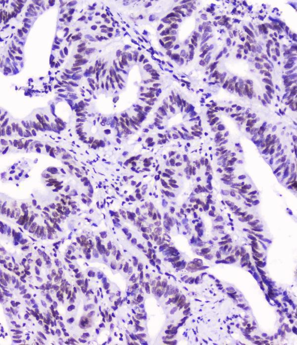
Real IHC data IHC analysis of SMC3 using anti-SMC3 antibody (PB9746). SMC3 was detected in a paraffin-embedded section of mouse intestine tissue. Heat mediated antigen retrieval was performed in EDTA buffer (pH 8.0, epitope retrieval solution). The tissue section was blocked with 10% goat serum. The tissue section was then incubated with 1 μg/ml rabbit anti-SMC3 Antibody (PB9746) overnight at 4°C. Biotinylated goat anti-rabbit IgG was used as secondary antibody and incubated for 30 minutes at 37°C. The tissue section was developed using Strepavidin-Biotin-Complex (SABC) (Catalog # SA1022) with DAB as the chromogen.
Anti-SMC3 Antibody ®
Cat # PB9746
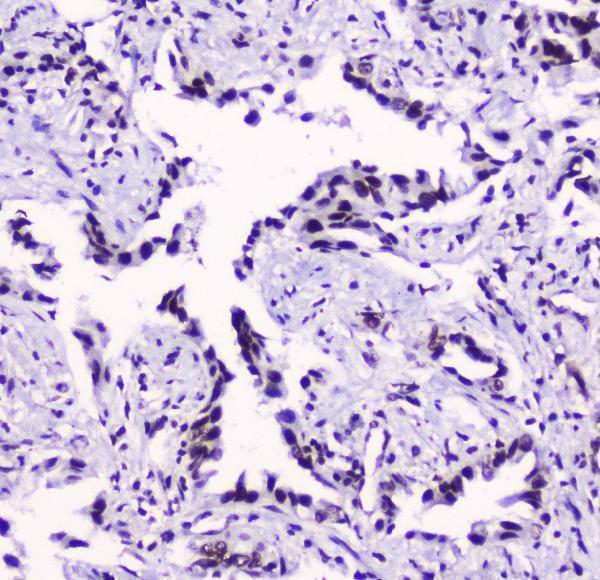
Real IHC data IHC analysis of SMC3 using anti-SMC3 antibody (M01930-1). SMC3 was detected in paraffin-embedded section of human lung cancer tissue. Heat mediated antigen retrieval was performed in citrate buffer (pH6, epitope retrieval solution) for 20 mins. The tissue section was blocked with 10% goat serum. The tissue section was then incubated with 2μg/ml mouse anti-SMC3 Antibody (M01930-1) overnight at 4°C. Biotinylated goat anti-mouse IgG was used as secondary antibody and incubated for 30 minutes at 37°C. The tissue section was developed using Strepavidin-Biotin-Complex (SABC)(Catalog # SA1021) with DAB as the chromogen.
Anti-SMC3 Antibody ® (monoclonal, 4C12)
Cat # M01930-1

PB9746 has IHC images from paraffin sections of mouse intestine, rat kidney, and human intestinal cancer (PB9746 image captions). M01930-1 has IHC images from paraffin sections of human lung and intestinal cancers (M01930-1 image captions).

Which to pick: For tissue IHC across species, choose PB9746: its rabbit antibody has paraffin-section images from human, mouse, and rat samples (catalog host; PB9746 image captions). M01930-1 is a mouse monoclonal option with human paraffin-section IHC images; for IF/ICC, choose M01930 because IF/ICC is listed among its applications, though no IF image is supplied (catalog clone, applications, and image captions). The IHC captions report paraffin sections but no fixative, so fixation is unreported (PB9746 and M01930-1 image captions).

Each figure is that product's own IHC / IF validation image from its datasheet.

References

  1. UniProt Consortium. UniProt entry Q9UQE7 (SMC3_HUMAN, Structural maintenance of chromosomes protein 3).
  2. Human Protein Atlas. SMC3 tissue IHC expression (reliability: Approved).
  3. Human Protein Atlas. SMC3 subcellular location (ICC-IF): Localized to the nucleoplasm..
  4. Human Protein Atlas. SMC3 antibody validation summary (1 antibodies).
  5. SMC3 Is Highly Expressed in Gliomas and Is Associated With Favorable Prognosis and B Cell-Related Immune Features. Technology in cancer research & treatment 2026 — PMC13219900.
  6. RIT1 regulates mitosis and promotes proliferation by interacting with SMC3 and PDS5 in hepatocellular carcinoma. Journal of experimental & clinical cancer research : CR 2023 — PMC10685607.
  7. cccDNA Surrogate MC-HBV-Based Screen Identifies Cohesin Complex as a Novel HBV Restriction Factor. Cellular and molecular gastroenterology and hepatology 2022 — PMC9579331.
  8. Decreased cohesin in the brain leads to defective synapse development and anxiety-related behavior. The Journal of experimental medicine 2017 — PMC5413336.
  9. PubMed PMID:9506951 — UniProt-cited evidence.
  10. PubMed PMID:14702039 — UniProt-cited evidence.
  11. PubMed PMID:15164054 — UniProt-cited evidence.