SMYD2 / N-lysine methyltransferase SMYD2 · IHC design guide

Design Immunohistochemistry for SMYD2

Plan SMYD2 paraffin IHC using the reported cytoplasmic pattern and high staining in glandular cells (HPA tissue IHC). Compare cell populations within each section and interpret the pattern cautiously because antibody staining and RNA expression show very low consistency (HPA tissue IHC).

Evidence assembled Oct 2026 · For research use; verify linked source records and product datasheet before use
Immunohistochemistry protocol sheet for SMYD2 (IHC for SMYD2): expected localisation Cytoplasmic tissue staining (HPA tissue IHC), antibody A04281, validated IHC image, and IHC protocol steps
Printable SMYD2 IHC protocol sheet — expected localisation Cytoplasmic tissue staining (HPA tissue IHC), antibody A04281, controls and protocol steps. Open the full SMYD2 IHC guide →

SMYD2 Immunohistochemistry Experimental Design Guide

Expected localisation, validated protocols, controls and antibodies — the at-a-glance facts below, then the full design guide.

Must know before staining
Expected localisation Cytoplasmic tissue staining (HPA tissue IHC)
Staining pattern Cytoplasmic staining in most tissues; high in glandular cells (HPA tissue IHC)
Antigen retrieval Tris-EDTA pH 9.0 HIER, heat-mediated (datasheet A04281)
Positive control ⓘ Adrenal gland+4 more · see all
Negative control ⓘ Adipose tissue+4 more · see all
Important caveats
Reasons your staining may differ from the expected pattern.
Fixation Keep fixation consistent across sections (standard IHC practice; not target-specific)
Caveat Tissue staining has very low RNA concordance (HPA tissue IHC)
Regulation Expression regulation is not annotated (UniProt)
Isoform / epitope 2 isoforms; epitope coverage is unspecified (UniProt)
Section 1

Recommended SMYD2 IHC & IF Protocols

The catalog antibody’s IHC-P protocol is accompanied by three published SMYD2 IHC examples (datasheet: IHC-P; PMC6749660; PMC13458550; PMC4453442).

Recommended immunohistochemistry (IHC-P) protocol parameters
SampleParaffin-embedded human liver cancer tissue; fixative not specified (datasheet A04281)
FixationImage fixative and duration unreported (datasheet A04281); verify before use.
Sectioning4–5 µm sections on charged slides (standard)
DeparaffinisationXylene, graded ethanol series to water (standard)
Antigen retrievalHeat retrieval: Tris-EDTA pH 9.0 (datasheet A04281); 20 min, 95–100 °C (standard)
Peroxidase block3% H2O2, 10 min, room temperature (standard)
Blocking10% normal serum of the secondary host, 30 min, room temperature (standard)
Primary antibodyRabbit anti-SMYD2, 1:50 recommended; image 1:200 (datasheet A04281)
Primary incubationOvernight at 4 °C (standard)
DetectionHRP-polymer secondary, DAB chromogen 5–10 min (standard)
CounterstainHematoxylin, blue, dehydrate and mount (standard)
Expected resultSMYD2-positive staining in glandular cells of adrenal gland (HPA tissue IHC: High). HPA tissue profile: Cytoplasmic expression in most tissues. No signal in the no-primary control.
💡Decision noteStart with Tris-EDTA pH 9.0 heat retrieval for the catalog antibody (datasheet: retrieval). The published examples use different retrieval methods (PMC13458550; PMC4453442).
Section 2

What Is the Expected SMYD2 Staining Pattern?

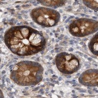
SMYD2 is reported in the cytosol and nucleus, with no transmembrane segment (UniProt Q9NRG4). In tissue IHC, expect chiefly cytoplasmic staining, including high staining in glandular cells of the colon and adrenal gland (HPA tissue IHC). Treat this as a provisional pattern: HPA rates tissue staining reliability Uncertain because antibody staining and RNA expression have very low consistency (HPA tissue IHC).

What am I looking at on my slide?
Cytoplasmic stain is strongest in glandular cells of colon, duodenum, or adrenal gland.This matches HPA's high glandular-cell staining and broadly cytoplasmic tissue profile (HPA tissue IHC). Score the named cells and their compartment separately; surrounding unstained cells do not invalidate a cell-specific result (general IHC practice). HPA's Uncertain reliability means a matching image alone does not establish antibody specificity (HPA tissue IHC).
Some cells show nuclear staining alongside cytoplasmic staining.Nuclear SMYD2 is compatible with UniProt's cytosol and nucleus annotation (UniProt Q9NRG4). Record nuclear and cytoplasmic scores separately (general IHC practice). HPA describes tissue staining chiefly as cytoplasmic, so predominantly nuclear tissue staining needs independent validation before it is called the expected IHC pattern (HPA tissue IHC).
Stain is confined to cell borders or another unexpected compartment.A membrane-only pattern conflicts with the reported cytosol and nucleus localization and lack of a transmembrane segment (UniProt Q9NRG4). Check section morphology, counterstain, and detection controls before assigning the signal to SMYD2 (general IHC practice).
Strong stain appears in adipocytes or ovarian stroma cells, or color covers the whole section.HPA reports SMYD2 as not detected in those cell types (HPA tissue IHC). Their staining warrants investigation of cross-reactivity or endogenous detection activity; broad color without cell boundaries also suggests background (general IHC practice). A not-detected HPA result is a comparison point, not proof of biological absence (HPA tissue IHC).
No signal appears in glandular cells of a well-preserved colon section.Colon glandular cells are reported High by HPA, so check antibody performance and the IHC workflow before interpreting the result as SMYD2 absence (HPA tissue IHC; general IHC practice). HPA rates the tissue profile Uncertain, so this reference cannot by itself prove that every colon specimen must stain (HPA tissue IHC).
💡Expected SMYD2 appearanceCall a result provisionally positive when glandular cells show clear, chiefly cytoplasmic staining consistent with HPA's High examples; membrane-only staining or widespread color outside identifiable cells is suspect (HPA tissue IHC; UniProt Q9NRG4; general IHC practice).
How each factor affects the staining
Tissue reference and validationHPA reports cytoplasmic expression in most tissues and rates the tissue profile Uncertain; antibody HPA029023 has Uncertain IHC status (HPA tissue IHC; HPA antibodies). Use high-staining cells as reference examples, then seek independent specificity evidence before treating intensity as definitive (general IHC practice).
Compartment and assay: what does IF/ICC add?HPA ICC-IF approves cytosol as the main location and mitochondria as an additional location; images include CACO-2, U-251MG, and U2OS (HPA subcellular). These cell-image findings can inform compartment assessment, but do not establish a mitochondrial tissue-IHC pattern (HPA subcellular; HPA tissue IHC).
Cell-type comparisonHPA reports High staining in several glandular-cell groups, Low in skin keratinocytes, and Not detected in adipocytes and ovarian stroma cells (HPA tissue IHC). Compare the scored cell type with its own HPA category; a whole-section average can obscure a cell-specific discrepancy (general IHC practice).
Isoforms and antigen identityUniProt lists 2 SMYD2 isoforms and a chain spanning residues 1–433, with no signal peptide or propeptide (UniProt Q9NRG4). These annotations alone do not show which isoforms an antibody recognizes; consult its documented immunogen before attributing a staining difference to isoforms (general IHC practice).
Why is my staining missing, weak or wrong?
SituationLikely causeNext action
Glandular reference cells are blank.The expected High reference pattern is missing, although that HPA classification is Uncertain (HPA tissue IHC).Check that the section contains intact glandular cells; review the antibody, detection reagents, retrieval, and a positive-control section using general IHC practice.
Every cell or empty space is colored.Diffuse signal may reflect background or endogenous detection activity rather than the cell-specific HPA pattern (HPA tissue IHC; general IHC practice).Inspect a no-primary control, blocking, washes, and chromogen development; assess endogenous enzyme activity for the detection system (general IHC practice).
Adipocytes stain strongly.HPA reports adipocytes as Not detected, making this a cell-type discrepancy rather than supporting evidence (HPA tissue IHC).Compare with glandular reference cells and no-primary controls; verify specificity independently before calling adipocyte signal SMYD2 (general IHC practice).
Only cell borders stain.A border-only pattern is inconsistent with annotated cytosol and nucleus localization and no transmembrane segment (UniProt Q9NRG4).Recheck morphology, detection controls, and whether the signal overlaps cells on the counterstained section (general IHC practice).
Nuclei stain more strongly than cytoplasm.Nuclear localization is annotated, while HPA describes tissue expression as cytoplasmic (UniProt Q9NRG4; HPA tissue IHC).Score both compartments, compare a glandular reference section, and seek independent validation before accepting a predominantly nuclear IHC pattern (general IHC practice).
Mitochondrial puncta are inferred from chromogenic tissue stain.The additional mitochondrial location comes from ICC-IF, while the tissue-IHC profile is chiefly cytoplasmic and Uncertain (HPA subcellular; HPA tissue IHC).Describe only the resolution actually visible in the tissue section; use the separate IF/ICC guide for compartment-resolved follow-up (general IHC practice).

Sample controls for SMYD2 IHC & IF

🧪Run colon first and require staining in its glandular cells (HPA: High in colon glandular cells). Run adipose tissue as the negative, with adipocytes expected to lack staining (HPA: Not detected in adipocytes); on the colon slide, use unstained stromal cells as internal negative candidates, while confirming their status because the supplied HPA row reports only glandular cells (HPA: colon glandular cells High).
Positive control tissue: Adrenal gland (Glandular cells, HPA High)
Negative control tissue: Adipose tissue (HPA Not detected)
ICC-IF cell lines (HPA subcellular resource): HPA ICC-IF images show SMYD2 in CACO-2, U-251MG, U2OS, with annotated localisation: Cytosol (approved) (HPA subcellular).
Technical controls: Include a no-primary, secondary-only control and an isotype control matched to the primary antibody’s host species and clonality (standard IHC practice), plus verified SMYD2 knockout material as a biological negative (standard IHC practice). For chromogenic detection in colon, block endogenous peroxidase and check background in the glandular compartment (standard IHC practice).
⚠️Feasibility: A target-specific fixation window or fixation effect is unreported in the supplied evidence, and the selected A04281 paraffin-section caption does not report a fixative (datasheet: fixative unreported). The caption uses Tris-EDTA at pH 9.0 for retrieval and a 1:200 primary dilution overnight at 4°C, but does not establish whether retrieval is required; the supplied evidence also does not establish that frozen sections or IF are easier (datasheet: paraffin-section caption). In colon, assess glandular staining against possible luminal mucus background (standard IHC practice).

HPA tissue IHC evidence for SMYD2

Comprehensive Human Protein Atlas IHC scoring per tissue (reliability: Uncertain — Very low consistency between antibody staining and RNA expression data. Pending external verification.). Rows are taken directly from the HPA tissue chart — click any row's HPA link to view the source.

Positive expression · recommended positive controls

TissueCell typeLevelEvidenceSource
Adrenal gland Glandular cells High Protein (IHC) HPA →
Appendix Glandular cells High Protein (IHC) HPA →
Cervix Glandular cells High Protein (IHC) HPA →
Colon Glandular cells High Protein (IHC) HPA →
Duodenum Glandular cells High Protein (IHC) HPA →

Undetected expression · recommended negative controls

TissueCell typeLevelEvidenceSource
Adipose tissue Adipocytes Not detected Protein (IHC) HPA →
Hippocampus Glial cells Not detected Protein (IHC) HPA →
Ovary Ovarian stroma cells Not detected Protein (IHC) HPA →
Spleen Cells in red pulp Not detected Protein (IHC) HPA →
Vagina Squamous epithelial cells Not detected Protein (IHC) HPA →
Section 3

Advanced SMYD2 IHC Tips

Use the catalog antibody’s paraffin-section image as a starting point, then verify compartment, cell type, and staining specificity before scoring SMYD2 (caption A04281; UniProt Q9NRG4).

Which retrieval condition should I start with for SMYD2 paraffin-section IHC?
Start with heat-mediated retrieval in Tris-EDTA at pH 9.0 (datasheet A04281). The selected paraffin-section image used this buffer, antibody at 1:200 overnight at 4 °C, and secondary antibody at 1:200 for 45 minutes at room temperature; its fixative was unreported (caption A04281). If staining is weak, compare a modest change in retrieval heating or duration on matched sections while keeping detection and exposure to chromogen constant (standard IHC practice). Include a no-primary control and inspect tissue preservation, because stronger retrieval can raise background or damage morphology (standard IHC practice).
How should I troubleshoot variable SMYD2 staining after tissue fixation?
Target-specific sensitivity of SMYD2 to fixation is unknown from the supplied evidence (caption A04281; HPA tissue IHC). The selected image identifies paraffin-embedded tissue but does not state its fixative or fixation time, so its staining cannot establish an optimal fixation condition (caption A04281). Record each specimen’s fixative, time before fixation, fixation duration, and processing history, then compare sections with matched retrieval and antibody incubation (standard IHC practice). If a batch stains unevenly, examine morphology and compare internal cell populations before changing the pH 9.0 retrieval condition (datasheet A04281; standard IHC practice).
Is cytoplasmic staining enough to call a section SMYD2 positive?
Cytoplasmic staining is plausible because SMYD2 is annotated in cytosol and nucleus, while the HPA tissue profile describes cytoplasmic expression in most tissues (UniProt Q9NRG4 subcellular; HPA tissue IHC). HPA subcellular imaging also reports approved cytosolic and additional mitochondrial localization, but those images come from ICC/IF rather than the scored tissue section (HPA subcellular). Score nuclear and cytoplasmic signal separately and identify the stained cell population on the counterstained section (standard IHC practice). Do not require nuclear staining in every positive cell, and do not treat diffuse chromogen over acellular areas as a compartment-specific signal (UniProt Q9NRG4 subcellular; standard IHC practice).
Could isoforms or epitope masking explain discordant SMYD2 IHC results?
SMYD2 has 2 annotated isoforms and a SET domain spanning residues 7–241, but the supplied caption does not identify the catalog antibody’s epitope (UniProt Q9NRG4 isoforms and domains; caption A04281). Consequently, staining cannot establish whether this antibody recognizes both isoforms or a particular domain (UniProt Q9NRG4 isoforms; caption A04281). Compare the antibody’s documented immunogen and isoform coverage, if available, before interpreting discordance between antibodies (standard IHC practice). A phosphoserine is annotated at residue 283; its presence alone does not establish a fixation, retrieval, or epitope-masking effect for this antibody (UniProt Q9NRG4 modified residues; caption A04281).
How can I check an SMYD2 IHC pattern by multiplex IF?
Use IF as a separate validation experiment and compare its cell and compartment pattern with the chromogenic section (standard IF/IHC practice). Multiplex SMYD2 with a marker for the cell type being evaluated; HPA reports high glandular-cell staining in colon and several other tissues, although its tissue IHC reliability is uncertain (HPA tissue IHC). Choose spectrally separated fluorophores after examining unstained tissue autofluorescence, and include single-stain controls for channel bleed-through (standard IF practice). Because SMYD2 has no transmembrane segment and is annotated in cytosol and nucleus, permeabilize fixed cells or sections as needed to access intracellular epitopes, then check whether nuclear and cytosolic signals remain distinguishable (UniProt Q9NRG4 topology and subcellular; standard IF practice).
What should I change when SMYD2 DAB staining looks diffuse?
First compare the stained section with a no-primary control and inspect whether pigment follows tissue structures or pools at folds and edges (standard IHC practice). Block endogenous peroxidase before chromogenic detection and control DAB development consistently across sections; these are general workflow steps, not SMYD2-specific findings (standard IHC practice). If background persists, titrate the primary antibody around the caption’s 1:200 starting dilution and review blocking, washes, and secondary-antibody specificity (caption A04281; standard IHC practice). Preserve the documented pH 9.0 retrieval condition during that comparison so a change in background can be attributed more clearly (datasheet A04281; standard IHC practice).
How should I quantify SMYD2 staining across paraffin sections? ⚠ ANSWER MARKED FOR VERIFICATION
Define the cell population and compartment before scoring, since SMYD2 is annotated in cytosol and nucleus and the HPA tissue profile is predominantly cytoplasmic (UniProt Q9NRG4 subcellular; HPA tissue IHC). For cellular staining, report the percentage of positive cells and an intensity-weighted H-score with its stated scoring range; score nuclear and cytoplasmic compartments separately (standard IHC practice). If measuring stained-cell density, divide positive-cell counts by analyzed viable tissue area in mm², rather than by the whole image area (standard IHC practice). Keep section selection, counterstain, DAB development, thresholds, and reviewer rules consistent, and normalize comparisons to the same eligible cell population or tissue area (standard IHC practice).
When should an apparent SMYD2-positive section be questioned?
Treat cytosolic or nuclear cellular staining as biologically plausible, while checking that the signal belongs to intact cells rather than extracellular debris (UniProt Q9NRG4 subcellular; standard IHC practice). Compare the stained cell type with an appropriate tissue reference: HPA reports high staining in several glandular-cell populations but no detection in adipocytes, and rates its tissue IHC reliability as uncertain (HPA tissue IHC). Question signal confined to section edges, folds, or necrotic areas, and investigate staining retained in a no-primary control for endogenous enzyme or detection background (standard IHC practice). Confirm consequential findings with independent evidence, because the HPA tissue staining has very low consistency with RNA expression and awaits external verification (HPA tissue IHC).
Boster reagents

Best SMYD2 / N-lysine methyltransferase SMYD2 IHC Antibodies

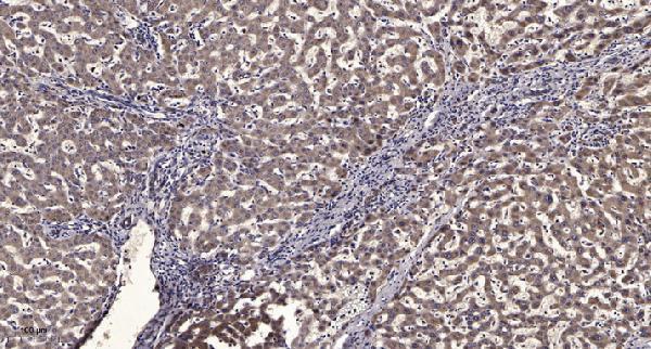
A04281 has a human liver cancer paraffin-section IHC image (A04281 image caption); both antibodies list human, mouse, and rat reactivity (catalog reactivity), while IF/ICC is listed only for A04281-2 (catalog applications).

Real IHC data Immunohistochemical analysis of paraffin-embedded human liver cancer. 1, Antibody was diluted at 1:200 (4° overnight). 2, Tris-EDTA,pH9.0 was used for antigen retrieval. 3,Secondary antibody was diluted at 1:200 (room temperature, 45min).
Anti-SMYD2/Kmt3C Antibody
Cat # A04281

A04281 will render with its paraffin-section human liver cancer IHC figure (A04281 image caption). A04281-2 lists IHC and IF/ICC for human, mouse, and rat, but has no IHC or IF figure supplied (catalog applications/reactivity/image alts).

Which to pick: Choose A04281 for tissue IHC when a documented paraffin-section example is useful; its caption does not report the fixative (A04281 image caption). Choose A04281-2 for IF/ICC because those applications are listed for that SKU (catalog applications). Both are rabbit polyclonal antibodies listing human, mouse, and rat reactivity, so either is a cross-species candidate within its listed applications (catalog host/dilution_raw/reactivity/applications).

Each figure is that product's own IHC / IF validation image from its datasheet.

References

  1. UniProt Consortium. UniProt entry Q9NRG4 (SMYD2_HUMAN, N-lysine methyltransferase SMYD2).
  2. Human Protein Atlas. SMYD2 tissue IHC expression (reliability: Uncertain).
  3. Human Protein Atlas. SMYD2 subcellular location (ICC-IF): Mainly localized to the cytosol. In addition localized to the mitochondria..
  4. Human Protein Atlas. SMYD2 antibody validation summary (1 antibodies).
  5. SMYD2 promotes cervical cancer growth by stimulating cell proliferation. Cell & bioscience 2019 — PMC6749660.
  6. A Novel Inhibitor of Methyltransferase SMYD2, AZ505 Protects Against Peritoneal Fibrosis in Mice. Clinical and experimental pharmacology & physiology 2025 — PMC12104009.
  7. SMYD2 regulates lymph node metastasis derived from intrahepatic cholangiocellular carcinoma through CCR7-dependent chemotaxis. Scientific reports 2026 — PMC13458550.
  8. Overexpression of SMYD2 contributes to malignant outcome in gastric cancer. British journal of cancer 2015 — PMC4453442.
  9. PubMed PMID:14702039 — UniProt-cited evidence.
  10. PubMed PMID:16710414 — UniProt-cited evidence.
  11. PubMed PMID:15489334 — UniProt-cited evidence.