This website uses cookies to ensure you get the best experience on our website.


- Table of Contents
Plan chromogenic IHC for SPTBN1 using its observed cytoplasmic and membranous tissue pattern (HPA tissue IHC). Compare staining across cell types with consistent fixation, and check which of its 3 isoforms the antibody recognizes (UniProt).

Expected localisation, validated protocols, controls and antibodies — the at-a-glance facts below, then the full design guide.
| Expected localisation | Cytoplasmic and membranous tissue staining (HPA tissue IHC) | |
| Staining pattern | Glandular, ciliated and endothelial cells; cytoplasmic/membranous (HPA tissue IHC) | |
| Antigen retrieval | Tris-EDTA pH 9.0 HIER, heat-mediated (datasheet A03164) | |
| Positive control | Adrenal gland+4 more · see all | |
| Negative control | None in HPA (detected in all 45 tissues); use no-primary + isotype controls |
| Fixation | Keep fixation consistent across sections. (standard IHC practice; not target-specific) | |
| Caveat | Broad tissue expression can limit cell-type contrast (HPA tissue IHC) | |
| Regulation | Regulation not reported (UniProt) | |
| Isoform / epitope | 3 isoforms; verify epitope coverage; no transmembrane segment (UniProt) |
The catalog antibody IHC-P protocol (datasheet A03164) is accompanied by three published SPTBN1 IHC protocols (PMC8017174; PMC6639966; PMC7716908).
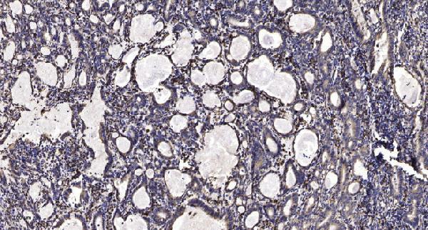
| Sample | Paraffin-embedded human Gastric adenocarcinoma tissue; fixative not specified (datasheet A03164) |
| Fixation | Image fixative and duration unreported (datasheet A03164); verify before use. |
| Sectioning | 4–5 µm sections on charged slides (standard) |
| Deparaffinisation | Xylene, graded ethanol series to water (standard) |
| Antigen retrieval | Heat retrieval: Tris-EDTA pH 9.0 (datasheet A03164); 20 min, 95–100 °C (standard) |
| Peroxidase block | 3% H2O2, 10 min, room temperature (standard) |
| Blocking | 10% normal serum of the secondary host, 30 min, room temperature (standard) |
| Primary antibody | Rabbit anti-SPTBN1, 1:100 - 1:300 (datasheet A03164) |
| Primary incubation | Overnight at 4 °C (standard) |
| Detection | HRP-polymer secondary, DAB chromogen 5–10 min (standard) |
| Counterstain | Hematoxylin, blue, dehydrate and mount (standard) |
| Expected result | SPTBN1-positive staining in glandular cells of adrenal gland (HPA tissue IHC: High). HPA tissue profile: General cytoplasmic and membranous expression. No signal in the no-primary control. |
SPTBN1 is a cytoskeletal protein reported in the cytoplasm, cytosol and at the cell membrane; it has no transmembrane segment (UniProt Q01082). In paraffin-section IHC, expect cytoplasmic and membranous staining in several cell populations, including glandular, ciliated and endothelial cells (HPA tissue IHC). HPA rates the tissue staining profile Supported, citing consistency between antibody staining and RNA expression (HPA tissue IHC).
| Cytoplasmic staining, sometimes with a membranous outline, appears in glandular, ciliated or endothelial cells. | This fits HPA’s general cytoplasmic and membranous IHC profile and UniProt’s cytoskeletal and membrane locations (HPA tissue IHC; UniProt Q01082). Read the signal in its tissue and cell context: HPA reports High staining in adrenal gland and appendix glandular cells, bronchial and fallopian-tube ciliated cell bodies, and cerebral-cortex and colon endothelial cells (HPA tissue IHC). |
| Strong staining is confined to nuclei, with little corresponding cytoplasmic signal. | A nuclear-only pattern does not match the supplied SPTBN1 locations or HPA’s tissue profile (UniProt Q01082; HPA tissue IHC). Treat it as suspect; review morphology and the detection controls before scoring it as SPTBN1 (standard IHC practice). |
| An unexpected cell population stains strongly while the expected cells in the same section do not. | HPA describes cell-specific differences, so identify the stained cells before interpreting the result (HPA tissue IHC). If the pattern cannot be reconciled with the reported cells, consider antibody cross-reactivity or endogenous detection activity; use appropriate controls to distinguish these possibilities (standard IHC practice). |
| Color spreads across stroma or much of the section without clear cell boundaries. | Diffuse background is difficult to assign to the cytoplasmic or membranous pattern reported for SPTBN1 (HPA tissue IHC). Check the no-primary control, blocking and washes, and whether chromogen development obscured the cells (standard IHC practice). |
| An apparently intact known-positive cell population shows no detectable signal. | Absence of signal in a population reported as High by HPA, such as adrenal gland glandular cells, raises a run-level concern but does not alone identify its cause (HPA tissue IHC). Check a positive control and the primary-antibody and detection steps before interpreting a test section as negative (standard IHC practice). |
| Location and topology (UniProt Q01082) | UniProt lists cytoplasm, cytoskeleton, cytosol and cell membrane, with no transmembrane segment. Interpret an apparent membrane outline alongside cytoplasmic staining and cell morphology; the record does not establish a required membrane-only pattern (UniProt Q01082; HPA tissue IHC). |
| Cell and tissue context (HPA tissue IHC) | HPA reports High staining in selected glandular, ciliated and endothelial cells, but Low staining in glial cells of hippocampus and caudate and squamous cells of oral mucosa, esophagus and vagina. Use the named cell population when comparing sections; a low-staining population is a weaker positive reference (HPA tissue IHC). |
| Antibody evidence (HPA antibody validation) | HPA012685 and HPA013149 each have Supported IHC status (HPA antibody validation). That supports the reported IHC patterns; it does not establish that every reagent or staining run will produce the same result (HPA antibody validation; standard IHC practice). |
| Isoforms and epitope coverage (UniProt Q01082) | UniProt lists 3 isoforms and reports isoform 2 protein in brain, lung and kidney (UniProt Q01082). The payload gives no epitope for the antibody used here, so isoform recognition and isoform-specific staining cannot be inferred from this record (UniProt Q01082). |
| IF/ICC Q&A: What pattern is reported? (HPA subcellular) | HPA reports cytosolic ICC-IF localisation but marks its main-location assignment uncertain; its image list includes A-431, U-251MG and U2OS (HPA subcellular). Use that observation as a cautious comparison for IF/ICC, not as a required pattern for chromogenic tissue IHC (HPA subcellular; HPA tissue IHC). |
| Situation | Likely cause | Next action |
|---|---|---|
| Known-positive cells are blank (HPA tissue IHC). | The run may have failed at primary-antibody incubation or detection; a blank section alone cannot distinguish these causes (standard IHC practice). | Inspect a positive control containing an HPA-reported High cell population, then check antibody preparation, incubation, retrieval and detection records (HPA tissue IHC; standard IHC practice). |
| Expected cells stain weakly, but the section has clear morphology (HPA tissue IHC). | HPA staining levels vary by cell population; a reported Low population offers a weak benchmark (HPA tissue IHC). | Compare the same named cell population with an HPA-reported High reference; review routine retrieval and primary-antibody conditions if that reference is also weak (HPA tissue IHC; standard IHC practice). |
| Nuclei dominate the staining (UniProt Q01082; HPA tissue IHC). | The result conflicts with the supplied cytoplasmic and membranous locations; its specific cause is undetermined (UniProt Q01082; HPA tissue IHC). | Check a no-primary control and section morphology; withhold a positive SPTBN1 call until a matching cellular pattern is demonstrated (standard IHC practice; HPA tissue IHC). |
| Broad, hazy chromogen covers cells and tissue spaces (standard IHC practice). | Nonspecific background or detection activity may obscure the reported cellular pattern (standard IHC practice; HPA tissue IHC). | Review the no-primary control, blocking, washes and chromogen development; score only signal that can be assigned to intact cells (standard IHC practice). |
| Unexpected cells stain more strongly than the HPA-reported cells (HPA tissue IHC). | Cross-reactivity or endogenous detection activity is possible; the supplied profile does not identify the unexpected signal (HPA tissue IHC; standard IHC practice). | Identify both cell populations on the counterstained section, compare a no-primary control and, where available, compare an independently validated antibody (standard IHC practice; HPA antibody validation). |
| A membrane-only rim is scored without cytoplasmic context (UniProt Q01082; HPA tissue IHC). | UniProt includes a membrane location, but HPA describes a general cytoplasmic and membranous tissue profile (UniProt Q01082; HPA tissue IHC). | Reassess the cell boundaries and cytoplasm in a reported positive population; document the observed compartment instead of treating a rim alone as definitive (HPA tissue IHC; standard IHC practice). |


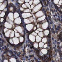


Comprehensive Human Protein Atlas IHC scoring per tissue (reliability: Supported — High consistency between antibody staining and RNA expression data.). Rows are taken directly from the HPA tissue chart — click any row's HPA link to view the source.
| Tissue | Cell type | Level | Evidence | Source |
|---|---|---|---|---|
| Adrenal gland | Glandular cells | High | Protein (IHC) | HPA → |
| Appendix | Glandular cells | High | Protein (IHC) | HPA → |
| Bronchus | Ciliated cells (cell body) | High | Protein (IHC) | HPA → |
| Cerebral cortex | Endothelial cells | High | Protein (IHC) | HPA → |
| Colon | Endothelial cells | High | Protein (IHC) | HPA → |
| Tissue | Cell type | Level | Evidence | Source |
|---|---|---|---|---|
| None in HPA: SPTBN1 is detected in all 45 scored tissues. Use a no-primary (secondary-only) and an isotype control instead. | ||||
Troubleshoot SPTBN1 staining in paraffin sections by checking retrieval, compartment, background and cell-specific scoring before interpreting chromogenic signal.
Two anti-SPTBN1 antibodies have IHC images from human gastric adenocarcinoma or human and rat kidney (catalog IHC captions). Both list IF and human, mouse and rat reactivity (catalog applications and reactivity).
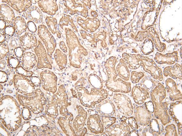
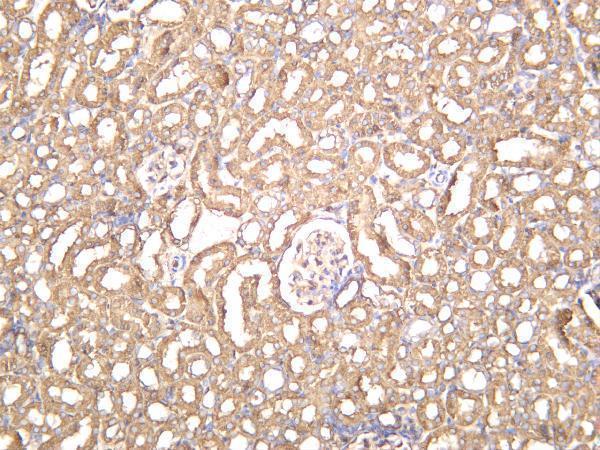
A03164 will render with IHC of paraffin-embedded human gastric adenocarcinoma and lists IF/ICC (A03164 IHC caption; catalog applications). M03164-1 will render with human kidney IHC; its catalog also documents rat kidney IHC and lists IF (M03164-1 IHC captions; catalog applications).
Which to pick: For paraffin-section IHC, choose A03164: its own image documents that preparation, although the fixative is unreported (A03164 IHC caption). For IF/ICC, choose A03164 when ICC is required (catalog applications); M03164-1 is a monoclonal IF option (catalog clonality and applications). For cross-species tissue IHC, M03164-1 has images from human and rat kidney; both antibodies list human, mouse and rat reactivity, but the M03164-1 captions do not report tissue processing or fixative (M03164-1 IHC captions; catalog reactivity).