SPTLC1 / Serine palmitoyltransferase 1 · IHC design guide

Design Immunohistochemistry for SPTLC1

Plan chromogenic SPTLC1 IHC on paraffin sections using tissue staining to choose controls and score cytoplasmic and membranous signal (HPA tissue IHC). This guide covers the expected ER membrane location (UniProt), fixation consistency, and strong lung macrophage staining (HPA tissue IHC).

Evidence assembled Oct 2026 · For research use; verify linked source records and product datasheet before use
Immunohistochemistry protocol sheet for SPTLC1 (IHC for SPTLC1): expected localisation Cytoplasmic and membranous staining; ER membrane expected (HPA tissue IHC; UniProt), antibody PA1777, validated IHC image, and IHC protocol steps
Printable SPTLC1 IHC protocol sheet — expected localisation Cytoplasmic and membranous staining; ER membrane expected (HPA tissue IHC; UniProt), antibody PA1777, controls and protocol steps. Open the full SPTLC1 IHC guide →

SPTLC1 Immunohistochemistry Experimental Design Guide

Expected localisation, validated protocols, controls and antibodies — the at-a-glance facts below, then the full design guide.

Must know before staining
Expected localisation Cytoplasmic and membranous staining; ER membrane expected (HPA tissue IHC; UniProt)
Staining pattern Most tissues show cytoplasmic and membranous staining (HPA tissue IHC)
Antigen retrieval EDTA pH 8.0 HIER, heat-mediated (datasheet PA1777)
Positive control ⓘ Esophagus+4 more · see all
Negative control ⓘ Smooth muscle
Important caveats
Reasons your staining may differ from the expected pattern.
Fixation Keep paraffin-section fixation consistent across samples and controls. (selected-SKU IHC image PA1777)
Caveat Strong lung macrophage staining may affect scoring (HPA tissue IHC)
Regulation Expression regulation unreported (UniProt)
Isoform / epitope 2 isoforms; cytoplasmic residues 37–473, no extracellular region (UniProt)
Section 1

Recommended SPTLC1 IHC & IF Protocols

The catalog antibody’s IHC-P protocol is paired with one published chromogenic IHC protocol for SPTLC1 in mouse tumor sections (PMC10302538).

Recommended immunohistochemistry (IHC-P) protocol parameters
SampleParaffin-embedded Human Mammary Cancer tissue; fixative not specified (datasheet PA1777)
FixationImage fixative and duration unreported (datasheet PA1777); verify before use.
Sectioning4–5 µm sections on charged slides (standard)
DeparaffinisationXylene, graded ethanol series to water (standard)
Antigen retrievalHeat retrieval: EDTA pH 8.0 (datasheet PA1777); 20 min, 95–100 °C (standard)
Peroxidase block3% H2O2, 10 min, room temperature (standard)
Blocking10% goat serum (datasheet PA1777)
Primary antibodyRabbit anti-SPTLC1, 0.5-1μg/ml (datasheet PA1777)
Primary incubationOvernight at 4 °C (datasheet PA1777)
DetectionHRP-conjugated secondary, DAB chromogen (datasheet PA1777)
CounterstainHematoxylin, blue, dehydrate and mount (standard)
Expected resultSPTLC1-positive staining in squamous epithelial cells of esophagus (HPA tissue IHC: High). HPA tissue profile: General cytoplasmic and membranous expression in most tissues. No signal in the no-primary control.
💡Decision noteStart with heat-mediated EDTA retrieval at pH 8.0 (datasheet: PA1777); the published protocol uses microwave retrieval but does not specify a buffer (PMC10302538).
Section 2

What Is the Expected SPTLC1 Staining Pattern?

SPTLC1 is an endoplasmic reticulum (ER) membrane protein with residues 37–473 facing the cytoplasm (UniProt O15269 topology). In paraffin-section IHC, expect cytoplasmic and membranous staining in many tissues, with high staining reported in esophageal squamous cells, gallbladder glandular cells, lung macrophages, and testicular Sertoli cells (HPA: tissue IHC). HPA rates the tissue pattern Supported, with medium consistency between staining and RNA expression (HPA: reliability).

What am I looking at on my slide?
Cytoplasmic and membranous signal in the expected cells, with little signal outside them.This fits the reported IHC pattern (HPA: tissue IHC) and ER membrane location (UniProt O15269). IHC may not resolve an ER network.
Predominantly nuclear or extracellular signal, with little cytoplasmic staining.That distribution conflicts with the reported ER location (UniProt O15269; HPA: subcellular). Check controls before interpreting it as SPTLC1.
Strong staining in smooth muscle cells beside an expected positive population.HPA reports smooth muscle cells as not detected (HPA: tissue IHC). Cross-reactivity or endogenous chromogenic activity is possible; one discrepant section is not proof.
Similar color across cells, stroma, and blank areas of the section.This lacks the reported cell-associated cytoplasmic and membranous pattern (HPA: tissue IHC). Assess background with a no-primary control (general IHC practice).
No signal in a section containing an expected high-staining cell population.That result warrants a workflow check, though HPA levels do not guarantee every specimen will stain (HPA: High in the listed cell populations).
💡Expected SPTLC1 appearanceCall a result consistent when the relevant cells show cytoplasmic and membranous chromogenic signal, strongest in HPA high-staining populations; isolated nuclear, extracellular, or uniform section-wide color is suspect (HPA: tissue IHC; UniProt O15269 location).
How each factor affects the staining
Compartment and topologySPTLC1 resides in the ER membrane; its large cytoplasmic region is residues 37–473 (UniProt O15269 topology). Antibody epitope location is unspecified, so topology alone cannot predict retrieval needs.
Cell populationHPA reports high staining in four named populations, medium staining in adipocytes and several glandular populations, and no detection in smooth muscle cells (HPA: tissue IHC). Score the relevant cells.
Strength of tissue evidenceThe HPA tissue profile is Supported, with medium staining–RNA consistency (HPA: reliability). Treat it as a reference pattern, allowing for specimen and assay differences.
Isoforms and antibody scopeUniProt lists 2 SPTLC1 isoforms (UniProt O15269). The supplied evidence gives no antibody epitope or isoform coverage, so an isoform-specific staining claim is unsupported.
IF/ICC Q&AWhat should IF/ICC show? ER localization is supported in HPA ICC-IF images (HPA: subcellular). That supports compartment interpretation, while the paraffin IHC pattern remains the guide here.
Why is my staining missing, weak or wrong?
SituationLikely causeNext action
Expected high-staining cells are blank.The stain may have failed; a high HPA rating alone cannot identify which step failed (HPA: tissue IHC).Confirm the positive tissue and cell population, then review retrieval, primary incubation, detection, and counterstain records (general IHC practice).
Signal is mostly nuclear.A nuclear-dominant pattern conflicts with the ER assignment (UniProt O15269; HPA: subcellular).Compare with a no-primary control and an expected positive tissue; reassess staining specificity before scoring (general IHC practice).
Color appears throughout the section.Diffuse deposition obscures the cell-associated HPA pattern (HPA: tissue IHC); background has several possible assay causes (general IHC practice).Check the no-primary control, blocking and washes, then adjust detection conditions according to the assay controls (general IHC practice).
Lung macrophages stain strongly, including in a control.Macrophages can be genuinely high for SPTLC1 (HPA: High in macrophages), while endogenous chromogenic activity can also contribute (general IHC practice).Inspect the no-primary control and address endogenous detection activity before attributing all macrophage color to SPTLC1 (general IHC practice).
Smooth muscle cells appear positive.HPA reports this population as not detected; the discrepancy may reflect assay background or a specimen difference (HPA: smooth muscle cells).Compare neighboring expected positive cells and controls, and report the discrepancy instead of using smooth muscle as a positive reference.
A weakly stained population seems negative.Several populations are reported at low staining levels, including kidney glomerular cells and pancreatic exocrine cells (HPA: tissue IHC).Check an HPA high-staining population on a suitable control section before treating weak signal as assay failure (HPA: tissue IHC; general IHC practice).

Sample controls for SPTLC1 IHC & IF

🧪Run esophagus first: its squamous epithelial cells should stain (HPA: High in esophageal squamous epithelial cells). Run smooth muscle as the negative tissue (HPA: Not detected in smooth muscle cells); cells outside the squamous epithelial compartment on the positive slide should show only background in this assay, but should not be assumed to be SPTLC1-negative (HPA: High in esophageal squamous epithelial cells).
Positive control tissue: Esophagus (Squamous epithelial cells, HPA High)
Negative control tissue: Smooth muscle (HPA Not detected)
ICC-IF cell lines (HPA subcellular resource): HPA ICC-IF images show SPTLC1 in A-431, U-251MG, U2OS, HaCaT, SiHa, with annotated localisation: Endoplasmic reticulum (supported) (HPA subcellular).
Technical controls: Include no-primary (secondary-only) and host-matched rabbit IgG isotype controls, matching clonality if known (PA1777 caption: rabbit primary and anti-rabbit secondary), plus SPTLC1 knockout material or a validated cognate-peptide block if available. For DAB detection, quench endogenous peroxidase and compare esophageal background with the no-primary control (PA1777 caption: HRP and DAB; HPA: High in esophageal squamous epithelial cells).
⚠️Feasibility: No matched evidence reports a target-specific fixation window or fixation effect, and the selected PA1777 paraffin-section caption does not report a fixative (PA1777 caption: fixative not stated). Heat retrieval in EDTA at pH 8.0 was used, but retrieval dependency was not established (PA1777 caption: EDTA retrieval). Whether frozen sections or IF are easier is unreported; for IF/ICC, an ER pattern is supported (HPA subcellular: endoplasmic reticulum), and esophageal epithelial edges should be checked for background staining against the no-primary control (HPA: High in esophageal squamous epithelial cells; standard IHC practice).

HPA tissue IHC evidence for SPTLC1

Comprehensive Human Protein Atlas IHC scoring per tissue (reliability: Supported — Medium consistency between antibody staining and RNA expression data.). Rows are taken directly from the HPA tissue chart — click any row's HPA link to view the source.

Positive expression · recommended positive controls

TissueCell typeLevelEvidenceSource
Esophagus Squamous epithelial cells High Protein (IHC) HPA →
Gallbladder Glandular cells High Protein (IHC) HPA →
Lung Macrophages High Protein (IHC) HPA →
Testis Sertoli cells High Protein (IHC) HPA →
Adipose tissue Adipocytes Medium Protein (IHC) HPA →

Undetected expression · recommended negative controls

TissueCell typeLevelEvidenceSource
Smooth muscle Smooth muscle cells Not detected Protein (IHC) HPA →
Section 3

Advanced SPTLC1 IHC Tips

Troubleshoot SPTLC1 chromogenic IHC by checking retrieval, cell type, and staining pattern against its ER localisation and documented tissue expression (UniProt O15269; HPA tissue IHC).

Which retrieval condition should I try first for SPTLC1 paraffin sections?
Start with heat mediated retrieval in EDTA at pH 8.0 for paraffin sections (datasheet PA1777). The documented tissue image used that condition before overnight incubation with 1 μg/ml catalog antibody at 4°C (datasheet PA1777). If signal is weak, compare a gentler or longer heating cycle on adjacent sections while holding antibody concentration, detection, and imaging conditions constant (standard IHC practice). Score cytoplasmic and membranous staining in comparable cells, and reject a retrieval change that mainly raises diffuse background or tissue damage (HPA tissue IHC profile; standard IHC practice). Include a no primary control with each detection run (standard IHC practice).
Could fixation explain weak or uneven SPTLC1 staining?
Target specific fixation sensitivity is unknown: the selected paraffin tissue caption does not report a fixative (datasheet PA1777). Record the actual fixative, fixation duration, tissue thickness, and processing history for each specimen before comparing staining intensity (standard IHC practice). If fixation histories differ, compare matched sections processed and stained together, using the documented EDTA pH 8.0 retrieval and 1 μg/ml antibody condition as a starting point (datasheet PA1777; standard IHC practice). Evaluate morphology alongside signal because poor preservation can make compartment assignment unreliable (standard IHC practice). Do not assign a target specific fixation effect from staining differences alone (standard IHC practice).
Where should convincing SPTLC1 chromogenic staining appear?
Expect staining associated with cytoplasm and membranes in tissue, consistent with SPTLC1 localisation to the endoplasmic reticulum membrane (HPA tissue IHC profile; UniProt O15269 subcellular). Its single transmembrane segment spans residues 16–36, with residues 37–473 on the cytoplasmic side (UniProt O15269 topology). The pattern need not resolve individual ER membranes in routine chromogenic sections, so judge localisation using cell morphology and matched controls (standard IHC practice). Strong isolated nuclear or extracellular signal warrants investigation before interpretation as SPTLC1 (UniProt O15269 subcellular; standard IHC practice). Compare the same cell type across sections to avoid mistaking changes in tissue composition for redistribution (standard IHC practice).
How can epitope location or isoforms affect the IHC result?
SPTLC1 has 2 annotated isoforms, but the supplied antibody evidence does not specify which epitopes or isoforms it detects (UniProt O15269 isoforms; datasheet PA1777). Obtain the immunogen sequence or mapped epitope before claiming isoform specificity, then check whether that sequence is retained in each isoform (standard IHC validation practice). Residues 1–15 are lumenal, residues 16–36 form the membrane segment, and residues 37–473 face the cytoplasm (UniProt O15269 topology). An antibody against a different region could respond differently to processing or retrieval, so treat that as a testable possibility rather than a demonstrated explanation for weak staining (standard IHC practice).
How should I check a multiplex IF result against this IHC pattern?
Use IF as a separate validation experiment and compare its cell level pattern with chromogenic staining in matched material (standard IF and IHC practice). Multiplex SPTLC1 with an independently validated marker for the expected cell type; lung macrophages are one documented high expression reference (HPA tissue IHC: High in lung macrophages). Choose fluorophores after inspecting unstained tissue autofluorescence, and include single stain controls to assess spectral bleed through (standard IF practice). Plan permeabilisation around the antibody's mapped epitope: residues 1–15 are lumenal, whereas residues 37–473 are cytoplasmic (UniProt O15269 topology). Confirm any apparent overlap against the supported ER localisation rather than treating fluorescence alone as proof of identity (HPA subcellular; standard IF practice).
What should I change when SPTLC1 DAB staining looks diffuse?
First compare the primary stained section with no primary and detection only controls to identify background from the detection workflow (standard IHC practice). The documented tissue image used 10% goat serum blocking, 1 μg/ml primary antibody overnight at 4°C, and DAB development (datasheet PA1777). Peroxidase blocking and careful control of DAB development are general chromogenic IHC steps, so evaluate them independently of SPTLC1 expression (standard IHC practice). If background persists, titrate the primary antibody and check wash conditions on adjacent sections while retaining the same retrieval condition (standard IHC practice). Judge improvements by preserved cytoplasmic or membranous cellular staining, rather than lower whole slide colour alone (HPA tissue IHC profile; standard IHC practice).
How should I quantify SPTLC1 staining across paraffin sections? ⚠ ANSWER MARKED FOR VERIFICATION
Define the anatomical region and cell population before scoring, then apply the same thresholds and imaging settings across sections (standard IHC quantification practice). An H score combines the percentage of cells at each intensity; alternatively report percent positive cells or positive cell density per mm² when segmentation supports it (standard IHC quantification practice). Normalise counts to viable tissue area or to the number of eligible cells in the defined compartment, and exclude damaged edges and necrosis consistently (standard IHC quantification practice). Report cell type alongside the score because HPA describes high staining in lung macrophages and absent staining in smooth muscle cells (HPA tissue IHC). Keep detection exposure and DAB development comparable across batches (standard IHC practice).
How can I distinguish true SPTLC1 staining from artefact?
A credible result follows a cellular cytoplasmic or membranous pattern compatible with ER localisation and occurs in an interpretable cell population (HPA tissue IHC profile; UniProt O15269 subcellular). HPA reports high staining in lung macrophages and no detected staining in smooth muscle cells, which can guide within tissue comparisons when those cells are present (HPA tissue IHC). Investigate predominantly nuclear or extracellular colour, staining concentrated at section edges or necrotic regions, and signal remaining in a no primary control (UniProt O15269 subcellular; standard IHC practice). Endogenous peroxidase can produce misleading DAB signal, so check the peroxidase block and detection controls before assigning positivity (standard IHC practice). HPA rates tissue staining support as medium consistency with RNA expression (HPA reliability description).
Boster reagents

Best SPTLC1 / Serine palmitoyltransferase 1 IHC Antibodies

SPTLC1 antibodies have paraffin-section IHC images from human mammary cancer and intestinal cancer tissues (catalog image captions); PB9894 also has a human-cell IF image (catalog IF caption).

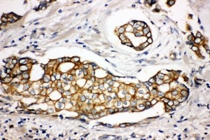
Real IHC data IHC analysis of SPTLC1 using antiSPTLC1 antibody (PA1777). SPTLC1 was detected in a paraffin-embedded section of Human Mammary Cancer tissue. Heat mediated antigen retrieval was performed in EDTA buffer (pH 8.0, epitope retrieval solution). The tissue section was blocked with 10% goat serum. The tissue section was then incubated with 1 μg/ml rabbit anti-SPTLC1 Antibody (PA1777) overnight at 4°C. Peroxidase Conjugated Goat Anti-rabbit IgG was used as secondary antibody and incubated for 30 minutes at 37°C. The tissue section was developed using HRP Conjugated Rabbit IgG Super Vision Assay Kit (Catalog # SV0002) with DAB as the chromogen.
Anti-Serine palmitoyltransferase 1 SPTLC1 Antibody ®
Cat # PA1777
Real IHC data IHC analysis of SPTLC1 using anti-SPTLC1 antibody (PB9894). SPTLC1 was detected in paraffin-embedded section of human intestinal cancer tissues. Heat mediated antigen retrieval was performed in citrate buffer (pH6, epitope retrieval solution) for 20 mins. The tissue section was blocked with 10% goat serum. The tissue section was then incubated with 1μg/ml rabbit anti-SPTLC1 Antibody (PB9894) overnight at 4°C. Biotinylated goat anti-rabbit IgG was used as secondary antibody and incubated for 30 minutes at 37°C. The tissue section was developed using Strepavidin-Biotin-Complex (SABC)(Catalog # SA1022) with DAB as the chromogen.
Anti-SPTLC1 Antibody ®
Cat # PB9894

PA1777 has an IHC image from a human mammary cancer paraffin section; its listed reactivity includes human, mouse and rat (PA1777 catalog). PB9894 has an IHC image from human intestinal cancer paraffin sections and an IF image from U20S cells; its listed reactivity is human (PB9894 catalog).

Which to pick: For human tissue IHC, choose PA1777 for its EDTA pH 8.0 retrieval example or PB9894 for its citrate pH 6 example; both images use paraffin sections, and neither caption reports the fixative (catalog IHC captions). For IF/ICC, choose PB9894 because those applications are listed and it has an IF image; its IF starting concentration is 2 μg/ml (PB9894 catalog). For mouse or rat work, PA1777 lists broader species reactivity, but its IHC caption documents only human tissue; neither product reports clonality (PA1777 catalog; catalog IHC captions).

Each figure is that product's own IHC / IF validation image from its datasheet.

References

  1. UniProt Consortium. UniProt entry O15269 (SPTC1_HUMAN, Serine palmitoyltransferase 1).
  2. Human Protein Atlas. SPTLC1 tissue IHC expression (reliability: Supported).
  3. Human Protein Atlas. SPTLC1 subcellular location (ICC-IF): Localized to the endoplasmic reticulum..
  4. Human Protein Atlas. SPTLC1 antibody validation summary (3 antibodies).
  5. Murine endothelial serine palmitoyltransferase 1 (SPTLC1) is required for vascular development and systemic sphingolipid homeostasis. eLife 2022 — PMC9578713.
  6. E2F2 serves as an essential prognostic biomarker and therapeutic target for human renal cell carcinoma by presenting "E2F2/miR-16-5p/SPTLC1" schema. Translational oncology 2023 — PMC10302538.
  7. ER stress in D1-MSNs mediates cocaine-induced behavioral plasticity via the ATF4-SPTLC1 axis. Frontiers in pharmacology 2025 — PMC12685851.
  8. PubMed PMID:9363775 — UniProt-cited evidence.
  9. PubMed PMID:11242114 — UniProt-cited evidence.
  10. PubMed PMID:14702039 — UniProt-cited evidence.