This website uses cookies to ensure you get the best experience on our website.


- Table of Contents
Use glandular cells in appendix as a medium-staining reference and oral squamous cells as a no-detection comparator (HPA tissue IHC). Interpret cytoplasmic tissue staining alongside STEAP3’s endosomal membrane location and check which side of the membrane the antibody recognizes (HPA tissue IHC; UniProt).

Expected localisation, validated protocols, controls and antibodies — the at-a-glance facts below, then the full design guide.
| Expected localisation | Mainly cytoplasmic in tissue sections (HPA tissue IHC) | |
| Staining pattern | Cytoplasmic staining in glandular and other cells (HPA tissue IHC) | |
| Antigen retrieval | Tris-EDTA pH 9.0 HIER, heat-mediated (datasheet A30509) | |
| Positive control | Adipose tissue+4 more · see all | |
| Negative control | Cerebral cortex+2 more · see all |
| Fixation | Keep fixation consistent across sections. (standard IHC practice; not target-specific) | |
| Caveat | Staining has low agreement with RNA expression (HPA tissue IHC) | |
| Regulation | Lower in hepatocellular carcinoma (UniProt) | |
| Isoform / epitope | 4 isoforms; check cytoplasmic vs vesicular epitope (UniProt) |
The catalog antibody’s IHC-P protocol is accompanied by one published STEAP3 tissue microarray protocol with sufficient method details (PMC9686107).
| Sample | Paraffin-embedded human Colon cancer tissue; fixative not specified (datasheet A30509) |
| Fixation | Image fixative and duration unreported (datasheet A30509); verify before use. |
| Sectioning | 4–5 µm sections on charged slides (standard) |
| Deparaffinisation | Xylene, graded ethanol series to water (standard) |
| Antigen retrieval | Heat retrieval: Tris-EDTA pH 9.0 (datasheet A30509); 20 min, 95–100 °C (standard) |
| Peroxidase block | 3% H2O2, 10 min, room temperature (standard) |
| Blocking | 10% normal serum of the secondary host, 30 min, room temperature (standard) |
| Primary antibody | Rabbit anti-STEAP3, 1:100 - 1:300 (datasheet A30509) |
| Primary incubation | Overnight at 4 °C (standard) |
| Detection | HRP-polymer secondary, DAB chromogen 5–10 min (standard) |
| Counterstain | Hematoxylin, blue, dehydrate and mount (standard) |
| Expected result | STEAP3-positive staining in adipocytes of adipose tissue (HPA tissue IHC: Medium). HPA tissue profile: Mainly cytoplasmic expression in most tissues. No signal in the no-primary control. |
In paraffin-section IHC, expect mainly cytoplasmic STEAP3 staining; adipocytes and several glandular or epithelial cell populations show medium staining (HPA tissue IHC). STEAP3 is a 6-pass endosome-membrane protein also found in vesicular-like structures near the plasma membrane and nucleus (UniProt Q658P3 topology and subcellular location). Treat the tissue pattern as provisional: HPA rates its IHC antibody Approved but reports low agreement between staining and RNA expression (HPA tissue IHC).
| Cytoplasmic staining in adipocytes or glandular cells of breast, appendix, cervix, duodenum, endometrium or epididymis (HPA tissue IHC). | This matches HPA's mainly cytoplasmic pattern and medium staining in those cells (HPA tissue IHC). Vesicular or perinuclear emphasis is compatible with UniProt's membrane-localisation annotation, but HPA IHC alone cannot identify the stained organelle (UniProt Q658P3; HPA tissue IHC). |
| A predominantly nuclear or sharply uniform cell-surface signal replaces the expected cytoplasmic pattern (HPA tissue IHC). | Treat this as a compartment mismatch and investigate staining artefact or antibody specificity (HPA tissue IHC; general IHC practice). HPA reports nucleolar localisation in ICC-IF, so an IF nucleolar signal alone should not be called an IHC failure (HPA subcellular ICC-IF). |
| Strong stain appears in cerebral-cortex endothelial cells, oral-mucosa squamous cells or smooth-muscle cells (HPA tissue IHC). | HPA reports STEAP3 as not detected in those cell populations; investigate cross-reactivity or endogenous chromogen-generating activity before calling them positive (HPA tissue IHC; general IHC practice). A negative HPA observation is a comparator, not proof that every specimen must be negative (HPA tissue IHC). |
| Diffuse chromogen covers tissue, including spaces or structures without a clear cellular pattern (general IHC practice). | The result cannot be scored confidently as STEAP3 because HPA describes a mainly cytoplasmic cellular pattern (HPA tissue IHC). Excess detection background, incomplete blocking or inadequate washing are general IHC possibilities; compare with a no-primary control (general IHC practice). |
| No cellular signal appears in an adipose, breast or appendix section expected to provide a positive comparator (HPA tissue IHC). | Check the run before interpreting absence as biology: HPA reports medium staining in adipocytes and the specified glandular cells (HPA tissue IHC). Verify that those cells are present, then review antibody dilution, retrieval and detection controls as general IHC steps (general IHC practice). |
| Membrane topology: 6 transmembrane segments with cytoplasmic and vesicular loops (UniProt Q658P3 topology). | The location of the antibody's epitope is not supplied, so topology cannot predict whether a specific retrieval or permeabilisation condition will expose it (UniProt Q658P3 topology; epitope information not supplied). Optimise those steps empirically under the antibody's IHC protocol (general IHC practice). |
| Tissue and cell selection: HPA lists medium staining in selected adipocytes and glandular or epithelial cells, with undetected cells elsewhere (HPA tissue IHC). | Include a listed positive cell population and a listed undetected population when assessing a run (HPA tissue IHC; general IHC practice). Preserve cell-level context: a tissue name alone does not specify which cells should stain (HPA tissue IHC). |
| Evidence strength: antibody HPA050510 is IHC Approved; HPA notes low consistency between antibody staining and RNA expression, with retesting pending (HPA tissue IHC; HPA antibodies). | Use the reported staining pattern as a working expectation, not definitive validation of an unexpected result (HPA tissue IHC). Confirm surprising compartment or cell-type staining with appropriate controls and independent evidence where available (general IHC practice). |
| Isoforms: UniProt lists 4 STEAP3 isoforms; the antibody epitope and isoform coverage are not supplied (UniProt Q658P3). | An absent or altered IHC signal cannot be assigned to a particular isoform from these records (UniProt Q658P3; epitope information not supplied). Interpret cell staining against the HPA IHC pattern and the run controls (HPA tissue IHC; general IHC practice). |
| Application-specific localisation: HPA ICC-IF reports mainly cytosol with additional nucleoli in imaged PC-3 and U2OS cells (HPA subcellular ICC-IF). | That IF observation can inform an IF/ICC question, but its cell lines and nucleolar annotation do not establish a nucleolar expectation in paraffin-section IHC (HPA subcellular ICC-IF; HPA tissue IHC). Use the separate IF/ICC guide for that application. |
| Situation | Likely cause | Next action |
|---|---|---|
| The positive comparator has no stain (HPA tissue IHC). | The expected cells may be absent, or the staining run may have failed; HPA reports medium signal only for specified cell populations (HPA tissue IHC; general IHC practice). | Confirm the relevant cells on the section, then check the catalog antibody's IHC-P instructions, retrieval step, dilution, detection reagents and run control (general IHC practice). Do not infer STEAP3-specific fixation sensitivity; none is supplied. |
| Every tissue compartment looks brown, including areas without defined cells (general IHC practice). | Diffuse detection background can obscure the mainly cytoplasmic pattern reported by HPA (general IHC practice; HPA tissue IHC). | Inspect a no-primary control, review blocking and washing, and adjust detection conditions according to the assay controls (general IHC practice). Score only interpretable cellular staining against the HPA pattern (HPA tissue IHC). |
| A listed undetected cell population stains strongly (HPA tissue IHC). | Cross-reactivity or endogenous detection activity is possible; HPA reports no detection in the specified cerebral-cortex endothelial, oral-mucosa squamous or smooth-muscle cells (general IHC practice; HPA tissue IHC). | Compare adjacent sections and a no-primary control, check endogenous peroxidase blocking for chromogenic IHC, and review whether the signal tracks a plausible cell compartment (general IHC practice). |
| Signal is mainly nuclear or surface-rimmed instead of cytoplasmic (HPA tissue IHC). | This differs from HPA's tissue-IHC profile; compartment-specific artefact or antibody specificity needs assessment (HPA tissue IHC; general IHC practice). | Check morphology, detection controls and repeatability across sections (general IHC practice). Do not transfer HPA's nucleolar ICC-IF annotation to an IHC result without separate support (HPA subcellular ICC-IF). |
| Staining varies between two tissues selected as positives (HPA tissue IHC). | HPA reports staining by cell population, and its Approved IHC record has low consistency with RNA expression (HPA tissue IHC). Different proportions of the reported cells can change the apparent field-wide signal (HPA tissue IHC; general IHC interpretation). | Compare the same annotated cell types at matched staining conditions, record their intensity separately, and treat a disagreement as unresolved until controls support interpretation (HPA tissue IHC; general IHC practice). |
| An IF/ICC image shows nucleolar signal while the IHC section looks cytoplasmic (HPA subcellular ICC-IF; HPA tissue IHC). | HPA reports additional nucleolar localisation for ICC-IF and mainly cytoplasmic staining for tissue IHC; these are different application observations (HPA subcellular ICC-IF; HPA tissue IHC). | Evaluate each result against its own HPA application record and controls; use the dedicated IF/ICC guide for IF interpretation (HPA subcellular ICC-IF; HPA tissue IHC; general IHC practice). |


Comprehensive Human Protein Atlas IHC scoring per tissue (reliability: Approved — Low consistency between antibody staining and RNA expression data. Pending retesting.). Rows are taken directly from the HPA tissue chart — click any row's HPA link to view the source.
| Tissue | Cell type | Level | Evidence | Source |
|---|---|---|---|---|
| Adipose tissue | Adipocytes | Medium | Protein (IHC) | HPA → |
| Appendix | Glandular cells | Medium | Protein (IHC) | HPA → |
| Breast | Glandular cells | Medium | Protein (IHC) | HPA → |
| Bronchus | Respiratory epithelial cells | Medium | Protein (IHC) | HPA → |
| Cervix | Glandular cells | Medium | Protein (IHC) | HPA → |
Troubleshoot STEAP3 staining in paraffin sections by checking retrieval, cellular pattern, controls and scoring before interpreting chromogenic signal.
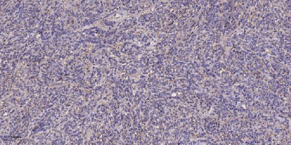
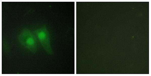
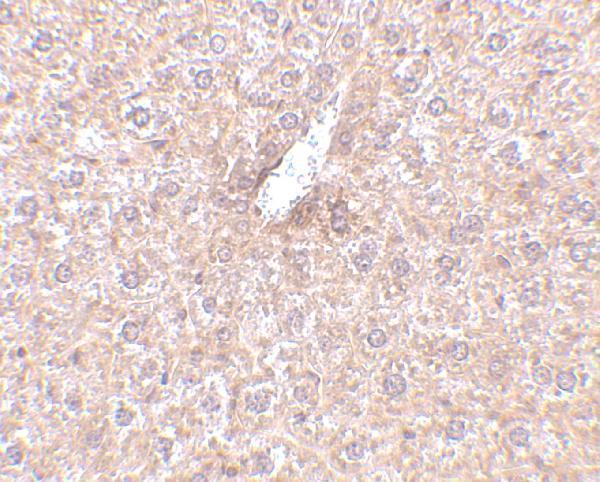
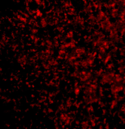
Two STEAP3 antibodies have IHC images from human colon cancer paraffin sections and mouse liver tissue, plus IF images from cells (catalog image captions).
A30509 lists IHC and IF applications, with images from human colon cancer paraffin sections and HeLa cells (A30509 applications and image captions). A05568-1 lists IHC-P and IF applications, with images from mouse liver tissue and mouse liver cells (A05568-1 applications and image captions).
Which to pick: For tissue IHC, choose A30509 for human colon cancer paraffin sections (A30509 IHC image caption: 1:200; fixative unreported) or A05568-1 for mouse liver tissue (A05568-1 IHC image caption: 2.5 μg/mL). For IF/ICC, A30509 lists both applications and has a HeLa cell IF image; A05568-1 lists IF and has a mouse liver cell IF image (catalog applications and IF image captions). For work across species, both list Human, Mouse and Rat reactivity; the supplied IHC images show human tissue for A30509 and mouse tissue for A05568-1 (catalog reactivity and IHC image captions).