This website uses cookies to ensure you get the best experience on our website.


- Table of Contents
Plan chromogenic STIM1 IHC in paraffin sections with cell-specific tissue controls and an IHC-validated antibody (HPA tissue IHC; datasheet A00312-1). Interpret variable cytoplasmic staining in light of STIM1’s ER and cell membrane localization (HPA tissue IHC; UniProt).

Expected localisation, validated protocols, controls and antibodies — the at-a-glance facts below, then the full design guide.
| Expected localisation | Variable cytoplasmic staining (HPA tissue IHC); ER and cell membrane localization (UniProt) | |
| Staining pattern | Variable cytoplasmic staining in most tissues (HPA tissue IHC) | |
| Antigen retrieval | EDTA pH 8.0 HIER, heat-mediated (datasheet A00312-1) | |
| Positive control | Cerebellum+4 more · see all | |
| Negative control | Adipose tissue+3 more · see all |
| Fixation | Keep fixation conditions consistent across sections. (standard IHC practice; not target-specific) | |
| Caveat | Calcium-store depletion can shift STIM1 to ER–membrane junctions (UniProt) | |
| Regulation | Intensity varies by cell type (HPA tissue IHC) | |
| Isoform / epitope | 2 isoforms; map the epitope to the extracellular or cytoplasmic region (UniProt) |
The catalog antibody protocol uses heat-mediated EDTA pH 8.0 retrieval (datasheet A00312-1). The published options below report STIM1 staining in human oral tissue (PMC8739746) and tumor tissue sections (PMC10964627).
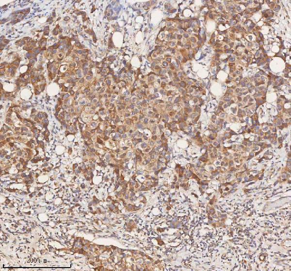
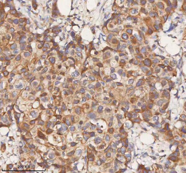
| Sample | Paraffin-embedded human breast cancer tissue; fixative not specified (datasheet A00312-1) |
| Fixation | Image fixative and duration unreported (datasheet A00312-1); verify before use. |
| Sectioning | 4–5 µm sections on charged slides (standard) |
| Deparaffinisation | Xylene, graded ethanol series to water (standard) |
| Antigen retrieval | Heat retrieval: EDTA pH 8.0 (datasheet A00312-1); 20 min, 95–100 °C (standard) |
| Peroxidase block | 3% H2O2, 10 min, room temperature (standard) |
| Blocking | 10% goat serum (datasheet A00312-1) |
| Primary antibody | Rabbit anti-STIM1, 2-5 μg/ml (datasheet A00312-1) |
| Primary incubation | Overnight at 4 °C (datasheet A00312-1) |
| Detection | HRP-conjugated secondary, DAB chromogen (datasheet A00312-1) |
| Counterstain | Hematoxylin, blue, dehydrate and mount (standard) |
| Expected result | STIM1-positive staining in purkinje cells - cytoplasm/membrane of cerebellum (HPA tissue IHC: High). HPA tissue profile: Cytoplasmic expression at variable levels in most tissues. No signal in the no-primary control. |
STIM1 is an endoplasmic reticulum membrane protein that can cluster near the cell membrane after calcium store depletion (UniProt Q13586). In paraffin sections, expect mainly cytoplasmic staining across many tissues, including high staining in esophageal squamous cells, kidney collecting ducts, and lung macrophages (HPA tissue IHC). HPA rates its tissue staining reliability Enhanced, with medium consistency between staining and RNA expression data (HPA tissue IHC).
| Cytoplasmic staining in esophageal squamous cells, kidney collecting ducts, or lung macrophages. | These are high-staining cell populations in HPA tissue IHC; compare intensity within the relevant cells, not across the entire section (HPA tissue IHC; general IHC practice). |
| Cytoplasmic or membrane-associated staining in cerebellar Purkinje cells. | Both compartments are reported for these cells; membrane-associated signal is also compatible with STIM1 topology (HPA: High in Purkinje cells; UniProt Q13586 topology). |
| Strong nuclear-only staining with little cytoplasmic signal. | This conflicts with the reported endoplasmic reticulum, cytoplasmic, and membrane locations; investigate staining artefact or antibody specificity (UniProt Q13586; HPA tissue IHC; general IHC practice). |
| Signal confined to cardiomyocytes or adipocytes, without staining in an included known-positive cell population. | HPA reports STIM1 as not detected in those cells. Check cell identification, nonspecific binding, and endogenous chromogenic activity before assigning the signal to STIM1 (HPA tissue IHC; general IHC practice). |
| Uniform stain over cells and surrounding tissue, obscuring cell boundaries. | Diffuse background cannot establish STIM1 localisation; assess blocking, detection reagents, and staining in a control lacking primary antibody (general IHC practice). |
| Tissue and cell choice | HPA reports variable cytoplasmic expression in most tissues. Use a named high-staining cell population to judge sensitivity; a low-staining population is a weaker benchmark (HPA tissue IHC). |
| Subcellular topology | STIM1 spans a membrane at residues 214–234, with residues 23–213 on the luminal or extracellular side and 235–685 cytoplasmic. Epitope location may matter, but the tested antibody's epitope is unspecified here (UniProt Q13586 topology). |
| Calcium-dependent distribution | Store depletion can move STIM1 into puncta at endoplasmic reticulum–plasma membrane junctions. A routine tissue section does not establish the calcium state or require visible puncta for a positive call (UniProt Q13586; general IHC practice). |
| Antibody evidence | HPA lists HPA011088 and HPA012123 as Enhanced for IHC, and CAB037128 and CAB037288 as Supported. These ratings support interpretation of HPA patterns; they do not validate an unspecified catalog antibody (HPA antibodies). |
| Low or undetected populations | HPA reports low staining in skeletal myocytes and ovarian stromal cells, and no detection in adipocytes or cardiomyocytes. Score the named cell type; other cells in those tissues can have different staining (HPA tissue IHC). |
| IF/ICC Q: What localisation is expected? | A: Predominantly endoplasmic reticulum signal is supported by HPA ICC-IF; UniProt also reports junctional puncta after calcium store depletion. The IF/ICC result does not prescribe an IHC-P workflow (HPA subcellular; UniProt Q13586). |
| Situation | Likely cause | Next action |
|---|---|---|
| No signal in esophageal squamous cells or kidney collecting ducts. | These cells are HPA high-staining; a failed run, inadequate detection, or antibody-specific performance is possible (HPA tissue IHC; general IHC practice). | Check the positive-control section, primary-antibody and detection steps, and the catalog antibody's IHC-P instructions; repeat with matched controls if needed (general IHC practice). |
| Signal appears only in nuclei. | Nuclear-only staining disagrees with reported STIM1 localisation (UniProt Q13586; HPA tissue IHC). | Review morphology and counterstain, then compare with a known-positive section and a control lacking primary antibody (general IHC practice). |
| Broad brown haze obscures cells. | Nonspecific reagent binding or chromogenic background may obscure the reported cytoplasmic pattern (HPA tissue IHC; general IHC practice). | Review blocking and detection steps; compare with a control lacking primary antibody before scoring (general IHC practice). |
| Strong signal is assigned to adipocytes or cardiomyocytes. | HPA reports STIM1 as not detected in those cell populations; cell misidentification, nonspecific staining, or endogenous detection activity may explain it (HPA tissue IHC; general IHC practice). | Confirm cell identity, inspect a control lacking primary antibody, and check endogenous enzyme blocking if using an enzyme-based detector (general IHC practice). |
| A low-staining tissue is scored as negative. | Weak signal can be difficult to distinguish from background; HPA reports low staining in hippocampal neuronal cells, ovarian stromal cells, and skeletal myocytes (HPA tissue IHC). | Judge the run against a named HPA high-staining cell population before interpreting a low-staining population (HPA tissue IHC; general IHC practice). |
| Expected calcium-dependent puncta are absent in a tissue section. | UniProt links junctional puncta to calcium store depletion; that state is not established by routine IHC appearance alone (UniProt Q13586; general IHC practice). | Use the reported cytoplasmic pattern and named positive cell populations for the IHC call; reserve puncta interpretation for an experiment that defines calcium state (HPA tissue IHC; UniProt Q13586). |


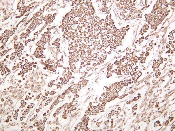


Comprehensive Human Protein Atlas IHC scoring per tissue (reliability: Enhanced — Medium consistency between antibody staining and RNA expression data.). Rows are taken directly from the HPA tissue chart — click any row's HPA link to view the source.
| Tissue | Cell type | Level | Evidence | Source |
|---|---|---|---|---|
| Cerebellum | Purkinje cells - cytoplasm/membrane | High | Protein (IHC) | HPA → |
| Endometrium | Glandular cells | Medium | Protein (IHC) | HPA → |
| Esophagus | Squamous epithelial cells | High | Protein (IHC) | HPA → |
| Kidney | Collecting ducts | High | Protein (IHC) | HPA → |
| Lung | Macrophages | High | Protein (IHC) | HPA → |
| Tissue | Cell type | Level | Evidence | Source |
|---|---|---|---|---|
| Adipose tissue | Adipocytes | Not detected | Protein (IHC) | HPA → |
| Caudate | Glial cells | Not detected | Protein (IHC) | HPA → |
| Heart muscle | Cardiomyocytes | Not detected | Protein (IHC) | HPA → |
| Smooth muscle | Smooth muscle cells | Not detected | Protein (IHC) | HPA → |
Troubleshoot STIM1 staining in paraffin sections by checking retrieval, compartment patterns, cell identity and assay controls.
IHC and IF image captions document anti-STIM1 staining in human, mouse, and rat tissues, plus A549 cells for IF (catalog image captions).
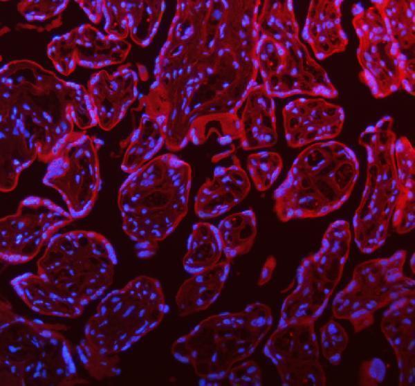
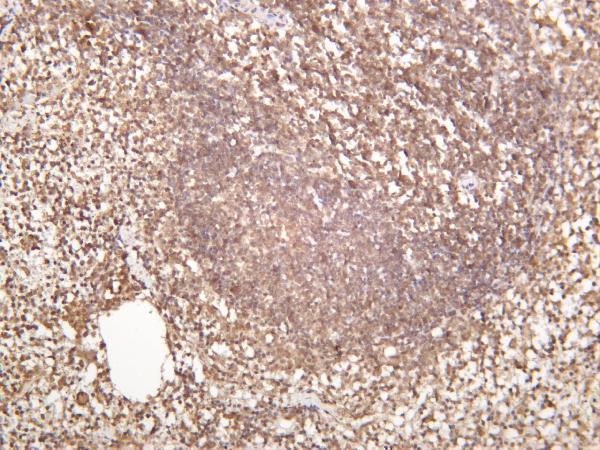
A00312-1 shows IHC in paraffin-embedded human breast cancer and IF in paraffin-embedded human placenta, mouse testis, and rat testis; PB9406 shows IHC in paraffin-embedded mouse intestine and IF in A549 cells (A00312-1 and PB9406 image captions). M00312-2 shows IHC in mouse spleen and lists IF as an application, but has no IF image (M00312-2 image captions; catalog applications).
Which to pick: For tissue IHC, choose A00312-1 for a human paraffin-section example at 2 μg/ml or PB9406 for a mouse intestine paraffin-section example at 1 μg/ml; both captions specify EDTA retrieval at pH 8.0, but neither reports the fixative (A00312-1 and PB9406 IHC image captions). For IF/ICC, PB9406 lists both applications and has an A549 IF image; A00312-1 has tissue IF images, while M00312-2 lists IF without an IF image (catalog applications; IF image captions). For cross-species IHC, PB9406 has paraffin-section images from human, mouse, and rat; recombinant rabbit monoclonal M00312-2 has human and mouse tissue IHC images and lists all three species, but its image captions do not report section processing or fixative (PB9406 IHC image captions; M00312-2 image captions; M00312-2 catalog description and reactivity).