STRAP / Serine-threonine kinase receptor-associated protein · IHC design guide

Design Immunohistochemistry for STRAP

Plan STRAP paraffin-section IHC around mainly cytoplasmic staining, with nuclear staining in several tissues (HPA tissue IHC). This guide identifies high-staining cell populations (HPA tissue IHC) and the catalog antibody’s 0.5–1 μg/ml IHC dilution range (datasheet PB9407).

Evidence assembled Oct 2026 · For research use; verify linked source records and product datasheet before use
Immunohistochemistry protocol sheet for STRAP (IHC for STRAP): expected localisation Predominantly cytoplasmic; nuclear in several tissues (HPA tissue IHC), antibody PB9407, validated IHC image, and IHC protocol steps
Printable STRAP IHC protocol sheet — expected localisation Predominantly cytoplasmic; nuclear in several tissues (HPA tissue IHC), antibody PB9407, controls and protocol steps. Open the full STRAP IHC guide →

STRAP Immunohistochemistry Experimental Design Guide

Expected localisation, validated protocols, controls and antibodies — the at-a-glance facts below, then the full design guide.

Must know before staining
Expected localisation Predominantly cytoplasmic; nuclear in several tissues (HPA tissue IHC)
Staining pattern High in adrenal glandular cells; mainly cytoplasmic across tissues, with some nuclear staining (HPA tissue IHC)
Antigen retrieval EDTA pH 8.0 HIER, heat-mediated (datasheet PB9407)
Positive control ⓘ Adrenal gland+4 more · see all
Negative control ⓘ None in HPA (detected in all 45 tissues); use no-primary + isotype controls
Important caveats
Reasons your staining may differ from the expected pattern.
Fixation Keep fixation consistent across paraffin sections (standard IHC practice; not target-specific)
Caveat Antibody staining and RNA show medium consistency (HPA tissue IHC)
Regulation Expression regulation is not specified (UniProt)
Isoform / epitope 2 isoforms; epitope coverage is unknown (UniProt)
Section 1

Recommended STRAP IHC & IF Protocols

The catalog antibody protocol uses EDTA pH 8.0 retrieval (datasheet: PB9407). The published IHC protocol below describes STRAP staining in mouse tumour sections (PMC5963096).

Recommended immunohistochemistry (IHC-P) protocol parameters
SampleParaffin-embedded human mammary cancer tissue; fixative not specified (datasheet PB9407)
FixationImage fixative and duration unreported (datasheet PB9407); verify before use.
Sectioning4–5 µm sections on charged slides (standard)
DeparaffinisationXylene, graded ethanol series to water (standard)
Antigen retrievalHeat retrieval: EDTA pH 8.0 (datasheet PB9407); 20 min, 95–100 °C (standard)
Peroxidase block3% H2O2, 10 min, room temperature (standard)
Blocking10% goat serum (datasheet PB9407)
Primary antibodyRabbit anti-STRAP, 0.5-1μg/ml (datasheet PB9407)
Primary incubationOvernight at 4 °C (datasheet PB9407)
DetectionStreptavidin-biotin complex (SABC), DAB chromogen (datasheet PB9407)
CounterstainHematoxylin, blue, dehydrate and mount (standard)
Expected resultSTRAP-positive staining in glandular cells of adrenal gland (HPA tissue IHC: High). HPA tissue profile: Mainly cytoplasmic expression but also nuclear expression in several tissues. No signal in the no-primary control.
💡Decision noteStart with heat-mediated EDTA pH 8.0 retrieval for the catalog antibody (datasheet: PB9407); the published mouse-tumour protocol specifies heat-induced retrieval without a buffer (PMC5963096).
Section 2

What Is the Expected STRAP Staining Pattern?

STRAP is predominantly cytoplasmic, with nuclear staining in some tissues (UniProt Q9Y3F4 localization; HPA tissue IHC). Look for staining in glandular, respiratory epithelial, myoepithelial, or endothelial cells in the listed positive tissues (HPA tissue IHC). STRAP has no transmembrane segment (UniProt Q9Y3F4 topology). HPA rates the tissue staining profile Approved, with medium consistency between antibody staining and RNA expression (HPA tissue IHC).

What am I looking at on my slide?
Cytoplasmic staining in adrenal glandular cells, with or without some nuclear staining.This fits the reported distribution. Adrenal glandular staining is High (HPA tissue IHC); cytoplasm predominates, while nuclear signal is possible (UniProt Q9Y3F4 localization; HPA tissue IHC). Judge the signal in the identified cells, rather than treating every stained area as equivalent.
Strong staining confined to cell borders or the cell surface, with little cytoplasmic signal.An exclusively membranous pattern conflicts with the predominantly cytoplasmic tissue pattern and the absence of a transmembrane segment (HPA tissue IHC; UniProt Q9Y3F4 topology). Check morphology and detection background before interpreting it as STRAP. HPA reports cell junction staining in ICC-IF as uncertain, so that observation alone does not validate membranous tissue IHC (HPA subcellular ICC-IF).
Prominent staining in cells outside the reported positive cell populations.First confirm cell identity against the counterstain and tissue morphology (standard IHC practice). The supplied HPA profile documents selected positive cells; it does not prove all other cells are negative (HPA tissue IHC). Unexpected signal can warrant checks for antibody cross-reactivity or endogenous detection activity (standard IHC practice).
Even chromogen haze across cells, stroma, and empty areas.A field-wide haze obscures the cell-resolved pattern expected from the HPA tissue observations (HPA tissue IHC). Review blocking, washes, detection reagents, and section handling as general IHC background checks (standard IHC practice); the supplied sources do not identify a STRAP-specific cause.
No convincing signal in adrenal glandular cells on a test section.That result conflicts with the reported High staining in those cells (HPA tissue IHC). Check that the expected cells are present, then review antibody dilution, retrieval, detection, and control-slide performance (standard IHC practice). One failed section does not establish absent STRAP expression.
💡Expected STRAP appearanceCall positive when the expected cells show clear, predominantly cytoplasmic chromogen, allowing some nuclear staining; adrenal glandular and bronchial respiratory epithelial cells are reported High (HPA tissue IHC). An exclusively surface-bound or uniform field-wide deposit needs investigation (UniProt Q9Y3F4 topology; standard IHC practice).
How each factor affects the staining
Cellular localizationCytoplasm is the main expected compartment; nuclear staining can occur in several tissues (UniProt Q9Y3F4 localization; HPA tissue IHC). Do not score all nuclear signal as artefact.
Choice of reference tissueAdrenal glandular, breast myoepithelial, bronchial respiratory epithelial, and colon endothelial cells are reported High (HPA tissue IHC). Caudate neurons and skeletal myocytes are reported Low, making them less useful as strong-signal references (HPA tissue IHC).
Antibody evidenceHPA027320 and HPA055557 have Approved IHC status (HPA antibodies). The supplied record gives no enhanced IHC validation or catalog-antibody dilution; compare a chosen reagent with its own IHC-P documentation before interpreting discrepancies.
Topology and processingSTRAP lacks a transmembrane segment, signal peptide, and annotated propeptide (UniProt Q9Y3F4 topology and processing). These annotations support an intracellular reading of staining; they do not establish how a particular epitope survives tissue preparation.
Isoforms and epitopeTwo isoforms are listed (UniProt Q9Y3F4 isoforms). The supplied sources do not map antibody epitopes or establish isoform-specific IHC staining, so a compartment difference cannot be assigned to an isoform from this record.
IF/ICC Q&AQ: Should IF/ICC show the same pattern? A: HPA supports mainly cytosolic ICC-IF staining and lists cell junction staining as uncertain (HPA subcellular ICC-IF). Tissue IHC also permits nuclear staining (HPA tissue IHC). Interpret each assay against its own evidence; IF/ICC methods belong in its separate guide.
Why is my staining missing, weak or wrong?
SituationLikely causeNext action
The expected positive cells are unstained.The result disagrees with the reported High signal in adrenal glandular cells (HPA tissue IHC); a failed antibody or detection step is possible (standard IHC practice).Verify tissue identity and intact cells, inspect the positive control, then review the antibody's IHC-P instructions, dilution, retrieval, and detection steps (standard IHC practice). The supplied sources give no STRAP-specific retrieval condition.
Signal appears only at cell surfaces.That distribution differs from the mainly cytoplasmic tissue profile and no-transmembrane topology (HPA tissue IHC; UniProt Q9Y3F4 topology).Examine the counterstained cells at higher magnification and run appropriate detection controls (standard IHC practice). Avoid calling an exclusively surface pattern positive solely because ICC-IF has an uncertain cell-junction annotation (HPA subcellular ICC-IF).
Nuclei stain along with cytoplasm.Nuclear STRAP is documented, although cytoplasm predominates (UniProt Q9Y3F4 localization; HPA tissue IHC).Assess whether nuclear signal tracks recognizable cells and accompanies a plausible cytoplasmic pattern (standard IHC practice; HPA tissue IHC). Investigate isolated, widespread nuclear staining against controls before scoring it as specific.
All tissue structures have similar brown haze.Poor contrast can arise from nonspecific background or detection activity (standard IHC practice); it is not the cell-resolved pattern described by HPA (HPA tissue IHC).Compare an appropriate negative reagent or detection control, then review blocking, washes, and chromogen development (standard IHC practice). Interpret cell-specific staining only after the background is controlled.
An unexpected cell population stains strongly.The reported positive-cell list is selective, while HPA rates antibody-versus-RNA consistency as medium (HPA tissue IHC). Cross-reactivity or endogenous detection activity also merit consideration (standard IHC practice).Confirm the cell type morphologically and compare controls and an independently validated antibody where available (standard IHC practice). Record the observation as unresolved if specificity cannot be established.
A low-signal reference tissue looks negative.Caudate neuronal cells, parathyroid and prostate glandular cells, and skeletal myocytes are listed Low (HPA tissue IHC); weak staining may be hard to distinguish from background (standard IHC practice).Check a reported High cell population on a suitable reference section before judging assay failure (HPA tissue IHC). Keep exposure or chromogen development within the validated workflow and compare controls (standard IHC practice).

Sample controls for STRAP IHC & IF

🧪Run adrenal gland first and score its glandular cells for STRAP staining (HPA: High in adrenal gland glandular cells). HPA detects STRAP in all 45 scored tissues, so there is no documented negative tissue; use no-primary and isotype controls, and expect any cells without specific staining on the positive slide to remain at background without assuming they are biologically STRAP-negative (HPA: no negative rows).
Positive control tissue: Adrenal gland (Glandular cells, HPA High)
Negative control tissue: None in HPA: STRAP is detected in all 45 scored tissues. Use a no-primary (secondary-only) and an isotype control instead.
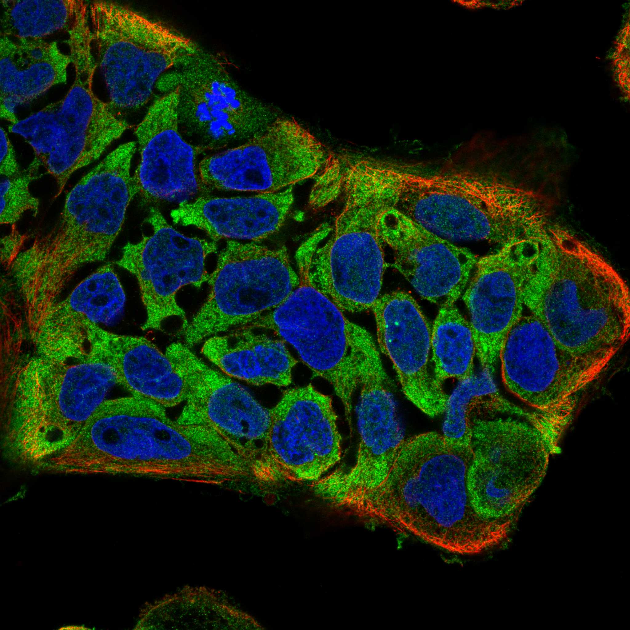
ICC-IF cell lines (HPA subcellular resource): HPA ICC-IF images show STRAP in U2OS, CACO-2, Rh30, KOLF2.1J, NIH 3T3, with annotated localisation: Cytosol (supported) (HPA subcellular).
Technical controls: Include a secondary-only slide, a rabbit IgG control matched to the primary antibody’s class and clonality once confirmed, and a STRAP knockout or validated peptide-block control (caption: rabbit primary antibody; standard IHC practice). For chromogenic detection, check endogenous peroxidase and endogenous biotin background because the supplied protocol uses biotin–streptavidin detection with DAB (caption: biotinylated secondary, SABC and DAB).
⚠️Feasibility: A target-specific fixation window or fixation effect is unreported, and the selected PB9407 paraffin-section caption does not state the fixative (caption: fixative unreported). That caption uses heat retrieval in EDTA at pH 8.0, but does not establish that retrieval is required or that frozen sections or IF are easier (caption: EDTA heat retrieval). No adrenal-specific artefact is documented in the supplied evidence; assess peroxidase and biotin background on the chosen tissue (standard IHC practice).

HPA tissue IHC evidence for STRAP

Comprehensive Human Protein Atlas IHC scoring per tissue (reliability: Approved — Medium consistency between antibody staining and RNA expression data.). Rows are taken directly from the HPA tissue chart — click any row's HPA link to view the source.

Positive expression · recommended positive controls

TissueCell typeLevelEvidenceSource
Adrenal gland Glandular cells High Protein (IHC) HPA →
Breast Myoepithelial cells High Protein (IHC) HPA →
Bronchus Respiratory epithelial cells High Protein (IHC) HPA →
Colon Endothelial cells High Protein (IHC) HPA →
Duodenum Glandular cells High Protein (IHC) HPA →

Undetected expression · recommended negative controls

TissueCell typeLevelEvidenceSource
None in HPA: STRAP is detected in all 45 scored tissues. Use a no-primary (secondary-only) and an isotype control instead.
Section 3

Advanced STRAP IHC Tips

Troubleshoot STRAP staining in paraffin sections by checking retrieval, cell compartment, tissue controls and scoring before interpreting biological differences.

What retrieval should I start with for STRAP in paraffin sections?
Start with heat-mediated retrieval in EDTA buffer at pH 8.0 (datasheet PB9407). This condition was used for STRAP staining in a paraffin-embedded human mammary cancer section, followed by 1 μg/ml primary antibody overnight at 4°C (datasheet PB9407). If staining is weak, compare a carefully controlled alternative retrieval condition on adjacent sections while keeping antibody concentration and detection constant (standard IHC practice). Score cytoplasmic signal separately from nuclear signal, because both are plausible locations for STRAP (UniProt Q9Y3F4 localisation; HPA tissue IHC profile). Excessively harsh retrieval can damage morphology and make apparent staining gains hard to trust (standard IHC practice).
How should I troubleshoot fixation-related loss of STRAP staining?
Target-specific fixation sensitivity is unknown: the selected paraffin-section caption does not state its fixative (datasheet PB9407 caption). Record the fixative, fixation duration and tissue thickness for each specimen, then compare sections processed together with the same EDTA pH 8.0 retrieval and detection steps (standard IHC practice; datasheet PB9407 retrieval). A weak result alone cannot establish that fixation masked STRAP, because primary antibody exposure and detection also affect signal (standard IHC practice). Use a consistently staining control section to judge whether a processing change helped, and retain morphology as a check against excessive retrieval (standard IHC practice).
Should STRAP staining be cytoplasmic, nuclear, or both?
Expect predominantly cytoplasmic staining, with nuclear staining possible in some tissues (UniProt Q9Y3F4 localisation; HPA tissue IHC profile). STRAP has no annotated transmembrane segment, so a sharp membrane-only pattern needs independent verification (UniProt Q9Y3F4 topology). Compare adjacent cells and compartments on the same section before changing the 1 μg/ml primary concentration used in the selected example (datasheet PB9407; standard IHC practice). Nuclear signal should be scored separately from cytoplasmic signal, with attention to counterstain overlap and local tissue damage (standard IHC practice). The HPA cell-junction assignment is uncertain and comes from subcellular imaging, so it does not establish a required IHC membrane pattern (HPA subcellular).
Could isoforms or modifications explain discordant STRAP staining?
STRAP has 2 annotated isoforms, while the supplied product caption gives no epitope sequence or isoform coverage (UniProt Q9Y3F4 isoforms; datasheet PB9407 caption). Therefore, staining with this antibody cannot establish which isoform is present without separate epitope and specificity evidence (standard IHC interpretation). Phosphoserines at residues 312, 335 and 338 are annotated, but their effect on this antibody’s staining is unknown (UniProt Q9Y3F4 modified residues; datasheet PB9407 caption). If sections disagree, first standardize EDTA pH 8.0 retrieval and overnight 4°C primary incubation, then assess antibody specificity with suitable controls (datasheet PB9407; standard IHC practice).
How can I use IF to clarify ambiguous STRAP IHC staining?
Use IF as a separate validation experiment and multiplex STRAP with a marker for the cell population being assessed, such as the relevant epithelial or endothelial population (HPA tissue IHC cell profiles; standard IF practice). Choose a fluorophore and filter set that separate its signal from tissue autofluorescence, and include a no-primary control (standard IF practice). Because STRAP is predominantly cytoplasmic and has no transmembrane segment, use permeabilisation appropriate for access to an intracellular epitope; the exact antibody epitope is unreported (UniProt Q9Y3F4 localisation and topology; datasheet PB9407 caption). Compare cytosolic and nuclear channels separately, since nuclear localisation is also reported (UniProt Q9Y3F4 localisation; HPA tissue IHC profile).
How do I separate STRAP signal from chromogenic background?
The selected paraffin-section example used 10% goat serum block, biotinylated secondary antibody and DAB detection (datasheet PB9407). Include no-primary and detection-only controls, and apply a peroxidase block appropriate to the tissue before DAB development (standard chromogenic IHC practice). Diffuse precipitate, staining at section edges or signal reproduced without primary antibody should prompt checks of washing, reagent carryover and detection chemistry (standard IHC practice). Compare suspected background with the expected mainly cytoplasmic STRAP pattern rather than calling every brown deposit positive (HPA tissue IHC profile; standard IHC interpretation). Keep development conditions matched across control and test sections (standard IHC practice).
How should I quantify STRAP IHC across specimens? ⚠ ANSWER MARKED FOR VERIFICATION
Define the cell population and compartment before scoring, then report cytoplasmic and nuclear staining separately (UniProt Q9Y3F4 localisation; HPA tissue IHC profile; standard IHC practice). For a cell-based readout, record the percentage of positive cells and intensity categories, or calculate an H-score from 0–300 using the same thresholds for every section (standard IHC practice). Normalize positive counts to the number of eligible cells, or positive area to the measured tissue area in mm², while excluding folds and damaged regions (standard IHC practice). Compare sections stained in the same run with matched retrieval, antibody exposure and DAB development (standard IHC practice).
When is an unusual STRAP pattern credible rather than artefact?
Treat mainly cytoplasmic staining, with possible nuclear signal, as the starting pattern for STRAP (UniProt Q9Y3F4 localisation; HPA tissue IHC profile). HPA reports high staining in breast myoepithelial cells and colon endothelial cells, so identify the stained cell population rather than assigning signal to an entire tissue (HPA tissue IHC). A membrane-only pattern conflicts with the absence of an annotated transmembrane segment and warrants specificity checks (UniProt Q9Y3F4 topology; standard IHC practice). Exclude section-edge deposits, necrotic regions and staining retained in no-primary controls; endogenous enzyme activity can also mimic chromogenic signal (standard IHC practice). HPA tissue IHC reliability is Approved with medium staining-to-RNA consistency, so interpret isolated differences cautiously (HPA tissue IHC reliability).
Boster reagents

Best STRAP / Serine-threonine kinase receptor-associated protein IHC Antibodies

PB9407 has real IHC data from a human mammary cancer paraffin section (IHC figure caption); its listed reactivity is Human, Mouse, and Rat (catalog: reactivity). No IF data are supplied (catalog: IF images).

Real IHC data IHC analysis of Unrip using anti-Unrip antibody (PB9407). Unrip was detected in a paraffin-embedded section of human mammary cancer tissue. Heat mediated antigen retrieval was performed in EDTA buffer (pH 8.0, epitope retrieval solution). The tissue section was blocked with 10% goat serum. The tissue section was then incubated with 1 μg/ml rabbit anti-Unrip Antibody (PB9407) overnight at 4°C. Biotinylated goat anti-rabbit IgG was used as secondary antibody and incubated for 30 minutes at 37°C. The tissue section was developed using Strepavidin-Biotin-Complex (SABC) (Catalog # SA1022) with DAB as the chromogen.
Anti-Unrip/STRAP Antibody ®
Cat # PB9407

PB9407 is the only card and shows IHC staining of a human mammary cancer paraffin section (PB9407 IHC figure caption). Its listed applications are IHC and WB, and its listed reactivity is Human, Mouse, and Rat (catalog: PB9407 applications and reactivity).

Which to pick: Choose PB9407 for paraffin section IHC: its own figure documents heat retrieval in EDTA at pH 8.0 and staining at 1 μg/ml (PB9407 IHC figure caption); the fixative is unreported (PB9407 IHC figure caption). No SKU in this payload is listed for IF/ICC, so there is no IF/ICC pick (catalog: applications). PB9407 is the only cross-species candidate because Human, Mouse, and Rat are listed, but the supplied IHC figure demonstrates Human tissue only (catalog: reactivity; PB9407 IHC figure caption).

Each figure is that product's own IHC / IF validation image from its datasheet.

References

  1. UniProt Consortium. UniProt entry Q9Y3F4 (STRAP_HUMAN, Serine-threonine kinase receptor-associated protein).
  2. Human Protein Atlas. STRAP tissue IHC expression (reliability: Approved).
  3. Human Protein Atlas. STRAP subcellular location (ICC-IF): Mainly localized to the cytosol. In addition localized to the cell junctions..
  4. Human Protein Atlas. STRAP antibody validation summary (3 antibodies).
  5. Multimodal fusion of radio-pathology and proteogenomics identify integrated glioma subtypes with prognostic and therapeutic opportunities. Nature communications 2025 — PMC11994800.
  6. Sanguinarine triggers intrinsic apoptosis to suppress colorectal cancer growth through disassociation between STRAP and MELK. BMC cancer 2018 — PMC5963096.
  7. STRAP promotes hepatocellular carcinoma progression through activation of an STRAP/Wnt-β-catenin/STRAP positive feedback loop. iScience 2026 — PMC13285651.
  8. Serine-Threonine Kinase Receptor-Associated Protein (STRAP) Knockout Decreases the Malignant Phenotype in Neuroblastoma Cell Lines. Cancers 2021 — PMC8268080.
  9. PubMed PMID:10049359 — UniProt-cited evidence.
  10. PubMed PMID:10646843 — UniProt-cited evidence.
  11. PubMed PMID:11230166 — UniProt-cited evidence.