This website uses cookies to ensure you get the best experience on our website.


- Table of Contents
Plan chromogenic IHC on paraffin sections around the cytoplasmic staining reported in most tissues (HPA tissue IHC). Use the reported high signal in gallbladder glandular cells and undetected signal in soft-tissue fibroblasts to inform control selection (HPA tissue IHC).

Expected localisation, validated protocols, controls and antibodies — the at-a-glance facts below, then the full design guide.
| Expected localisation | Cytoplasmic staining in most tissues (HPA tissue IHC) | |
| Staining pattern | Cytoplasmic staining across most tissues (HPA tissue IHC) | |
| Antigen retrieval | EDTA pH 8.0 HIER, heat-mediated (datasheet PB9826) | |
| Positive control | Gallbladder+4 more · see all | |
| Negative control | Soft tissue |
| Fixation | Keep fixation consistent across sections. (standard IHC practice; not target-specific) | |
| Caveat | Staining and RNA show medium consistency (HPA tissue IHC) | |
| Regulation | Low tissue specificity (HPA tissue RNA) | |
| Isoform / epitope | No isoforms annotated; no signal peptide or TM segment (UniProt) |
The catalog antibody’s IHC-P protocol (datasheet PB9826) is accompanied by published protocols for ovarian tissue (PMC8223286) and interspinous ligament samples (PMC8806538).
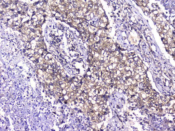
| Sample | Paraffin-embedded human ovary cancer tissue; fixative not specified (datasheet PB9826) |
| Fixation | Image fixative and duration unreported (datasheet PB9826); verify before use. |
| Sectioning | 4–5 µm sections on charged slides (standard) |
| Deparaffinisation | Xylene, graded ethanol series to water (standard) |
| Antigen retrieval | Heat retrieval: EDTA pH 8.0 (datasheet PB9826); 20 min, 95–100 °C (standard) |
| Peroxidase block | 3% H2O2, 10 min, room temperature (standard) |
| Blocking | 10% goat serum (datasheet PB9826) |
| Primary antibody | Rabbit anti-TCP1, 2-5μg/ml (datasheet PB9826) |
| Primary incubation | Overnight at 4 °C (datasheet PB9826) |
| Detection | HRP-conjugated secondary, DAB chromogen (datasheet PB9826) |
| Counterstain | Hematoxylin, blue, dehydrate and mount (standard) |
| Expected result | TCP1-positive staining in glandular cells of gallbladder (HPA tissue IHC: High). HPA tissue profile: Cytoplasmic expression in most tissues. No signal in the no-primary control. |
TCP1 is a cytosolic protein with a reported centrosome association and no transmembrane segment (UniProt P17987 localization and topology). In paraffin-section IHC, expect predominantly cytoplasmic staining across many tissues (HPA: cytoplasmic expression in most tissues). Gallbladder glandular cells, ovarian follicle cells and cells in testicular seminiferous ducts are reported as high-staining examples (HPA: High). HPA rates its tissue IHC evidence Approved, with medium consistency between staining and RNA data (HPA: reliability).
| Cytoplasmic staining is clear in gallbladder glandular cells, ovarian follicle cells or cells in seminiferous ducts. | This matches the reported compartment and high-staining cell populations (HPA: cytoplasmic expression; High in these cells). Compare staining within identifiable cells and against a negative control on the same run (general IHC practice). A focal centrosomal accent may be compatible with UniProt localization, but the supplied HPA IHC record does not establish a required centrosomal pattern (UniProt P17987 localization; HPA: tissue IHC profile). |
| Strong nuclear-only or membrane-rim staining dominates while cytoplasm is clear. | Treat this as discordant with the expected cytoplasmic pattern and lack of a transmembrane segment (HPA: cytoplasmic expression; UniProt P17987 topology). Review morphology and the detection controls before assigning biological meaning (general IHC practice). The supplied sources do not validate nuclear-only or membrane-rim staining as a TCP1 IHC result. |
| Soft-tissue fibroblasts stain strongly, especially if expected glandular or follicle cells are weak. | HPA reports TCP1 as not detected in soft-tissue fibroblasts, while selected glandular and follicle cells stain at medium or high levels (HPA: tissue IHC). Unexpected fibroblast signal raises possible antibody cross-reactivity or endogenous detection activity; the image alone cannot distinguish them (general IHC practice). Compare with a no-primary control and an independently validated antibody (general IHC practice). |
| Chromogen spreads across extracellular spaces, tissue edges or many unrelated cells without clear cytoplasmic borders. | This distribution is difficult to score as cellular TCP1 because the reported tissue pattern is cytoplasmic (HPA: tissue IHC profile). Diffuse background can arise from nonspecific reagent binding or detection activity (general IHC practice). Check the no-primary control and staining distribution before calling weak positive cells (general IHC practice). |
| No cytoplasmic staining appears in a chosen high-staining reference tissue. | Absence in gallbladder glandular cells, ovarian follicle cells or cells in seminiferous ducts conflicts with their reported high levels (HPA: High in these populations). Check that the relevant cells are present, then review the run controls and assay settings (general IHC practice). This result alone does not show that TCP1 is absent from the specimen. |
| Compartment and topology (UniProt P17987 localization and topology) | TCP1 is reported in cytosol and at the centrosome, with no transmembrane segment (UniProt P17987). Score the predominant cellular distribution against HPA's cytoplasmic IHC pattern; do not require visible centrosomal puncta in paraffin sections (HPA: tissue IHC profile; general IHC practice). |
| Cell-specific tissue distribution (HPA: tissue IHC) | High staining in selected gallbladder, ovary and testis cells contrasts with low staining in several listed cell populations and non-detection in soft-tissue fibroblasts (HPA: tissue IHC). Select a control by its documented cell population, then interpret weak signal in that cellular context. |
| Strength of tissue evidence (HPA: reliability and antibody records) | The tissue profile is Approved but has medium staining-to-RNA consistency; three listed antibodies have Approved IHC status (HPA: reliability; HPA: HPA027337, HPA031082, CAB017460). Those ratings support comparison with the reported pattern, without proving that every signal in a new specimen is specific. |
| IF/ICC evidence boundary (HPA: subcellular and antibody records) | HPA supplies no ICC/IF images or main IF location, and lists no ICC validation status for the three antibodies (HPA: subcellular record; HPA: antibody records). UniProt cytosolic and centrosomal localization can guide interpretation, but the supplied HPA record cannot establish an IF staining pattern (UniProt P17987 localization). |
| Situation | Likely cause | Next action |
|---|---|---|
| A high-staining reference tissue gives no signal. | The expected positive cell population may be absent from the section, or the IHC run may have failed (HPA: High in selected cells; general IHC practice). | Confirm the named cell population on the section; review positive and no-primary controls, antigen retrieval, primary incubation and detection steps for the run (general IHC practice). Do not infer TCP1 absence from a failed control. |
| Signal is faint in glia, thyroid glands or cardiomyocytes. | Low staining is reported for these cell populations (HPA: Low in caudate glial cells, thyroid glandular cells and heart cardiomyocytes). | Compare with an HPA high-staining cell population processed in the same run, and score the weak signal only if it is cellular and above the negative control (HPA: High in selected populations; general IHC practice). |
| Staining is nuclear-only or outlines cell membranes. | That pattern conflicts with reported cytoplasmic IHC expression and the absence of a transmembrane segment (HPA: tissue IHC profile; UniProt P17987 topology). | Check the no-primary control and cell morphology; repeat with an independently IHC-approved antibody if the discordant pattern persists (general IHC practice; HPA: listed antibodies have Approved IHC status). |
| Fibroblasts stain strongly. | Soft-tissue fibroblasts are reported as not detected; cross-reactivity or endogenous detection activity is possible, but the cause is unresolved (HPA: soft-tissue fibroblasts; general IHC practice). | Compare a no-primary control and the expected positive cells on matched sections; if needed, compare an independently IHC-approved antibody (general IHC practice; HPA: antibody records). |
| Diffuse chromogen obscures cellular boundaries. | Nonspecific binding or endogenous detection activity may contribute background (general IHC practice); HPA reports a cytoplasmic cellular pattern (HPA: tissue IHC profile). | Inspect the no-primary control, verify blocking and detection steps, and adjust assay conditions using controls before scoring weak cytoplasmic staining (general IHC practice). |
| Can the IHC result be used as an IF/ICC pattern or protocol? | The supplied HPA record has no ICC/IF images, main IF location or ICC validation status for the listed antibodies (HPA: subcellular and antibody records). | Interpret IF/ICC separately using appropriate modality controls; use cytosolic and centrosomal localization only as a provisional expectation, not an HPA-validated IF pattern (UniProt P17987 localization; HPA: subcellular record; general IF practice). |




Comprehensive Human Protein Atlas IHC scoring per tissue (reliability: Approved — Medium consistency between antibody staining and RNA expression data.). Rows are taken directly from the HPA tissue chart — click any row's HPA link to view the source.
| Tissue | Cell type | Level | Evidence | Source |
|---|---|---|---|---|
| Gallbladder | Glandular cells | High | Protein (IHC) | HPA → |
| Ovary | Follicle cells | High | Protein (IHC) | HPA → |
| Testis | Cells in seminiferous ducts | High | Protein (IHC) | HPA → |
| Adrenal gland | Glandular cells | Medium | Protein (IHC) | HPA → |
| Appendix | Glandular cells | Medium | Protein (IHC) | HPA → |
| Tissue | Cell type | Level | Evidence | Source |
|---|---|---|---|---|
| Soft tissue | Fibroblasts | Not detected | Protein (IHC) | HPA → |
Troubleshoot TCP1 staining in paraffin sections by checking retrieval, compartment, tissue controls and detection conditions before interpreting signal.
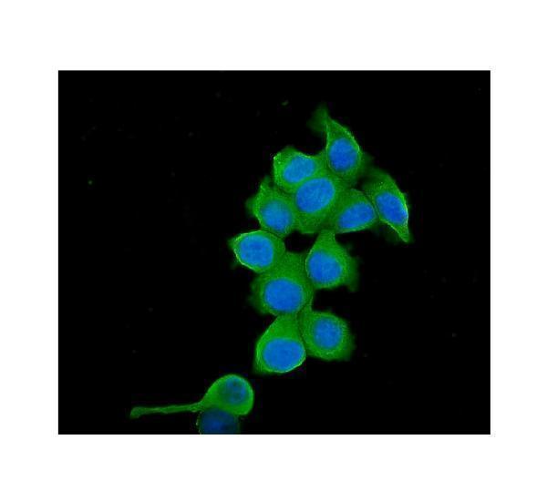
IHC images show TCP1 staining in paraffin sections of human ovary cancer, lung cancer, placenta and testis, plus mouse and rat testis (PB9826 and M02389 IHC captions). IF is documented in MCF7 cells (M02389 IF caption).
PB9826 will render with its human ovary cancer IHC figure; its other captions show human, mouse and rat testis paraffin sections (PB9826 IHC captions). M02389 will render with its human lung cancer IHC figure; its other captions show human placenta IHC and MCF7 cell IF (M02389 IHC and IF captions).
Which to pick: For paraffin section IHC across human, mouse and rat samples, choose rabbit PB9826 (PB9826 applications, reactivity and IHC captions); for human tissue IHC, mouse monoclonal M02389 also has paraffin section images (M02389 clone, reactivity and IHC captions). For IF/ICC, prefer M02389 because its own IF caption documents MCF7 cells; PB9826 lists IF/ICC, but its supplied IF caption names PB9077 (M02389 IF caption; PB9826 applications and IF caption). Both IHC captions omit the fixative, so fixation conditions are unreported (PB9826 and M02389 IHC captions).