TFF1 / Trefoil factor 1 · IHC design guide

Design Immunohistochemistry for TFF1

Plan paraffin-section TFF1 IHC with catalog antibody PA1689 at 0.5–1 μg/mL (datasheet). Assess selective cytoplasmic staining in superficial gastric mucosa and goblet cells while accounting for TFF1 secretion (HPA tissue IHC; UniProt).

Evidence assembled Oct 2026 · For research use; verify linked source records and product datasheet before use
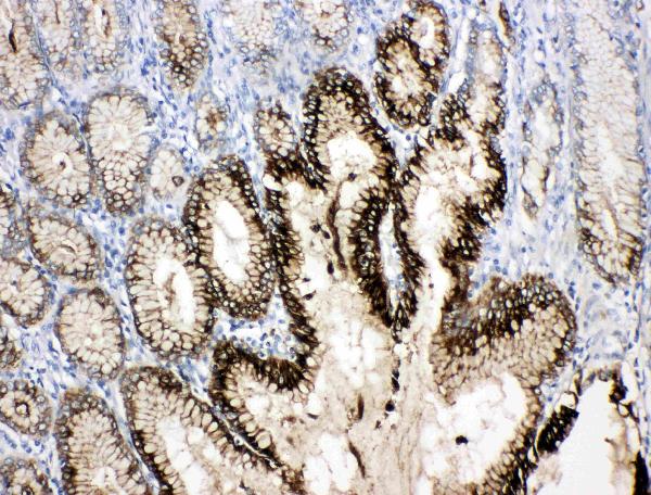
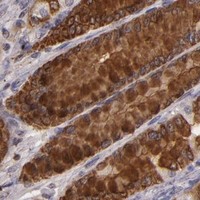
Immunohistochemistry protocol sheet for TFF1 (IHC for TFF1): expected localisation Cytoplasmic in superficial gastric mucosa and goblet cells (HPA tissue IHC), antibody PA1689, validated IHC image, and IHC protocol steps
Printable TFF1 IHC protocol sheet — expected localisation Cytoplasmic in superficial gastric mucosa and goblet cells (HPA tissue IHC), antibody PA1689, controls and protocol steps. Open the full TFF1 IHC guide →

TFF1 Immunohistochemistry Experimental Design Guide

Expected localisation, validated protocols, controls and antibodies — the at-a-glance facts below, then the full design guide.

Must know before staining
Expected localisation Cytoplasmic in superficial gastric mucosa and goblet cells (HPA tissue IHC)
Staining pattern Selective cytoplasmic staining in gastric surface cells and goblet cells (HPA tissue IHC)
Antigen retrieval Citrate pH 6.0 HIER, 95–98 °C, 20 min (rule: cytoplasmic / membrane antigen)
Positive control ⓘ Appendix+4 more · see all
Negative control ⓘ Adipose tissue+4 more · see all
Important caveats
Reasons your staining may differ from the expected pattern.
Fixation Matched tissue-IHC evidence does not establish the fixation claim. Validate the specimen-specific method before use. (selected-SKU IHC image PA1689)
Caveat Secreted TFF1 may stain away from producing cells (UniProt; HPA tissue IHC)
Regulation Estrogen-regulated in MCF-7 cells (UniProt)
Isoform / epitope No listed isoforms; secreted mature chain spans residues 25–84 (UniProt)
Section 1

Recommended TFF1 IHC & IF Protocols

The catalog antibody’s IHC-P protocol is accompanied by two published TFF1 IHC methods: one for breast cancer tissue (PMC12891483) and one for paraffin sections (PMC3141110).

Recommended immunohistochemistry (IHC-P) protocol parameters
SampleTissue sections; selected-image fixative not specified (standard IHC workflow)
FixationImage fixative and duration unreported (datasheet PA1689); verify before use.
Sectioning4–5 µm sections on charged slides (standard)
DeparaffinisationXylene, graded ethanol series to water (standard)
Antigen retrievalHeat-induced epitope retrieval in citrate buffer, pH 6.0, 20 min at 95–98 °C (standard rule: cytoplasmic / membrane antigen)
Peroxidase block3% H2O2, 10 min, room temperature (standard)
Blocking10% normal serum of the secondary host, 30 min, room temperature (standard)
Primary antibodyRabbit anti-TFF1, 0.5-1μg/ml (datasheet PA1689)
Primary incubationOvernight at 4 °C (standard)
DetectionHRP-polymer secondary, DAB chromogen 5–10 min (standard)
CounterstainHematoxylin, blue, dehydrate and mount (standard)
Expected resultTFF1-positive staining in endocrine cells of appendix (HPA tissue IHC: High). HPA tissue profile: Selective cytoplasmic expression in the superficial layer of gastric mucosa and goblet cells. No signal in the no-primary control.
💡Decision noteStart with citrate pH 6.0 retrieval at 95–98 °C for 20 min (page retrieval); the breast cancer study used microwave retrieval at pH 6 for 20 min (PMC12891483).
Section 2

What Is the Expected TFF1 Staining Pattern?

TFF1 is secreted and has no transmembrane segment (UniProt P04155). In IHC, expect selective cytoplasmic staining in superficial gastric mucosa and goblet cells (HPA tissue IHC). Stomach glandular cells stain at high levels, and HPA rates the tissue pattern Enhanced (HPA tissue IHC).

What am I looking at on my slide?
Strong cytoplasmic stain in superficial gastric glandular cells (HPA tissue IHC)Expected positive pattern (HPA tissue IHC).
Predominantly nuclear stainUnexpected for a secreted protein; assess artefact (UniProt P04155; general IHC practice).
Stain in adipocytesUnexpected: HPA reports no detection; check cross-reactivity or endogenous activity (HPA tissue IHC).
Diffuse stain across the sectionPossible background; review blocking and detection controls (general IHC practice).
No stain in stomach glandular cellsUnexpected for a high-level positive; check the IHC run and antibody (HPA tissue IHC).
💡Expected TFF1 appearanceHigh cytoplasmic stain in superficial gastric glandular cells is expected; nuclear stain is suspect (HPA tissue IHC).
How each factor affects the staining
Secretion and processing (UniProt P04155)TFF1 has a signal peptide at residues 1–24 and a mature chain at 25–84 (UniProt P04155).
Antibody validation (HPA antibodies)HPA003425 and CAB002170 each have Enhanced IHC validation (HPA antibodies).
ICC-IF expected pattern?TFF1 is secreted; HPA provides no ICC-IF images or main location (HPA subcellular).
Why is my staining missing, weak or wrong?
SituationLikely causeNext action
No stomach signalStomach glandular cells are high-level positives (HPA tissue IHC).Check positive control, retrieval and detection steps (general IHC practice).
Weak gastric signalThe expected gastric glandular signal is high (HPA tissue IHC).Review antibody dilution and detection conditions (general IHC practice).
Nuclear-dominant stainTFF1 is secreted, while HPA reports cytoplasmic tissue stain (UniProt P04155; HPA tissue IHC).Review controls and scoring before calling it positive (general IHC practice).
Adipocytes stainHPA reports no adipocyte detection; cross-reactivity is possible (HPA tissue IHC; general IHC practice).Check antibody specificity controls (general IHC practice).
Diffuse brown backgroundEndogenous activity or nonspecific binding is possible (general IHC practice).Check blocking and no-primary controls (general IHC practice).

Sample controls for TFF1 IHC & IF

🧪Run colon first and expect staining in its endocrine cells (HPA: High in colon endocrine cells). Use adipose tissue as the negative, with adipocytes at background (HPA: Not detected in adipose adipocytes); compare other cells on the colon slide with background, but do not assume they are TFF1-negative from the supplied HPA row.
Positive control tissue: Appendix (Endocrine cells, HPA High)
Negative control tissue: Adipose tissue (HPA Not detected)
ICC-IF cell lines (HPA subcellular resource): HPA carries no ICC-IF cell line for TFF1; derive a cell-line control from the positive tissue's cell type (Endocrine cells) and confirm it by RNA or western blot first.
Technical controls: Include a no-primary (secondary-only) control, an isotype control matched to the primary antibody’s host species and immunoglobulin class, and a TFF1 knockout or validated peptide-block control. For chromogenic colon IHC, quench endogenous peroxidase and assess background from any biotin-based detection system; for IF, check tissue autofluorescence.
⚠️Feasibility: A target-specific fixation window and retrieval effect are unreported in the supplied evidence; the selected PA1689 tissue-IHC caption also leaves the fixative unreported (PA1689 caption: fixative not stated). Optimize antigen retrieval empirically, and do not assume frozen sections or IF will be easier: TFF1 is secreted (UniProt P04155: Secreted), while the supplied HPA record provides no ICC-IF cell-line images (HPA subcellular record: empty cell-line list). In colon, assess luminal or mucus-associated signal separately from endocrine-cell staining to avoid misassigning extracellular signal (UniProt P04155: secreted; HPA: High in colon endocrine cells).

HPA tissue IHC evidence for TFF1

Comprehensive Human Protein Atlas IHC scoring per tissue (reliability: Enhanced — High consistency between antibody staining and RNA expression data. Secreted protein, tissue location of RNA and protein is expected to differ.). Rows are taken directly from the HPA tissue chart — click any row's HPA link to view the source.

Positive expression · recommended positive controls

TissueCell typeLevelEvidenceSource
Appendix Endocrine cells High Protein (IHC) HPA →
Colon Endocrine cells High Protein (IHC) HPA →
Duodenum Endocrine cells High Protein (IHC) HPA →
Rectum Endocrine cells High Protein (IHC) HPA →
Small intestine Endocrine cells High Protein (IHC) HPA →

Undetected expression · recommended negative controls

TissueCell typeLevelEvidenceSource
Adipose tissue Adipocytes Not detected Protein (IHC) HPA →
Adrenal gland Glandular cells Not detected Protein (IHC) HPA →
Bone marrow Hematopoietic cells Not detected Protein (IHC) HPA →
Breast Adipocytes Not detected Protein (IHC) HPA →
Bronchus Respiratory epithelial cells Not detected Protein (IHC) HPA →
Section 3

Advanced TFF1 IHC Tips

Use the expected gastric epithelial pattern, a documented retrieval condition, and matched controls to troubleshoot TFF1 staining in chromogenic paraffin-section IHC.

How should I troubleshoot weak TFF1 staining after antigen retrieval?
Start with citrate pH 6.0 heat-induced antigen retrieval at 95–98 °C for 20 min (page retrieval rule). Compare sections processed in the same run, including a stomach control with superficial mucosal glands expected to stain (HPA: stomach glandular cells High; HPA: superficial gastric mucosal expression). If the control is weak, check that the retrieval bath reached the specified temperature and that sections stayed immersed throughout the 20 min hold (standard IHC practice). Change one retrieval variable per trial and keep primary-antibody incubation and chromogen development constant so the effect can be interpreted (standard IHC practice). Judge improvement by clearer epithelial signal without increased staining across unrelated tissue compartments (standard IHC practice; UniProt P04155: secreted).
Could fixation explain variable TFF1 staining between paraffin blocks?
Target-specific fixation sensitivity is unknown: the supplied tissue-IHC caption identifies human gastric cancer tissue but does not state its fixative (PA1689 tissue-IHC caption). Record each block’s fixative, fixation duration, and processing history before comparing stain intensity, because these preanalytical differences can affect paraffin-section IHC (standard IHC practice). Run blocks with comparable processing alongside the same positive control and keep retrieval at citrate pH 6.0, 95–98 °C for 20 min (page retrieval rule). If staining differs, review tissue preservation and control performance before assigning the difference to TFF1 abundance (standard IHC practice). The gastric expression pattern establishes an expected comparison site, not a TFF1-specific fixation response (HPA: superficial gastric mucosal expression).
Where should TFF1 staining appear in a convincing gastric section?
Look for staining associated with superficial gastric mucosal epithelial cells; the HPA profile describes selective cytoplasmic expression there and in goblet cells (HPA: tissue IHC profile). TFF1 is secreted and has no transmembrane segment, so a crisp membrane-only outline is unexpected from its annotated topology (UniProt P04155: subcellular location and topology). Secreted material may also appear near the producing epithelium, but interpret extracellular colour with the cellular pattern and controls rather than treating it as proof alone (UniProt P04155: secreted; standard IHC practice). Compare glands within the same section for anatomical context and check a negative-primary control for nonspecific colour (standard IHC practice). Do not demand nuclear staining as evidence of TFF1 (UniProt P04155: secreted; HPA: cytoplasmic profile).
How do processing and epitope location affect TFF1 IHC interpretation?
TFF1 is annotated as an 84-amino-acid precursor with a signal peptide at residues 1–24 and a mature chain at 25–84 (UniProt P04155: processing). Its P-type domain spans residues 29–72, while the supplied record lists 0 isoforms and 0 glycosylation sites (UniProt P04155: domains, isoforms, glycosylation). The antibody’s precise epitope is not supplied, so do not assume that staining distinguishes precursor from secreted mature TFF1 (supplied antibody evidence: epitope unspecified; UniProt P04155: processing). When results disagree across antibodies, obtain each epitope specification and compare staining in the same positive tissue under matched IHC conditions (standard IHC practice). Assess cellular pattern as well as intensity before proposing a processing-related explanation (HPA: cytoplasmic gastric profile; standard IHC practice).
How can I assess TFF1 by IF alongside a gastric cell marker?
Treat IF as a separate application requiring its own validation; the supplied PA1689 tissue image documents IHC(P), not IF performance (PA1689 tissue-IHC caption). Multiplex TFF1 with a separately validated marker of superficial gastric epithelial cells, then assess overlap at the cell level (HPA: superficial gastric mucosal expression; standard IF practice). Select fluorophores in channels with low background from the specimen, and inspect unstained tissue to identify autofluorescence before setting exposures (standard IF practice). Because TFF1 has a signal peptide and no transmembrane segment, do not assume a membrane-facing epitope; obtain the antibody epitope information before choosing permeabilisation conditions (UniProt P04155: processing and topology; standard IF practice). Include single-label and secondary-only controls to separate bleed-through from antibody background (standard IF practice).
What should I check when TFF1 DAB staining looks diffuse?
First compare the diffuse colour with a section lacking primary antibody; persistent colour points to detection or tissue background that needs investigation (standard IHC practice). Include a peroxidase block before chromogenic detection and inspect its control, because endogenous enzyme activity can produce DAB signal independently of the primary antibody (standard chromogenic IHC practice). Optimise blocking, antibody concentration, washes, and DAB development one variable at a time while preserving the documented retrieval condition (standard IHC practice; page retrieval rule). Secreted TFF1 can complicate assignment of extracellular colour to a particular cell, so prioritise a reproducible superficial epithelial pattern (UniProt P04155: secreted; HPA: superficial gastric mucosal expression). Check section edges and damaged regions separately from intact glands when judging background (standard IHC practice).
How should I quantify TFF1 across gastric IHC sections? ⚠ ANSWER MARKED FOR VERIFICATION
Define the superficial gastric epithelial compartment before scoring, because TFF1 expression is reported there and gastric glandular cells show high staining (HPA: tissue IHC profile; HPA: stomach glandular cells High). Score epithelial intensity and percentage of positive epithelial cells, or calculate an H-score from the percentages at each intensity category (standard IHC practice). Normalise counts and stained area to the amount of evaluable epithelium, such as epithelial cells or epithelial area, rather than whole-section area (standard IHC practice). Apply the same staining run, image settings, and scoring thresholds to compared specimens, and exclude folds, necrosis, and missing mucosa using predefined rules (standard IHC practice). Report epithelial staining separately from luminal material because TFF1 is secreted (UniProt P04155: subcellular location).
How can I distinguish true TFF1 positivity from IHC artefact?
A convincing positive has a reproducible superficial gastric epithelial pattern with cytoplasmic staining, supported by a working positive control (HPA: tissue IHC profile; standard IHC practice). Question isolated nuclear or sharply membrane-only staining, since TFF1 is annotated as secreted without a transmembrane segment and HPA describes cytoplasmic tissue staining (UniProt P04155: topology and location; HPA: tissue IHC profile). Examine edge-only colour, necrotic areas, and colour persisting without primary antibody as possible artefacts (standard IHC practice). If DAB signal remains in the negative-primary control, reassess the peroxidase block and detection reagents before assigning positivity (standard chromogenic IHC practice). Interpret tumour staining against nearby evaluable epithelium, since gastric cancer can show lower TFF1 expression than normal gastric mucosa (UniProt P04155: tissue specificity).
Boster reagents

Best TFF1 / Trefoil factor 1 IHC Antibodies

These anti-TFF1 antibodies have human tissue IHC data (PA1689 image caption) or human MCF-7 cell IF data (A01391-4 and M01391-2 image captions).

Real IHC data Anti-Estrogen Inducible Protein Ps2 antibody, PA1689, IHC(P) IHC(P): Human Gastric Cancer Tissue
Anti-Estrogen Inducible Protein pS2/TFF1 Antibody ®
Cat # PA1689
Real IF data IF analysis of TFF1 using anti-TFF1 antibody (A01391-4). TFF1 was detected in an immunocytochemical section of MCF-7 cells. Enzyme antigen retrieval was performed using IHC enzyme antigen retrieval reagent (AR0022) for 15 mins. The cells were blocked with 10% goat serum. And then incubated with 5 μg/mL rabbit anti-TFF1 Antibody (A01391-4) overnight at 4°C. DyLight®488 Conjugated Goat Anti-Rabbit IgG (BA1127) was used as secondary antibody at 1:100 dilution and incubated for 30 minutes at 37°C. The section was counterstained with DAPI. Visualize using a fluorescence microscope and filter sets appropriate for the label used.
Anti-TFF1 Antibody ®
Cat # A01391-4
Real IF data Immunofluorescent analysis of 4% paraformaldehyde-fixed, 0.1% Triton X-100 permeabilized MCF-7 (human breast cancer cell line) cells labeling TFF1 with M01391-2 at 1/25 dilution, followed by Dylight® 488-conjugated goat anti-rabbit IgG secondary antibody at 1/200 dilution (green). Immunofluorescence image showing secreted staining on MCF-7 cell line. The nuclear counter stain is DAPI (blue).
Anti-TFF1 Antibody (Center)
Cat # M01391-2

PA1689 is listed for human IHC and has a paraffin-section image of human gastric cancer tissue (catalog applications; PA1689 image caption). A01391-4 is listed for human ICC/IF with an MCF-7 image, while M01391-2 is listed for human IF with an MCF-7 image (catalog applications; respective image captions).

Which to pick: Choose PA1689 for paraffin-section tissue IHC: its own image shows human gastric cancer tissue, and the fixative is unreported (PA1689 image caption). For cell IF/ICC, choose A01391-4 for its ICC/IF listing or M01391-2 for its IF listing; both have MCF-7 images (catalog applications; respective image captions). No cross-species choice is supported: all three list human reactivity only (catalog reactivity).

Each figure is that product's own IHC / IF validation image from its datasheet.

References

  1. UniProt Consortium. UniProt entry P04155 (TFF1_HUMAN, Trefoil factor 1).
  2. Human Protein Atlas. TFF1 tissue IHC expression (reliability: Enhanced).
  3. Human Protein Atlas. TFF1 subcellular location (ICC-IF): Secreted.
  4. Human Protein Atlas. TFF1 antibody validation summary (2 antibodies).
  5. Expression of MUC5AC and Trefoil Peptide 1 (TFF1) in the Subtypes of Intestinal Metaplasia. Clinical endoscopy 2012 — PMC3401619.
  6. Serum TFF1 and TFF3 but not TFF2 are higher in women with breast cancer than in women without breast cancer. Scientific reports 2017 — PMC5501858.
  7. Low tumoral Trefoil Factor 1 expression relates to aggressive tumor features and poor survival in young women with breast cancer. Scientific reports 2026 — PMC12891483.
  8. Deficiency in trefoil factor 1 (TFF1) increases tumorigenicity of human breast cancer cells and mammary tumor development in TFF1-knockout mice. Oncogene 2011 — PMC3141110.
  9. PubMed PMID:6324130 — UniProt-cited evidence.
  10. PubMed PMID:3838275 — UniProt-cited evidence.
  11. PubMed PMID:3822834 — UniProt-cited evidence.