This website uses cookies to ensure you get the best experience on our website.


- Table of Contents
Plan TNFAIP6 IHC around extracellular deposits and medium Leydig cell staining (HPA tissue IHC). The catalog antibody has an IHC dilution range of 1:100–1:300 (datasheet A30469); interpret tissue staining cautiously because its reliability is rated uncertain (HPA tissue IHC).

Expected localisation, validated protocols, controls and antibodies — the at-a-glance facts below, then the full design guide.
| Expected localisation | Extracellular deposits in most tissues; uncertain (HPA tissue IHC) | |
| Staining pattern | Extracellular deposits; medium Leydig cell staining (HPA tissue IHC) | |
| Antigen retrieval | Tris-EDTA pH 9.0 HIER, heat-mediated (datasheet A30469) | |
| Positive control | Testis | |
| Negative control | Adipose tissue+4 more · see all |
| Fixation | Keep fixation consistent across paraffin sections. (standard IHC practice; not target-specific) | |
| Caveat | Secreted protein may stain away from its source cells (HPA tissue IHC) | |
| Regulation | TNF-inducible (UniProt) | |
| Isoform / epitope | No isoforms reported; mature secreted chain is aa 18–277 (UniProt) |
The catalog antibody protocol (datasheet A30469) and a published breast-tissue IHC report (PMC11673214) provide starting points for TNFAIP6 staining.
| Sample | Paraffin-embedded human tonsil tissue; fixative not specified (datasheet A30469) |
| Fixation | Image fixative and duration unreported (datasheet A30469); verify before use. |
| Sectioning | 4–5 µm sections on charged slides (standard) |
| Deparaffinisation | Xylene, graded ethanol series to water (standard) |
| Antigen retrieval | Heat retrieval: Tris-EDTA pH 9.0 (datasheet A30469); 20 min, 95–100 °C (standard) |
| Peroxidase block | 3% H2O2, 10 min, room temperature (standard) |
| Blocking | 10% normal serum of the secondary host, 30 min, room temperature (standard) |
| Primary antibody | Rabbit anti-TNFAIP6, 1:100 - 1:300 (datasheet A30469) |
| Primary incubation | Overnight at 4 °C (standard) |
| Detection | HRP-polymer secondary, DAB chromogen 5–10 min (standard) |
| Counterstain | Hematoxylin, blue, dehydrate and mount (standard) |
| Expected result | TNFAIP6-positive staining in leydig cells of testis (HPA tissue IHC: Medium). HPA tissue profile: Extracellular deposits in most tissues and expression in kidney. No signal in the no-primary control. |
TNFAIP6 is a secreted protein without a transmembrane segment, so extracellular deposits are the main expected tissue pattern (UniProt P98066 topology; HPA: extracellular deposits in most tissues). HPA reports medium staining in testicular Leydig cells and expression in kidney, but its tissue IHC reliability is Uncertain; neither observation establishes a universally positive cell type (HPA: Leydig cells Medium; HPA: kidney expression; HPA: reliability Uncertain).
| Extracellular staining, with medium signal around Leydig cells in a testis section (HPA: extracellular deposits; HPA: Leydig cells Medium). | This is compatible with the reported pattern, but a deposit beside a cell does not prove that cell produced TNFAIP6: the protein is secreted (UniProt P98066: Secreted). Treat the match as provisional because HPA rates tissue IHC reliability Uncertain (HPA: reliability Uncertain). |
| Predominantly crisp nuclear staining, with little extracellular signal (UniProt P98066: Secreted; HPA: extracellular deposits). | A nuclear dominant pattern conflicts with the reported location and warrants an artefact check (UniProt P98066: Secreted; HPA: extracellular deposits). Review the no primary control and detection reagents before assigning this signal to TNFAIP6 (general IHC practice). |
| Strong cell restricted staining in a type reported as not detected, such as adipocytes (HPA: adipocytes Not detected). | Check for antibody cross reactivity or endogenous detection activity (general IHC practice). The HPA result is a reference observation, not proof of absence in every specimen, and its tissue IHC reliability is Uncertain (HPA: reliability Uncertain). |
| Broad, even color over tissue and empty slide areas, obscuring cell boundaries (general IHC practice). | Consider nonspecific background from detection or blocking conditions (general IHC practice). It should not be scored as the extracellular deposits described by HPA; compare tissue with a no primary control (HPA: extracellular deposits; general IHC practice). |
| No convincing signal in a testis section expected to show medium Leydig cell staining (HPA: Leydig cells Medium). | Review tissue preservation, retrieval, antibody conditions and detection with controls (general IHC practice). A negative result does not by itself disprove TNFAIP6 expression because the reference staining has Uncertain reliability (HPA: reliability Uncertain). |
| Secretion and matrix association (UniProt P98066: Secreted; UniProt P98066: extracellular matrix organization). | Expect signal outside cells as well as possible cell associated staining; nearby deposits cannot identify the producing cell on their own (UniProt P98066: Secreted; HPA: extracellular deposits). Score compartment and cell association separately (general IHC practice). |
| Signal peptide and mature chain (UniProt P98066: signal 1–17; chain 18–277). | The recorded protein is processed after its signal peptide (UniProt P98066: signal 1–17; chain 18–277). Without the antibody epitope, this record cannot predict whether staining detects precursor, mature protein, or both (UniProt P98066: processing; antibody epitope not supplied). |
| Tissue reference and reliability (HPA: Leydig cells Medium; HPA: reliability Uncertain). | Use testis as a tentative reference for a detectable signal, not an absolute pass or fail control (HPA: Leydig cells Medium; HPA: reliability Uncertain). HPA notes that secreted protein location can differ from RNA location (HPA: reliability description). |
| Airway evidence differs by source (UniProt P98066: airway epithelium and submucosal gland; HPA: bronchus respiratory epithelial cells Not detected). | Report the specimen and assay context when interpreting airway staining; neither source resolves the discrepancy for a particular section (UniProt P98066: tissue specificity; HPA: bronchus Not detected). Do not treat bronchus as a guaranteed negative control (HPA: reliability Uncertain). |
| Antibody validation and retrieval (HPA: HPA050884 and CAB032719 IHC Uncertain). | Neither listed antibody has an enhanced IHC validation status in the supplied record (HPA: both IHC Uncertain). No target specific antigen retrieval or fixation effect is supplied, so optimize retrieval using controlled comparisons rather than predicting a TNFAIP6 specific response (general IHC practice). |
| Situation | Likely cause | Next action |
|---|---|---|
| Extracellular staining is weak or absent in the testis reference section (HPA: Leydig cells Medium; HPA: extracellular deposits). | The assay may have low sensitivity, or the reference may vary; HPA rates this tissue pattern Uncertain (general IHC practice; HPA: reliability Uncertain). | Check tissue quality, detection controls and the antibody's IHC conditions; compare retrieval conditions systematically without assuming a known TNFAIP6 fixation effect (general IHC practice; target specific fixation effect not supplied). |
| Nuclei stain more strongly than extracellular spaces (UniProt P98066: Secreted; HPA: extracellular deposits). | This compartment pattern conflicts with the secreted protein annotation and may reflect nonspecific detection (UniProt P98066: Secreted; general IHC practice). | Inspect the no primary control, blocking and counterstain; withhold a positive call unless a distinct extracellular pattern remains (general IHC practice; HPA: extracellular deposits). |
| Color covers most of the section without discrete deposits (general IHC practice; HPA: extracellular deposits). | Diffuse detection background can obscure the reported extracellular pattern (general IHC practice; HPA: extracellular deposits). | Compare the no primary control, review blocking and washing, and adjust detection conditions before scoring intensity (general IHC practice). |
| Unexpected strong staining appears in adipocytes or bronchial respiratory epithelium (HPA: both Not detected). | Cross reactivity or endogenous detection activity is possible, while sample differences remain possible because HPA reliability is Uncertain (general IHC practice; HPA: reliability Uncertain). | Check no primary and detection controls, then document compartment and specimen context; interpret airway results alongside UniProt's airway protein report (general IHC practice; UniProt P98066: airway tissue specificity). |
| A kidney section stains, but the positive cell type is unclear (HPA: kidney expression). | HPA reports kidney expression without identifying a kidney cell type in this payload, and secreted protein may appear away from its source (HPA: kidney expression; UniProt P98066: Secreted). | Record the observed compartment and distribution without assigning a kidney cell source; compare with controls and the reported extracellular profile (general IHC practice; HPA: extracellular deposits). |
| Can IF/ICC use the IHC pattern as its expected cellular location? (HPA: subcellular summary Secreted). | HPA lists no ICC/IF image cell lines or main subcellular location, so a finer cellular pattern is unverified here (HPA: ICC/IF images none; HPA: main location Not available). | Use the separate IF/ICC guide for assay design; interpret any IF signal against controls and the secreted annotation without assigning an unsupported organelle location (general IF practice; UniProt P98066: Secreted). |


Comprehensive Human Protein Atlas IHC scoring per tissue (reliability: Uncertain — No tissue staining or RNA expression data available for comparison. Secreted protein, tissue location of RNA and protein is expected to differ. Pending external verification.). Rows are taken directly from the HPA tissue chart — click any row's HPA link to view the source.
| Tissue | Cell type | Level | Evidence | Source |
|---|---|---|---|---|
| Testis | Leydig cells | Medium | Protein (IHC) | HPA → |
| Tissue | Cell type | Level | Evidence | Source |
|---|---|---|---|---|
| Adipose tissue | Adipocytes | Not detected | Protein (IHC) | HPA → |
| Adrenal gland | Glandular cells | Not detected | Protein (IHC) | HPA → |
| Appendix | Glandular cells | Not detected | Protein (IHC) | HPA → |
| Bone marrow | Hematopoietic cells | Not detected | Protein (IHC) | HPA → |
| Breast | Adipocytes | Not detected | Protein (IHC) | HPA → |
Troubleshooting chromogenic TNFAIP6 IHC requires attention to retrieval, extracellular staining, and controls for a secreted protein (UniProt P98066; HPA tissue IHC).
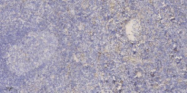
A30469 has real IHC data from human paraffin-embedded tonsil (IHC image caption). IF/ICC applications and human/mouse reactivity are listed, without an IF figure (catalog: A30469).
A30469 is listed for IHC, IF and ICC, with human and mouse reactivity (catalog: A30469 applications/reactivity). Its IHC figure shows human paraffin-embedded tonsil stained at 1:200 after Tris–EDTA retrieval at pH 9.0; no IF figure is supplied (A30469 IHC image caption; catalog: A30469 IF images).
Which to pick: For tissue IHC, choose A30469 because its own figure documents staining in human paraffin-embedded tonsil; the fixative is unreported (A30469 IHC image caption). For IF/ICC, A30469 lists both applications and a 1:200–1:1000 dilution range, but provides no IF figure (catalog: A30469 applications/dilution/IF images). For work involving human and mouse samples, A30469 is listed as a rabbit polyclonal reactive with both species; its IHC figure documents human tissue only (catalog: A30469 host/clonality/reactivity; A30469 IHC image caption).