TREX1 / Three-prime repair exonuclease 1 · IHC design guide

Design Immunohistochemistry for TREX1

Plan chromogenic TREX1 IHC around the ubiquitous nuclear tissue pattern (HPA tissue IHC), with skin melanocytes and testis preleptotene spermatocytes as high-staining references (HPA tissue IHC). The catalog antibody has an IHC starting concentration of 2.5 μg/mL (datasheet: A00940); DNA damage can alter nuclear localisation (UniProt).

Evidence assembled Oct 2026 · For research use; verify linked source records and product datasheet before use
Immunohistochemistry protocol sheet for TREX1 (IHC for TREX1): expected localisation Nuclear in tissue IHC (HPA tissue IHC); cytosol and ER membrane (UniProt), antibody A00940, validated IHC image, and IHC protocol steps
Printable TREX1 IHC protocol sheet — expected localisation Nuclear in tissue IHC (HPA tissue IHC); cytosol and ER membrane (UniProt), antibody A00940, controls and protocol steps. Open the full TREX1 IHC guide →

TREX1 Immunohistochemistry Experimental Design Guide

Expected localisation, validated protocols, controls and antibodies — the at-a-glance facts below, then the full design guide.

Must know before staining
Expected localisation Nuclear in tissue IHC (HPA tissue IHC); cytosol and ER membrane (UniProt)
Staining pattern Ubiquitous nuclear staining across tissue cell types (HPA tissue IHC)
Antigen retrieval Tris-EDTA pH 9.0 HIER, 95–98 °C, 20 min (rule: nuclear antigen)
Positive control ⓘ Skin+4 more · see all
Negative control ⓘ Bone marrow+2 more · see all
Important caveats
Reasons your staining may differ from the expected pattern.
Fixation Keep formalin fixation consistent across sections (standard IHC practice; not target-specific)
Caveat DNA damage can shift TREX1 into nuclei (UniProt)
Regulation Expression regulation is unreported (UniProt)
Isoform / epitope 3 isoforms; antibody epitope coverage unspecified (UniProt; datasheets)
Section 1

Recommended TREX1 IHC & IF Protocols

Compare the catalog antibody's IHC-P protocol with three published TREX1 IHC protocols (PMC6404532; PMC12651403; PMC5122164).

Recommended immunohistochemistry (IHC-P) protocol parameters
SampleTissue sections; selected-image fixative not specified (standard IHC workflow)
FixationImage fixative and duration unreported (datasheet A00940); verify before use.
Sectioning4–5 µm sections on charged slides (standard)
DeparaffinisationXylene, graded ethanol series to water (standard)
Antigen retrievalHeat-induced epitope retrieval in Tris-EDTA buffer, pH 9.0, 20 min at 95–98 °C (standard rule: nuclear antigen)
Peroxidase block3% H2O2, 10 min, room temperature (standard)
Blocking10% normal serum of the secondary host, 30 min, room temperature (standard)
Primary antibodyRabbit anti-TREX1, 2.5 μg/mL (datasheet A00940)
Primary incubationOvernight at 4 °C (standard)
DetectionHRP-polymer secondary, DAB chromogen 5–10 min (standard)
CounterstainHematoxylin, blue, dehydrate and mount (standard)
Expected resultTREX1-positive staining in melanocytes of skin (HPA tissue IHC: High). HPA tissue profile: Ubiquitous nuclear expression. No signal in the no-primary control.
💡Decision noteStart with Tris-EDTA pH 9.0 at 95–98 °C for 20 min (page retrieval: nuclear antigen); consider boiled citrate pH 6.0 for jejunal sections (PMC12651403: Immunostaining).
Section 2

What Is the Expected TREX1 Staining Pattern?

TREX1 can localise to the nucleus and cytosol; its C-terminal region helps retain it in the cytoplasm, and no transmembrane segment is annotated (UniProt Q9NSU2 topology and subcellular location). In paraffin-section IHC, expect predominantly nuclear staining across many cell types, with particularly strong staining in skin melanocytes and testis preleptotene spermatocytes (HPA tissue IHC). HPA rates the tissue pattern Supported, with medium consistency against RNA expression data (HPA tissue IHC).

What am I looking at on my slide?
Distinct nuclear staining appears in several cell types, with strong melanocyte or preleptotene spermatocyte staining.This fits the reported ubiquitous nuclear IHC profile and High staining in those two cell populations (HPA tissue IHC). Judge the pattern within identified cells; adjacent cells need not have equal intensity, and one strongly stained cell type does not make the entire section uniformly positive.
Cytoplasmic staining accompanies nuclear staining, or an apparently positive section shows only cytoplasmic staining.Cytoplasmic TREX1 is biologically plausible (UniProt Q9NSU2 subcellular location). Yet a cytoplasm-only paraffin IHC result departs from HPA’s nuclear tissue profile (HPA tissue IHC). Compare a known-positive section and a matched detection control before assigning that result to TREX1; compartment alone cannot establish antibody specificity.
Strong stain falls in cells reported as Not detected, such as bone-marrow hematopoietic cells or liver cholangiocytes.This conflicts with the listed cell-specific HPA observations (HPA tissue IHC). Check cell identification and staining controls. Cross-reactivity or endogenous chromogen-generating activity is possible in chromogenic IHC (general IHC practice); a dark structure in the right tissue is not evidence that the intended cell expressed TREX1.
Weak color spreads through stroma, empty spaces, or most of the section without clear cell boundaries.This is a background pattern to resolve before scoring (general IHC practice). Review the no-primary control, blocking, washes and chromogen development. The HPA profile describes cellular nuclear staining, so diffuse deposit cannot be counted as the expected TREX1 pattern (HPA tissue IHC).
No signal appears in a section chosen because HPA reports High staining in its target cell population.First confirm that the relevant melanocytes or preleptotene spermatocytes are present (HPA tissue IHC). If present, review the IHC-validated antibody, detection reagents and positive-control run (general IHC practice). A failed control supports a technical problem; a single negative section does not establish absent TREX1 expression.
💡Expected TREX1 appearanceCall paraffin IHC positive when identifiable cells show clear nuclear staining, ideally High in melanocytes or preleptotene spermatocytes (HPA tissue IHC); widespread cell-free color or strong staining in an HPA Not detected cell population warrants investigation before scoring.
How each factor affects the staining
Assay and compartment: why might IF look different from IHC?HPA tissue IHC describes ubiquitous nuclear staining, whereas HPA ICC-IF supports cytosolic localisation (HPA tissue IHC; HPA subcellular). UniProt also records nucleus, cytosol and ER membrane, including nuclear translocation after DNA damage (UniProt Q9NSU2 subcellular location). Interpret each assay against its own evidence; the IF/ICC workflow belongs in its separate guide.
Cell population selected for the positive controlHPA lists High staining in skin melanocytes and testis preleptotene spermatocytes, but Not detected in bone-marrow hematopoietic cells, liver cholangiocytes and parathyroid glandular cells (HPA tissue IHC). Record the scored cell type, since a tissue label alone does not identify the expected positive or negative cells.
Antibody validation and scopeThe tissue IHC profile is Supported with medium RNA–staining consistency; HPA035437 is Supported for IHC, while HPA046721 is Supported for ICC-IF (HPA tissue IHC; HPA antibodies). These assay-specific ratings support pattern comparison, but do not establish identical staining across antibodies or prove every individual signal is specific.
Retrieval and target-specific sensitivityNo TREX1-specific fixation or antigen-retrieval effect is established by the supplied HPA and UniProt records. Use the catalog antibody’s validated IHC-P conditions as the starting point and assess retrieval changes against matched controls (general IHC practice); do not infer an epitope-masking effect from localisation or staining level.
Why is my staining missing, weak or wrong?
SituationLikely causeNext action
Expected nuclear staining is absent from a positive-control section.The scored cell population may be missing, or an IHC detection step may have failed (HPA tissue IHC; general IHC practice).Confirm the relevant cells are present, then check the catalog antibody’s IHC-P conditions, detection reagents and control run (general IHC practice). Repeat scoring only after the control works.
Only cytoplasmic color appears in paraffin IHC.Cytosolic TREX1 is documented, but the HPA tissue IHC profile is nuclear; the discrepancy has no single established cause (UniProt Q9NSU2; HPA tissue IHC).Compare an HPA-positive cell population, no-primary control and cell boundaries. Report the compartment discrepancy if it persists; do not reclassify a cytoplasm-only IHC pattern as the HPA nuclear pattern.
An HPA Not detected cell population stains strongly.Cell misidentification, antibody cross-reactivity or endogenous chromogenic activity may explain the result (HPA tissue IHC; general IHC practice).Verify cell identity on the counterstained section and inspect no-primary and detection controls. Treat the signal as unresolved until the unexpected cell-specific pattern is checked.
Diffuse color obscures nuclei throughout the section.Background from detection, insufficient washing or excessive chromogen development is possible (general IHC practice).Inspect the no-primary control and review blocking, washes and development time under the catalog IHC-P workflow (general IHC practice). Score TREX1 only where cell boundaries and nuclear localisation remain readable.
Different areas or cell types within one tissue vary in intensity.HPA reports cell-specific levels, including Low appendix glandular cells and Low spleen red-pulp cells; a uniform tissue-wide intensity is not established (HPA tissue IHC).Annotate the cell type and compartment for each scored area. Compare like cells with like cells and avoid calling an entire tissue negative because its listed positive population is sparse.
IHC and ICC-IF appear to disagree on the main compartment.HPA reports nuclear tissue IHC and supported cytosolic ICC-IF; UniProt records both compartments (HPA tissue IHC; HPA subcellular; UniProt Q9NSU2).Keep assay, specimen type and antibody identity with each observation. Use the separate IF/ICC guide to assess IF images, and judge paraffin IHC against its tissue control and nuclear reference pattern.

Sample controls for TREX1 IHC & IF

🧪Run skin first and assess melanocytes, which should stain (HPA: High in skin melanocytes). Use parathyroid glandular cells as the negative tissue (HPA: Not detected in parathyroid glandular cells); on the skin slide, compare melanocytes with neighboring cells lacking signal as a background reference, without treating those cells as validated biological negatives.
Positive control tissue: Skin (Melanocytes, HPA High)
Negative control tissue: Bone marrow (HPA Not detected)
ICC-IF cell lines (HPA subcellular resource): HPA ICC-IF images show TREX1 in HeLa, U-251MG, U2OS, with annotated localisation: Cytosol (supported) (HPA subcellular).
Technical controls: Include a no-primary (secondary-only) control, an isotype control matched to the primary antibody’s host species and clonality, and TREX1-knockout tissue or cells as a biological negative (standard IHC practice). Quench endogenous peroxidase for chromogenic detection and distinguish pigment in skin melanocytes from chromogen signal (standard IHC practice; HPA: High in skin melanocytes).
⚠️Feasibility: A target-specific fixation window or fixation effect is unreported in the supplied evidence, and the selected A00940 spleen IHC caption does not state a fixative (A00940 tissue-IHC caption). Retrieval dependence is unreported; optimize antigen retrieval empirically for paraffin sections (standard IHC practice). The supplied evidence does not establish whether frozen sections or IF are easier; HPA supports cytosolic ICC-IF localization, while endogenous pigment can complicate chromogenic assessment of skin melanocytes (HPA: Cytosol, supported; HPA: High in skin melanocytes; standard IHC practice).

HPA tissue IHC evidence for TREX1

Comprehensive Human Protein Atlas IHC scoring per tissue (reliability: Supported — Medium consistency between antibody staining and RNA expression data.). Rows are taken directly from the HPA tissue chart — click any row's HPA link to view the source.

Positive expression · recommended positive controls

TissueCell typeLevelEvidenceSource
Skin Melanocytes High Protein (IHC) HPA →
Testis Preleptotene spermatocytes High Protein (IHC) HPA →
Adipose tissue Adipocytes Medium Protein (IHC) HPA →
Adrenal gland Glandular cells Medium Protein (IHC) HPA →
Breast Adipocytes Medium Protein (IHC) HPA →

Undetected expression · recommended negative controls

TissueCell typeLevelEvidenceSource
Bone marrow Hematopoietic cells Not detected Protein (IHC) HPA →
Liver Cholangiocytes Not detected Protein (IHC) HPA →
Parathyroid gland Glandular cells Not detected Protein (IHC) HPA →
Section 3

Advanced TREX1 IHC Tips

Use matched controls and compartment-aware scoring to troubleshoot TREX1 staining in paraffin sections; treat the IF guidance as a separate application.

What retrieval should I use when TREX1 staining is weak?
Use Tris-EDTA pH 9.0 heat-induced antigen retrieval at 95–98 °C for 20 min for TREX1 paraffin-section IHC (page retrieval setting). Let sections cool in retrieval buffer, then wash consistently before blocking and applying the primary antibody (standard IHC practice). Compare retrieved and untreated serial sections with the same chromogen exposure, since retrieval can change both signal and background (standard IHC practice). If staining remains weak, test citrate pH 6.0 as a fallback on matched sections, keeping detection conditions constant (standard IHC practice). Interpret nuclear and cytoplasmic signal separately because both compartments are reported for TREX1 (UniProt Q9NSU2 subcellular location).
Could fixation explain weak or uneven TREX1 staining?
Target-specific fixation sensitivity is unknown from the supplied evidence; the selected A00940 spleen caption does not state a fixative (A00940 tissue-IHC caption). Record the actual fixative, fixation duration, section thickness, and time before processing for each specimen (standard IHC practice). On matched paraffin sections, hold retrieval and antibody conditions constant while comparing specimens with documented processing histories (standard IHC practice). Check whether staining loss follows folds, poorly processed areas, or section edges before attributing it to TREX1 biology (standard IHC practice). Do not use the reported tissue staining pattern to infer a preferred fixative (HPA tissue IHC; A00940 tissue-IHC caption).
How should I assess nuclear versus cytoplasmic TREX1 staining?
Score nuclear and cytoplasmic staining separately: TREX1 is reported in both compartments, with endoplasmic-reticulum membrane association also described (UniProt Q9NSU2 subcellular location). HPA tissue IHC describes a ubiquitous nuclear pattern, while its ICC/IF summary supports cytosolic localisation (HPA tissue IHC; HPA subcellular). Use a nuclear counterstain and inspect the same cells at high magnification to assign chromogen to a compartment (standard IHC practice). Nuclear enrichment can depend on context, including DNA damage or GZMA-mediated cell death (UniProt Q9NSU2 subcellular location). Confirm an unexpected compartment pattern across matched sections and controls before calling it biological relocalisation (standard IHC practice).
Could an isoform or masked epitope explain inconsistent staining?
TREX1 has 3 listed isoforms, but the supplied evidence does not map the catalog antibody’s epitope to any of them (UniProt Q9NSU2 isoforms; supplied antibody evidence). Obtain the immunogen coordinates before assigning isoform-specific staining, then compare those coordinates with the relevant sequences (standard antibody validation practice). The record lists phosphoserines at residues 78, 167, and 261, without establishing whether they affect antibody binding (UniProt Q9NSU2 modified residues). TREX1 has no annotated transmembrane segment, despite its reported endoplasmic-reticulum membrane association (UniProt Q9NSU2 topology; UniProt Q9NSU2 subcellular location). Compare retrieval conditions on adjacent sections before interpreting a staining difference as altered isoform abundance (standard IHC practice).
How can I check a TREX1 pattern by multiplex IF?
Treat IF/ICC as a separate application and validate the antibody’s fluorescence signal with appropriate controls (standard IF practice). For a cell-type comparison, multiplex TREX1 with a melanocyte marker such as SOX10 in skin, where HPA reports high melanocyte staining (HPA tissue IHC; standard marker practice). Choose a far-red TREX1 fluorophore when tissue autofluorescence interferes with shorter-wavelength channels, and inspect single-stain controls (standard IF practice). Permeabilise sufficiently for an antibody targeting a cytosolic epitope; if epitope position is unknown, optimise permeabilisation empirically rather than assuming membrane accessibility (HPA subcellular; standard IF practice). Use a nuclear dye to distinguish nuclear from cytosolic signal during imaging (standard IF practice).
What should I check when chromogenic background obscures TREX1?
Inspect a no-primary control beside the stained section to identify signal from detection reagents or endogenous activity (standard IHC practice). Include a peroxidase block before HRP/DAB detection, particularly when interpreting chromogen in blood-rich areas such as spleen red pulp (standard IHC practice; HPA tissue IHC). If diffuse staining persists, titrate the primary antibody and strengthen protein blocking while keeping retrieval fixed (standard IHC practice). The selected A00940 spleen image used 2.5 µg/mL, which documents that image’s condition rather than a validated range for every specimen (A00940 tissue-IHC caption). Examine folds, edges, and precipitate at high magnification before scoring cells as positive (standard IHC practice).
How should I quantify TREX1 across paraffin sections? ⚠ ANSWER MARKED FOR VERIFICATION
Define the cell population and compartment before scoring because TREX1 has reported nuclear and cytoplasmic locations (UniProt Q9NSU2 subcellular location). Within comparable regions, record the percentage of positive cells and staining intensity, or calculate an H-score from intensity categories 0–3 for a possible range of 0–300 (standard IHC scoring practice). Normalise counts to the number of eligible cells, or report positive-cell density per mm² of analysed tissue (standard quantitative IHC practice). Exclude folds, necrosis, and damaged edges using the same rule for every section (standard IHC practice). Report nuclear and cytoplasmic results separately, with controls and exposure or development conditions documented (standard IHC practice).
How do I distinguish true TREX1 staining from artefact?
A plausible TREX1 signal may be nuclear or cytoplasmic, depending on context, so compartment alone cannot establish specificity (UniProt Q9NSU2 subcellular location; HPA tissue IHC; HPA subcellular). Compare candidate positive cells with the documented distribution: HPA reports high staining in skin melanocytes and low staining in spleen red-pulp cells (HPA tissue IHC). Treat staining confined to cut edges, folds, or necrotic regions as suspect and inspect a no-primary control for detection background (standard IHC practice). In HRP/DAB assays, residual endogenous peroxidase can mimic a positive deposit, so verify the peroxidase block (standard IHC practice). Seek reproducible cellular staining across matched sections before making a biological claim (standard IHC practice).
Boster reagents

Best TREX1 / Three-prime repair exonuclease 1 IHC Antibodies

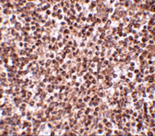
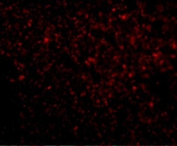
A00940 has IHC and IF images from human spleen (A00940 image captions); its listed reactivity covers human, mouse, and rat (catalog reactivity).

Real IHC data Immunohistochemistry of TREX1 in human spleen tissue with TREX1 antibody at 2.5 μg/mL.
Anti-TREX1 Antibody
Cat # A00940

A00940 is the only SKU with a rendered card (cards payload). Its IHC and IF images show human spleen (A00940 image captions); IHC-P and IF are listed applications (catalog applications).

Which to pick: Choose A00940 for paraffin-section tissue IHC: IHC-P is listed, and its own IHC caption shows human spleen at 2.5 μg/mL; the fixative is unreported (catalog applications; A00940 IHC caption). For IF, A00940 has a human spleen image; for human ICC, M00940-1 is a rabbit monoclonal with ICC/IF listed but no supplied IHC or IF image (A00940 IF caption; M00940-1 catalog applications and image alts). For mouse or rat work, choose A00940 on listed reactivity, while recognizing that the supplied images show human tissue (A00940 catalog reactivity and image captions).

Each figure is that product's own IHC / IF validation image from its datasheet.

References

  1. UniProt Consortium. UniProt entry Q9NSU2 (TREX1_HUMAN, Three-prime repair exonuclease 1).
  2. Human Protein Atlas. TREX1 tissue IHC expression (reliability: Supported).
  3. Human Protein Atlas. TREX1 subcellular location (ICC-IF): Localized to the cytosol..
  4. Human Protein Atlas. TREX1 antibody validation summary (2 antibodies).
  5. Carbon ion irradiation combined with PD-1 inhibitor trigger abscopal effect in Lewis lung cancer via a threshold dose. Journal of Cancer 2024 — PMC10937268.
  6. TREX1 is expressed by microglia in normal human brain and increases in regions affected by ischemia. Brain pathology (Zurich, Switzerland) 2018 — PMC6404532.
  7. cGAS/STING Pathway Mediates Accelerated Intestinal Cell Senescence and SASP After GCR Exposure in Mice. Cells 2025 — PMC12651403.
  8. Verification of TREX1 as a promising indicator of judging the prognosis of osteosarcoma. Journal of orthopaedic surgery and research 2016 — PMC5122164.
  9. PubMed PMID:10393201 — UniProt-cited evidence.
  10. PubMed PMID:10391904 — UniProt-cited evidence.
  11. PubMed PMID:11278605 — UniProt-cited evidence.