This website uses cookies to ensure you get the best experience on our website.


- Table of Contents
Plan chromogenic TSHZ3 IHC-P using the catalog antibody’s 2.5 μg/mL starting concentration (datasheet). Assess nuclear tissue staining while accounting for low concordance between antibody staining and RNA expression (HPA tissue IHC).

Expected localisation, validated protocols, controls and antibodies — the at-a-glance facts below, then the full design guide.
| Expected localisation | Ubiquitous nuclear tissue staining (HPA tissue IHC) | |
| Staining pattern | Nuclear staining across tissue cell types (HPA tissue IHC) | |
| Antigen retrieval | Tris-EDTA pH 9.0 HIER, 95–98 °C, 20 min (rule: nuclear antigen) | |
| Positive control | Adipose tissue+4 more · see all | |
| Negative control | None in HPA (detected in all 45 tissues); use no-primary + isotype controls |
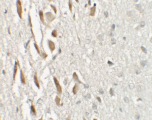
| Fixation | Matched tissue-IHC evidence does not establish the fixation claim. Validate the specimen-specific method before use. (selected-SKU IHC image A08453) | |
| Caveat | Staining has low concordance with RNA expression (HPA tissue IHC) | |
| Regulation | Reduced in elderly Alzheimer disease brain (UniProt) | |
| Isoform / epitope | No isoforms annotated; one 1–1081 chain (UniProt) |
Compare the catalog antibody’s IHC-P protocol (datasheet) with published TSHZ3 staining methods for esophageal carcinoma and ureteropelvic junction tissue (PMC13265166; PMC5018636).
| Sample | Tissue sections; selected-image fixative not specified (standard IHC workflow) |
| Fixation | Image fixative and duration unreported (datasheet A08453); verify before use. |
| Sectioning | 4–5 µm sections on charged slides (standard) |
| Deparaffinisation | Xylene, graded ethanol series to water (standard) |
| Antigen retrieval | Heat-induced epitope retrieval in Tris-EDTA buffer, pH 9.0, 20 min at 95–98 °C (standard rule: nuclear antigen) |
| Peroxidase block | 3% H2O2, 10 min, room temperature (standard) |
| Blocking | 10% normal serum of the secondary host, 30 min, room temperature (standard) |
| Primary antibody | Rabbit anti-TSHZ3, 2.5 μg/mL (datasheet A08453) |
| Primary incubation | Overnight at 4 °C (standard) |
| Detection | HRP-polymer secondary, DAB chromogen 5–10 min (standard) |
| Counterstain | Hematoxylin, blue, dehydrate and mount (standard) |
| Expected result | TSHZ3-positive staining in adipocytes of adipose tissue (HPA tissue IHC: High). HPA tissue profile: Ubiquitous nuclear expression. No signal in the no-primary control. |
TSHZ3 is expected mainly in nuclei: HPA describes ubiquitous nuclear tissue staining, while UniProt places the protein in the nucleus and reports no transmembrane segment (HPA: tissue IHC; UniProt Q63HK5: subcellular location, topology). HPA reports high staining in several epithelial, neural, hematopoietic, and adipose cell populations, but its tissue IHC reliability is Approved with low consistency between staining and RNA expression (HPA: tissue IHC).
| Distinct nuclear staining in the sampled cell population, with nuclei easier to identify against the counterstain. | This fits the reported tissue pattern and nuclear localization (HPA: ubiquitous nuclear expression; UniProt Q63HK5: nucleus). Judge intensity against the same run's controls; HPA's High labels describe particular cell populations, not a universal intensity threshold (HPA: tissue IHC). |
| Predominantly cytoplasmic, surface, or extracellular staining with little nuclear signal. | For tissue IHC, this departs from the expected dominant pattern and warrants an artefact or nonspecific staining check (HPA: ubiquitous nuclear expression). A membrane signal alone is not proof of error: HPA also reports plasma-membrane localization in ICC-IF, which is a different assay (HPA: subcellular ICC-IF). |
| Strong staining in a cell population expected to be low, or a pattern that tracks pigment or reaction deposits rather than nuclei. | Compare with morphology and controls before assigning TSHZ3: cross-reactivity or endogenous detection activity can produce misleading signal (general IHC practice). Liver cholangiocytes are reported Low, not absent, and HPA calls overall tissue staining ubiquitous (HPA: tissue IHC). |
| Haze across tissue, stroma, or empty spaces obscures nuclear boundaries. | Diffuse background limits interpretation even when some nuclei appear positive (general IHC practice). Review blocking, wash steps, detection chemistry, and exposure or development time before scoring; the supplied sources do not identify a TSHZ3-specific background mechanism (general IHC practice). |
| No convincing nuclear signal in a run that includes an HPA-reported High population. | First consider assay failure or poor control performance, then biological variation (general IHC practice). HPA reports High staining in cerebral-cortex glial cells and cerebellar granular-layer cells, among others, but its Approved tissue IHC has low staining–RNA consistency (HPA: tissue IHC). |
| Which compartment anchors IHC scoring? | Nuclear staining anchors the call (HPA: ubiquitous nuclear tissue expression; UniProt Q63HK5: nucleus). UniProt also lists growth cones by similarity, so localization is context dependent (UniProt Q63HK5: subcellular location). |
| Which populations can serve as observed positives? | HPA reports High staining in adipocytes, bone-marrow hematopoietic cells, bronchial respiratory epithelial cells, and cerebral-cortex glial cells, among others (HPA: tissue IHC). These are observed patterns, not guaranteed assay controls (HPA: Approved; low staining–RNA consistency). |
| How strong is the tissue IHC evidence? | HPA labels tissue IHC Approved and notes low consistency with RNA expression; the listed HPA008834 antibody is IHC Approved, while HPA071010 has no listed IHC status (HPA: tissue IHC; HPA: antibody validation). Do not treat Approved as IHC Enhanced (HPA: antibody validation). |
| Do topology or processing predict a separate tissue compartment? | UniProt reports no transmembrane segment, signal peptide, propeptide, or annotated isoforms, and lists one 1–1081 chain (UniProt Q63HK5: topology, processing, isoforms). These annotations do not establish retrieval requirements or fixation sensitivity (UniProt Q63HK5: record scope). |
| What does IF/ICC add? | HPA reports enhanced nucleoplasmic localization and an approved additional plasma-membrane location in ICC-IF (HPA: subcellular ICC-IF). Use this as a localization cross-check; IF/ICC assay design belongs to its own guide (HPA: subcellular ICC-IF). |
| Situation | Likely cause | Next action |
|---|---|---|
| No nuclear staining in an expected-positive area. | Detection, retrieval, or antibody conditions may have failed; an HPA High label is not a guarantee for every section (general IHC practice; HPA: tissue IHC). | Check the run's positive and detection controls, then verify retrieval and antibody conditions against the chosen assay instructions (general IHC practice). Compare with an HPA High population only after controls pass (HPA: tissue IHC). |
| Strong colour appears mainly outside nuclei. | The IHC pattern conflicts with HPA's ubiquitous nuclear description; staining chemistry or specificity may be contributing (HPA: tissue IHC; general IHC practice). | Review matched negative controls and cellular morphology, then repeat with adjusted detection conditions if controls implicate background (general IHC practice). Consider the separate ICC-IF membrane observation as context, not as IHC validation (HPA: subcellular ICC-IF). |
| Broad brown background obscures nuclei. | Nonspecific binding, incomplete washing, or excessive chromogen development can obscure chromogenic IHC (general IHC practice). | Check the negative control; review blocking, washing, and development time, then score only nuclei distinguishable from background (general IHC practice). |
| Signal follows blood-rich or pigmented areas rather than cell nuclei. | Endogenous detection activity or native pigment can mimic a chromogenic reaction (general IHC practice). | Compare controls and morphology; assess endogenous-enzyme blocking when the chosen detection chemistry requires it (general IHC practice). Do not count deposits without a cellular nuclear pattern (HPA: ubiquitous nuclear expression). |
| Liver cholangiocytes appear strongly positive. | This differs from HPA's Low cholangiocyte entry, although Low does not mean negative and HPA reports low staining–RNA consistency (HPA: tissue IHC). | Confirm cell identity and control behaviour, then compare nuclear signal with other populations in the same run (general IHC practice). Treat the discrepancy as a reason to investigate, not automatic proof of cross-reactivity (HPA: tissue IHC). |
| IF/ICC shows membrane signal alongside nuclear signal. | HPA lists an additional plasma-membrane location in ICC-IF, with nucleoplasm as the main location (HPA: subcellular ICC-IF). | Record both compartments and assess IF/ICC controls within that assay (general IF practice). Use the separate IF/ICC guide for its procedure; do not transfer its membrane result directly into chromogenic tissue IHC scoring (HPA: subcellular ICC-IF; HPA: tissue IHC). |


Comprehensive Human Protein Atlas IHC scoring per tissue (reliability: Approved — Low consistency between antibody staining and RNA expression data.). Rows are taken directly from the HPA tissue chart — click any row's HPA link to view the source.
| Tissue | Cell type | Level | Evidence | Source |
|---|---|---|---|---|
| Adipose tissue | Adipocytes | High | Protein (IHC) | HPA → |
| Appendix | Glandular cells | High | Protein (IHC) | HPA → |
| Bone marrow | Hematopoietic cells | High | Protein (IHC) | HPA → |
| Bronchus | Respiratory epithelial cells | High | Protein (IHC) | HPA → |
| Caudate | Glial cells | High | Protein (IHC) | HPA → |
| Tissue | Cell type | Level | Evidence | Source |
|---|---|---|---|---|
| None in HPA: TSHZ3 is detected in all 45 scored tissues. Use a no-primary (secondary-only) and an isotype control instead. | ||||
Use nuclear staining as the primary IHC readout for TSHZ3, and interpret tissue patterns alongside the reported limits of antibody evidence (UniProt Q63HK5; HPA tissue IHC).
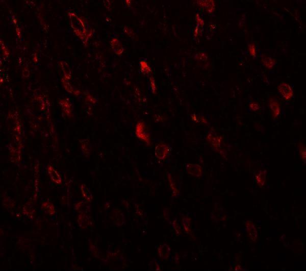
A08453 has IHC and IF images from mouse brain tissue (catalog image captions) and lists Human, Mouse and Rat reactivity (catalog: reactivity).
A08453 is listed for IHC-P and IF (catalog: applications), with a mouse brain tissue IHC image at 2.5 μg/mL (catalog: IHC image caption). Its IF image also shows mouse brain tissue (catalog: IF image caption); Human, Mouse and Rat are listed as reactive species (catalog: reactivity).
Which to pick: Choose A08453 for paraffin-section IHC because IHC-P is listed (catalog: applications); its IHC image shows mouse brain tissue, but the fixative is unreported (catalog: IHC image caption). For IF, A08453 has a mouse brain tissue image (catalog: IF image caption); ICC validation and clonality are unreported (catalog: applications and clone). For Human or Rat samples, A08453 lists reactivity (catalog: reactivity), while the pictured IHC and IF data are from mouse tissue (catalog image captions).