TUBB3 / Tubulin beta-3 chain · IHC design guide

Design Immunohistochemistry for TUBB3

Plan TUBB3 staining in paraffin sections using peripheral nerves and CNS neurons as positive tissue references (HPA tissue IHC). This guide covers fixation, controls and interpretation of cytoplasmic staining, including the reported specificity concern (HPA tissue IHC).

Evidence assembled Oct 2026 · For research use; verify linked source records and product datasheet before use
Immunohistochemistry protocol sheet for TUBB3 (IHC for TUBB3): expected localisation Cytoplasmic staining in tissue sections (HPA tissue IHC), antibody M01857-1, validated IHC image, and IHC protocol steps
Printable TUBB3 IHC protocol sheet — expected localisation Cytoplasmic staining in tissue sections (HPA tissue IHC), antibody M01857-1, controls and protocol steps. Open the full TUBB3 IHC guide →

TUBB3 Immunohistochemistry Experimental Design Guide

Expected localisation, validated protocols, controls and antibodies — the at-a-glance facts below, then the full design guide.

Must know before staining
Expected localisation Cytoplasmic staining in tissue sections (HPA tissue IHC)
Staining pattern Peripheral nerves: high; CNS neurons: medium, cytoplasmic (HPA tissue IHC)
Antigen retrieval EDTA pH 8.0 HIER, heat-mediated (datasheet M01857-1)
Positive control ⓘ Soft tissue+3 more · see all
Negative control ⓘ Adipose tissue+4 more · see all
Important caveats
Reasons your staining may differ from the expected pattern.
Fixation Keep fixation consistent across sections (standard IHC practice; not target-specific); Matched tissue-IHC evidence does not establish the fixation claim. Validate the specimen-specific method before use. (selected-SKU IHC image M01857-1)
Caveat Cross-reactivity may complicate staining interpretation (HPA tissue IHC)
Regulation Increased in many cancers (UniProt)
Isoform / epitope 2 isoforms; epitope impact is undetermined (UniProt)
Section 1

Recommended TUBB3 IHC & IF Protocols

Compare the catalog antibody’s EDTA pH 8.0 IHC-P protocol (datasheet M01857-1) with published TUBB3 protocols for breast cancer TMAs (PMC4774425) and leiomyosarcoma sections (PMC6033248).

Recommended immunohistochemistry (IHC-P) protocol parameters
SampleParaffin-embedded human testicular seminoma tissue; fixative not specified (datasheet M01857-1)
FixationImage fixative and duration unreported (datasheet M01857-1); verify before use.
Sectioning4–5 µm sections on charged slides (standard)
DeparaffinisationXylene, graded ethanol series to water (standard)
Antigen retrievalHeat retrieval: EDTA pH 8.0 (datasheet M01857-1); 20 min, 95–100 °C (standard)
Peroxidase block3% H2O2, 10 min, room temperature (standard)
Blocking10% goat serum (datasheet M01857-1)
Primary antibodyRabbit monoclonal (clone CC-20) anti-TUBB3, 1:50 (datasheet M01857-1)
Primary incubationOvernight at 4 °C (datasheet M01857-1)
DetectionHRP-conjugated secondary, DAB chromogen (datasheet M01857-1)
CounterstainHematoxylin, blue, dehydrate and mount (standard)
Expected resultTUBB3-positive staining in peripheral nerve of soft tissue (HPA tissue IHC: High). HPA tissue profile: Cytoplasmic expression in most tissues, most abundant in CNS, and peripheral nerves. No signal in the no-primary control.
💡Decision noteStart with EDTA pH 8.0 heat retrieval for the catalog antibody (datasheet M01857-1); use the published retrieval conditions when adapting either tissue protocol (PMC4774425; PMC6033248).
Section 2

What Is the Expected TUBB3 Staining Pattern?

TUBB3 is a cytoplasmic microtubule protein with no transmembrane segment (UniProt Q13509: location and topology). In paraffin sections, expect staining in neuronal cells and peripheral nerves, with variable staining elsewhere (HPA: tissue IHC). HPA rates its tissue staining profile Approved, but reports medium consistency with RNA data and cautions that the antibodies may detect proteins from more than one gene (HPA: tissue IHC reliability).

What am I looking at on my slide?
Peripheral nerves stain strongly; neuronal cells in cerebral cortex or hippocampus stain at a moderate level.This matches the reported cell and tissue pattern: peripheral nerve is High, while neuronal cells in cerebral cortex and hippocampus are Medium (HPA: tissue IHC). Interpret the chromogenic signal in the cytoplasm, consistent with TUBB3’s cytoskeletal location (UniProt Q13509: subcellular location).
The dominant signal is confined to nuclei or appears as a membrane outline.Neither pattern matches the reported cytoplasmic tissue profile or the protein’s cytoskeletal location and lack of a transmembrane segment (HPA: tissue IHC; UniProt Q13509: location and topology). Review the staining and detection controls before assigning the signal to TUBB3 (general IHC practice).
A cell type listed as Not detected stains prominently, such as adipocytes or bone marrow hematopoietic cells.Those cell types are reported as Not detected (HPA: tissue IHC). Consider cross-reactivity or endogenous detection activity and compare with controls (general IHC practice). The HPA profile itself cautions about presumed off-target binding and antibodies targeting proteins from more than one gene (HPA: tissue IHC reliability).
Brown color is diffuse across the section and does not resolve into stained cells or structures.A uniform haze is hard to reconcile with the reported cell-specific differences, including High peripheral nerves and Not detected adipocytes (HPA: tissue IHC). Check background controls, blocking, washing and detection chemistry before scoring the section (general IHC practice).
A known-positive peripheral nerve has no detectable signal.Peripheral nerve is reported High and is a useful positive reference for this pattern (HPA: tissue IHC). First confirm that the positive reference is present on the section, then check the staining workflow and detection controls (general IHC practice). A negative run alone cannot establish absence of TUBB3.
💡Expected TUBB3 appearanceCall a result positive when cytoplasmic staining is strong in peripheral nerve or moderate in cerebral cortex or hippocampal neuronal cells; isolated nuclear staining or similarly strong staining of HPA Not detected cell types warrants investigation (HPA: tissue IHC; UniProt Q13509: location and topology).
How each factor affects the staining
Tissue and cell selectionPeripheral nerve is High; cerebral cortex and hippocampal neuronal cells are Medium; pachytene spermatocytes are also High (HPA: tissue IHC). Select controls by the named cell type, since a tissue label alone does not define which cells should stain (general IHC practice).
Interpretation of weak stainingHPA describes cytoplasmic expression in most tissues and lists Low staining in several epithelial and glial cell types (HPA: tissue IHC). Weak cytoplasmic staining outside the strongest neural examples is therefore not automatically an artefact; assess its cell type and controls (general IHC practice).
Antibody specificityThe tissue profile is Approved, with medium consistency against RNA data; HPA cautions about presumed off-target binding and detection of proteins from more than one gene (HPA: tissue IHC reliability). Treat an unexpected positive cell type as provisional until controls support it (general IHC practice).
Isoforms and modified residuesUniProt lists two isoforms and modified residues at positions 172, 438 and 444 (UniProt Q13509: isoforms and modified residues). No antibody epitope is supplied, so these annotations do not establish isoform preference, staining intensity or a retrieval requirement.
IF/ICC Q: What localisation can corroborate the IHC pattern?A: HPA supports microtubules as the main IF/ICC location and also reports structures including the mitotic spindle and cytokinetic bridge (HPA: subcellular). Its IF/ICC summary cautions that the antibodies target proteins from multiple genes; use that pattern as a localisation cross-check, not proof of TUBB3 specificity.
Why is my staining missing, weak or wrong?
SituationLikely causeNext action
No staining in peripheral nerveThe expected High control is absent or the staining workflow failed (HPA: peripheral nerve High; general IHC practice).Confirm peripheral nerve is represented on the section and review the run’s positive control, reagent steps and chromogen development (general IHC practice).
Signal appears nuclear onlyThe compartment disagrees with cytoplasmic tissue staining and cytoskeletal localisation (HPA: tissue IHC; UniProt Q13509: location).Check counterstain and detection controls, then compare with a section containing an expected cytoplasmic positive cell type (general IHC practice; HPA: tissue IHC).
Prominent staining in adipocytes or hematopoietic cellsBoth are reported Not detected; non-specific antibody binding or endogenous detection activity is possible (HPA: tissue IHC; general IHC practice).Inspect appropriate background controls and review blocking and detection steps; keep the finding provisional given HPA’s off-target caution (general IHC practice; HPA: tissue IHC reliability).
Diffuse brown background obscures cellsExcess background from staining or detection can prevent cell-level interpretation (general IHC practice).Check background controls and review blocking, washes, antibody dilution and chromogen development before scoring (general IHC practice).
Weak neuronal staining beside strong peripheral nerve stainingDifferent reported levels may explain the contrast: neuronal cells in cortex and hippocampus are Medium; peripheral nerve is High (HPA: tissue IHC).Score the named cell types separately and compare each with its expected level; check controls if neuronal staining is absent (HPA: tissue IHC; general IHC practice).
An unexpected positive population persists across runsThe antibody may detect proteins from more than one gene, and HPA reports presumed off-target binding (HPA: tissue IHC reliability).Record the cell type and compartment, compare with HPA’s reported pattern, and seek independent specificity evidence before attributing the signal to TUBB3 (HPA: tissue IHC; general IHC practice).

Sample controls for TUBB3 IHC & IF

🧪Run testis first and require staining in pachytene spermatocytes (HPA: High in pachytene spermatocytes); use adipose tissue as the negative control, with adipocytes lacking detectable signal (HPA: Not detected in adipocytes). On the testis slide, compare spermatocyte staining with background in other cell populations, without assuming those cells are proven TUBB3-negative (HPA: positive designation is specific to pachytene spermatocytes).
Positive control tissue: Soft tissue (Peripheral nerve, HPA High)
Negative control tissue: Adipose tissue (HPA Not detected)
ICC-IF cell lines (HPA subcellular resource): HPA ICC-IF images show TUBB3 in A-549, HEL, U2OS, HaCaT, MCF-7, ASC52telo, RPTEC/TERT1, hTERT-RPE1 (serum starved), Sperm, with annotated localisation: Microtubules (supported) (HPA subcellular).
Technical controls: Include a no-primary, secondary-only section and a matched rabbit IgG isotype control (caption: rabbit primary antibody); confirm specificity with a validated TUBB3 knockout sample or peptide competition if the immunizing peptide is available (standard IHC practice). Quench endogenous peroxidase and inspect the no-primary testis section for background before interpreting DAB staining (caption: HRP/DAB detection).
⚠️Feasibility: No target-specific fixation window or fixation effect is reported, and the exact M01857-1 tissue-IHC caption leaves the fixative unreported (caption: fixative not stated). Heat retrieval in EDTA at pH 8.0 is documented for a paraffin section, but its necessity has not been established; the caption uses a 1:50 primary dilution overnight at 4°C (caption: retrieval and incubation conditions). The supplied evidence cannot establish whether frozen sections or IF are easier; the illustrated specimen is testicular seminoma, so its staining should not be treated as a map of normal testis cell populations (caption: testicular seminoma; HPA: High in pachytene spermatocytes).

HPA tissue IHC evidence for TUBB3

Comprehensive Human Protein Atlas IHC scoring per tissue (reliability: Approved — Medium consistency between antibody staining and RNA expression data. Caution, targets protein from more than one gene. Presumed off target binding observed and disregarded.). Rows are taken directly from the HPA tissue chart — click any row's HPA link to view the source.

Positive expression · recommended positive controls

TissueCell typeLevelEvidenceSource
Soft tissue Peripheral nerve High Protein (IHC) HPA →
Testis Pachytene spermatocytes High Protein (IHC) HPA →
Cerebral cortex Neuronal cells Medium Protein (IHC) HPA →
Hippocampus Neuronal cells Medium Protein (IHC) HPA →

Undetected expression · recommended negative controls

TissueCell typeLevelEvidenceSource
Adipose tissue Adipocytes Not detected Protein (IHC) HPA →
Adrenal gland Glandular cells Not detected Protein (IHC) HPA →
Appendix Glandular cells Not detected Protein (IHC) HPA →
Bone marrow Hematopoietic cells Not detected Protein (IHC) HPA →
Cerebellum Cells in granular layer Not detected Protein (IHC) HPA →
Section 3

Advanced TUBB3 IHC Tips

Use the documented paraffin-section workflow as the starting point for TUBB3 chromogenic IHC, then assess staining by cell type and compartment (datasheet M01857-1; HPA tissue IHC).

Which retrieval conditions should I start with for paraffin-section TUBB3 IHC?
Start with heat-mediated antigen retrieval in EDTA at pH 8.0 for paraffin sections (datasheet M01857-1). The documented image used a 1:50 primary dilution overnight at 4°C after retrieval, so hold those conditions constant while assessing retrieval (datasheet M01857-1). If staining is weak, compare a second retrieval condition on adjacent sections while keeping detection and imaging consistent (standard IHC practice). Judge the result by clearer cytoplasmic staining in expected cells and lower background in negative compartments, rather than by signal intensity alone (UniProt Q13509 subcellular location; HPA tissue IHC; standard IHC practice).
How should I troubleshoot weak TUBB3 staining when fixation history varies?
The selected paraffin-section caption does not report a fixative, so target-specific TUBB3 sensitivity to fixation is unknown (datasheet M01857-1). Record each specimen’s fixative and fixation duration when available, and compare sections processed with the same retrieval and detection conditions (standard IHC practice). Use the documented EDTA pH 8.0 retrieval and 1:50 overnight primary incubation as a consistent starting point for this antibody (datasheet M01857-1). If similarly processed expected-positive cells stain differently, investigate handling and fixation records before attributing the difference to TUBB3 abundance (HPA tissue IHC; standard IHC practice).
What staining pattern fits TUBB3, and which compartments should raise concern?
Expect staining in the cytoplasm and cytoskeleton, potentially extending into neuronal projections where those structures are present (UniProt Q13509 subcellular location; HPA tissue IHC). TUBB3 contributes to microtubules and is reported at growth cones, lamellipodia and filopodia, so assess fine processes when section quality permits (UniProt Q13509 function and subcellular location). HPA also reports microtubule localisation, but its subcellular evidence carries a warning about antibodies targeting proteins from multiple genes (HPA subcellular). Predominantly crisp nuclear or membrane-rim chromogen without convincing cytoplasmic staining warrants a review of antibody specificity and section artefacts (UniProt Q13509 topology and subcellular location; standard IHC practice).
Could isoforms or modified residues explain an inconsistent TUBB3 IHC signal?
The record lists 2 TUBB3 isoforms, but the supplied antibody caption does not identify its epitope or establish isoform coverage (UniProt Q13509 isoforms; datasheet M01857-1). It also lists modification sites at residues 172, 438 and 444; their effect on this antibody’s staining is unknown (UniProt Q13509 modified residues; datasheet M01857-1). Because TUBB3 has no annotated transmembrane segment, interpret the expected signal as intracellular rather than as a cell-surface pattern (UniProt Q13509 topology and subcellular location). When results disagree across specimens, compare matched processing and consult epitope documentation before assigning the difference to an isoform or modification (standard IHC practice; UniProt Q13509 isoforms).
How can I check an ambiguous IHC pattern with multiplex IF?
For a separate IF experiment, pair TUBB3 with a marker for the expected cell type so that apparent colocalisation can be assessed cell by cell (standard IF practice; HPA tissue IHC). Choose fluorophores after checking unstained tissue autofluorescence, and use channels that separate the marker signal from background and from each other (standard IF practice). TUBB3 is intracellular and has no transmembrane segment, so include a controlled permeabilisation step when the antibody must reach its cytoskeletal epitope (UniProt Q13509 topology and subcellular location; standard IF practice). Optimise fixation and permeabilisation for IF independently: the selected paraffin-section caption reports neither IF conditions nor a fixative (datasheet M01857-1).
What should I check when TUBB3 DAB staining is diffuse or widespread?
First compare expected cytoplasmic staining with a no-primary control and a matched negative cell population on sections processed together (UniProt Q13509 subcellular location; HPA tissue IHC; standard IHC practice). The documented workflow used 10% goat serum blocking, a 1:50 primary dilution and peroxidase-based DAB detection (datasheet M01857-1). If nonspecific colour persists, check the general peroxidase block, secondary-only background, wash quality and chromogen development time (standard IHC practice). Treat widespread low-level colour cautiously: HPA reports presumed off-target binding and warns that its antibody evidence may involve proteins from more than one gene (HPA tissue IHC).
How should I score TUBB3 IHC across specimens with different tissue composition? ⚠ ANSWER MARKED FOR VERIFICATION
Define the cell population and cytoplasmic compartment before scoring, since TUBB3 is reported in the cytoskeleton and expression varies by cell type (UniProt Q13509 subcellular location; HPA tissue IHC). For a cellular readout, record the percentage of positive viable target cells or an H-score using the same intensity thresholds across sections (standard IHC practice). For nerve-rich regions, density of positive profiles per mm² may be useful if profiles can be identified consistently (HPA tissue IHC; standard IHC practice). Normalise to the counted viable target cells or measured tissue area, and exclude necrosis, folds and section edges by a prespecified rule (standard IHC practice).
How do I distinguish convincing TUBB3 positivity from staining artefact?
A convincing result places chromogen in the expected cytoplasmic or process-rich compartment of identifiable cells, with limited signal in matched negative cells (UniProt Q13509 subcellular location; HPA tissue IHC; standard IHC practice). Peripheral nerves show high HPA staining, while adipocytes are reported as not detected, providing useful cell-type context when present in a specimen (HPA tissue IHC). Review staining confined to tissue edges or necrotic areas as possible processing artefact, and compare it with a no-primary control (standard IHC practice). Also check endogenous peroxidase background before interpreting DAB deposits; HPA’s approved tissue profile still notes medium RNA–protein consistency and presumed off-target binding (standard IHC practice; HPA tissue IHC).
Boster reagents

Best TUBB3 / Tubulin beta-3 chain IHC Antibodies

Three anti-TUBB3 rabbit monoclonal antibodies have human tissue IHC images; two also have IF image captions. All list human, mouse and rat reactivity (catalog: image captions and reactivity).

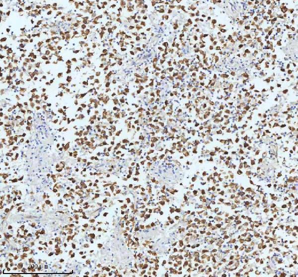
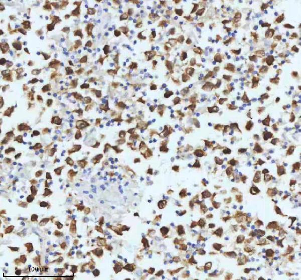
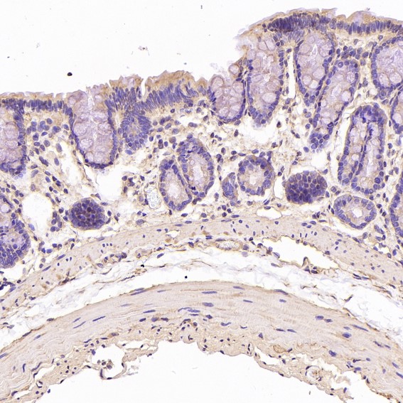
Real IHC data IHC analysis of TUBB3 using anti-TUBB3 antibody (M01857-1). TUBB3 was detected in a paraffin-embedded section of human testicular seminoma tissue. Heat mediated antigen retrieval was performed in EDTA buffer (pH 8.0, epitope retrieval solution). The tissue section was blocked with 10% goat serum. The tissue section was then incubated with 1:50 rabbit anti-TUBB3 Antibody (M01857-1) overnight at 4°C. Peroxidase Conjugated Goat Anti-rabbit IgG was used as secondary antibody and incubated for 30 minutes at 37°C. The tissue section was developed using HRP Conjugated Rabbit IgG Super Vision Assay Kit (Catalog # SV0002) with DAB as the chromogen.
Anti-beta III Tubulin TUBB3 Rabbit Monoclonal Antibody
Cat # M01857-1
Real IHC data Immunohistochemical analysis of paraffin-embedded human ovarian cancer, using beta III Tubulin Antibody(M01857) TUBB3 was detected in paraffin-embedded tissue section. Heat mediated antigen retrieval was performed in citrate buffer (pH6, epitope retrieval solution) for 20 mins. The tissue section was blocked with 10% goat serum. The tissue section was then incubated with 1ug/ml rabbit anti-TUBB3 Antibody (M01857)overnight at 4 Biotinylated goat anti-rabbit IgG was used as secondary antibody and incubated for 30 minutes at 37 The tissue section was developed using Strepavidin-Biotin-Complex (SABC)(Catalog # SA1022) with DAB as the chromogen.
Anti-beta III Tubulin TUBB3 Rabbit Monoclonal Antibody
Cat # M01857
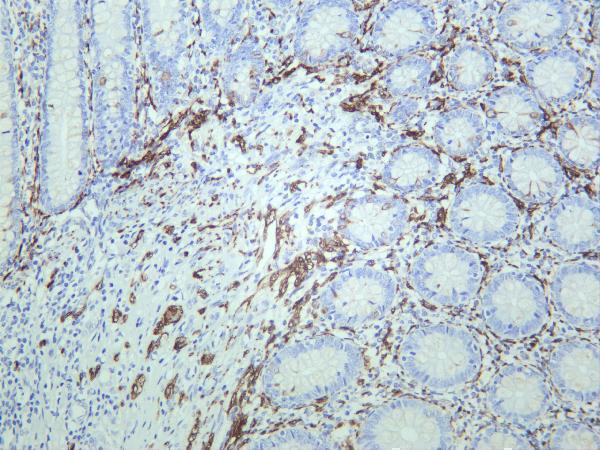
Real IHC data Human appendix was stained with anti-β3 Tubulin rabbit antibody
Anti-β3 Tubulin Rabbit Monoclonal Antibody
Cat # M01857-6

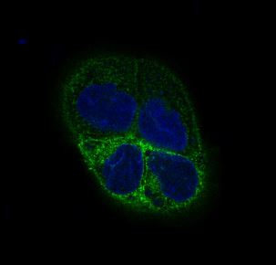
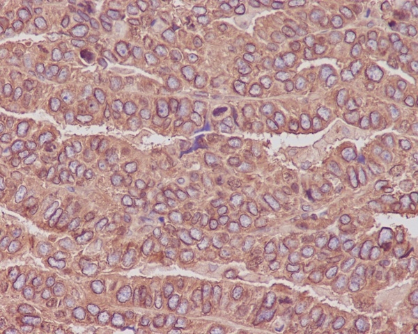
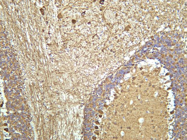
M01857-1 has IHC images of paraffin-embedded human testicular seminoma and thyroid papillary carcinoma, plus an IF image of A431 cells (catalog: M01857-1 image captions). M01857 has an IHC image of paraffin-embedded human ovarian cancer and an IF image caption; M01857-6 has IHC images of human appendix and mouse brain (catalog: M01857 and M01857-6 image captions).

Which to pick: For paraffin-section IHC, choose M01857-1 for its human tissue images using EDTA retrieval at pH 8.0 and a 1:50 primary dilution, or M01857 for its ovarian cancer image using citrate retrieval at pH 6 (catalog: each SKU’s IHC image caption). For IF/ICC, choose M01857-1: both applications are listed and its IF image caption identifies A431 cells (catalog: M01857-1 applications and IF image caption). For cross-species tissue IHC, M01857-6 has human appendix and mouse brain images and lists rat reactivity; its captions give no processing details, and the fixative is unreported for all three SKUs (catalog: image captions and reactivity).

Each figure is that product's own IHC / IF validation image from its datasheet.

References

  1. UniProt Consortium. UniProt entry Q13509 (TBB3_HUMAN, Tubulin beta-3 chain).
  2. Human Protein Atlas. TUBB3 tissue IHC expression (reliability: Approved).
  3. Human Protein Atlas. TUBB3 subcellular location (ICC-IF): Mainly localized to the microtubules. In addition localized to the cytokinetic bridge, mitotic spindle, primary cilium, primary cilium tip, basal body, flagellar centriole, principal piece and end piece. Caution: Based on antibodies targeting proteins from multiple genes..
  4. Human Protein Atlas. TUBB3 antibody validation summary (2 antibodies).
  5. High protein and mRNA expression levels of TUBB3 (class III ß-tubulin) are associated with aggressive tumor features in esophageal adenocarcinomas. Oncotarget 2017 — PMC5777763.
  6. High levels of class III β-tubulin expression are associated with aggressive tumor features in breast cancer. Oncology letters 2016 — PMC4774425.
  7. Trabecular Bone Microarchitecture Improvement Is Associated With Skeletal Nerve Increase Following Aerobic Exercise Training in Middle-Aged Mice. Frontiers in physiology 2021 — PMC8902445.
  8. Class III β-Tubulin Overexpression Induces Chemoresistance to Eribulin in a Leiomyosarcoma Cell Line. Analytical cellular pathology (Amsterdam) 2018 — PMC6033248.
  9. PubMed PMID:9473684 — UniProt-cited evidence.
  10. PubMed PMID:14702039 — UniProt-cited evidence.
  11. PubMed PMID:15616553 — UniProt-cited evidence.