TUBG1 / Tubulin gamma-1 chain · IHC design guide

Design Immunohistochemistry for TUBG1

Plan TUBG1 chromogenic IHC on paraffin sections using testis pachytene spermatocytes as a high-staining control (HPA tissue IHC). This guide covers expected cytoplasmic staining (HPA tissue IHC), a 1:50 starting dilution (datasheet: IHC 1:50), and cell-type-specific interpretation (HPA tissue IHC).

Evidence assembled Oct 2026 · For research use; verify linked source records and product datasheet before use
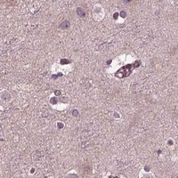
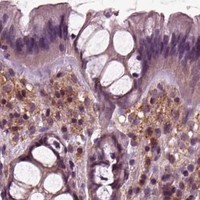
Immunohistochemistry protocol sheet for TUBG1 (IHC for TUBG1): expected localisation Cytoplasmic staining (HPA tissue IHC); centrosome/spindle expected (UniProt), antibody M06313, validated IHC image, and IHC protocol steps
Printable TUBG1 IHC protocol sheet — expected localisation Cytoplasmic staining (HPA tissue IHC); centrosome/spindle expected (UniProt), antibody M06313, controls and protocol steps. Open the full TUBG1 IHC guide →

TUBG1 Immunohistochemistry Experimental Design Guide

Expected localisation, validated protocols, controls and antibodies — the at-a-glance facts below, then the full design guide.

Must know before staining
Expected localisation Cytoplasmic staining (HPA tissue IHC); centrosome/spindle expected (UniProt)
Staining pattern Cytoplasmic staining; strongest in testis spermatocytes (HPA tissue IHC)
Antigen retrieval Citrate pH 6.0 HIER, 95–98 °C, 20 min (rule: cytoplasmic / membrane antigen)
Positive control ⓘ Testis+4 more · see all
Negative control ⓘ Esophagus+4 more · see all
Important caveats
Reasons your staining may differ from the expected pattern.
Fixation Keep fixation conditions consistent across sections (standard IHC practice; not target-specific)
Caveat Staining and RNA show medium consistency (HPA tissue IHC)
Regulation Testis-enhanced RNA expression (HPA tissue RNA)
Isoform / epitope No annotated isoforms; full-length chain, no TM segment (UniProt)
Section 1

Recommended TUBG1 IHC & IF Protocols

The catalog antibody’s IHC-P protocol (datasheet) is accompanied by one published paraffin-section workflow for TUBG1 (PMC9850107 methods).

Recommended immunohistochemistry (IHC-P) protocol parameters
SampleParaffin-embedded human breast cancer tissue; fixative not specified (datasheet M06313)
FixationImage fixative and duration unreported (datasheet M06313); verify before use.
Sectioning4–5 µm sections on charged slides (standard)
DeparaffinisationXylene, graded ethanol series to water (standard)
Antigen retrievalHeat-induced epitope retrieval in citrate buffer, pH 6.0, 20 min at 95–98 °C (standard rule: cytoplasmic / membrane antigen)
Peroxidase block3% H2O2, 10 min, room temperature (standard)
Blocking10% normal serum of the secondary host, 30 min, room temperature (standard)
Primary antibodyRabbit monoclonal (clone DAE-20) anti-TUBG1, 1:50 (datasheet M06313)
Primary incubationOvernight at 4 °C (standard)
DetectionHRP-polymer secondary, DAB chromogen 5–10 min (standard)
CounterstainHematoxylin, blue, dehydrate and mount (standard)
Expected resultTUBG1-positive staining in pachytene spermatocytes of testis (HPA tissue IHC: High). HPA tissue profile: General cytoplasmic expression, most abundant in testis. No signal in the no-primary control.
💡Decision noteStart with citrate pH 6.0 retrieval at 95–98 °C for 20 min (page retrieval rule); the paper used citric acid at 105 °C for 10 min (PMC9850107 methods).
Section 2

What Is the Expected TUBG1 Staining Pattern?

TUBG1 is a cytoplasmic, centrosomal protein that also localizes to mitotic spindle microtubules; it has no transmembrane segment (UniProt P23258). In paraffin-section IHC, expect general cytoplasmic staining, strongest in testicular pachytene spermatocytes (HPA tissue IHC: High). HPA rates its tissue IHC reliability Enhanced, while reporting medium consistency between staining and RNA expression (HPA tissue IHC).

What am I looking at on my slide?
Strong cytoplasmic staining in pachytene spermatocytes; staining in several other cell populations is less intense (HPA tissue IHC).This fits the reported pattern: High in pachytene spermatocytes and Medium in the listed adrenal, appendix, breast, bronchus, marrow and brain cell populations (HPA tissue IHC). Judge each named population separately.
Cytoplasmic staining with a small perinuclear focus, or signal associated with a mitotic spindle (UniProt P23258).A focus can fit centrosomal localization, and spindle-associated signal can fit mitotic localization (UniProt P23258). HPA describes tissue staining more broadly as cytoplasmic; visible puncta are not required to call a chromogenic section positive (HPA tissue IHC; standard IHC interpretation).
Predominantly nuclear or membrane-outline staining, with little cytoplasmic signal.That distribution conflicts with the annotated cytoplasmic, centrosomal and spindle locations and the absence of a transmembrane segment (UniProt P23258). Treat it as suspect; review controls and morphology before assigning specificity (standard IHC practice).
Strong signal in esophageal squamous cells, cardiomyocytes or skeletal myocytes (HPA tissue IHC: Not detected).HPA reports no detection in those specific cell populations, so unexpected strong staining merits a cross-reactivity or detection-background check (HPA tissue IHC; standard IHC practice). These entries do not make every cell in those tissues a negative control.
Diffuse color across tissue and blank areas, or no signal in testicular pachytene spermatocytes.Diffuse color makes localization hard to score and can reflect assay background; an unstained known-positive population makes a negative test section inconclusive (standard IHC practice; HPA tissue IHC: pachytene spermatocytes High).
💡Expected TUBG1 appearanceCall a convincing positive result cytoplasmic staining in the relevant cells, especially High signal in testicular pachytene spermatocytes; strong isolated nuclear, membrane-outline or blank-area color is suspect (HPA tissue IHC; UniProt P23258; standard IHC practice).
How each factor affects the staining
Localization and topologyTUBG1 is cytoplasmic, centrosomal and associated with mitotic spindle microtubules, with no transmembrane segment (UniProt P23258). Score cytoplasmic signal in its cellular context; do not use a membrane pattern as the expected readout.
Tissue and cell selectionTesticular pachytene spermatocytes are High, while listed populations in other tissues are Medium, Low or Not detected (HPA tissue IHC). Use the named cell population when selecting and reading controls; a tissue name alone is insufficient.
IHC antibody evidenceCAB004608 has Enhanced IHC validation; HPA describes the tissue profile as Enhanced with medium consistency against RNA expression (HPA antibodies; HPA tissue IHC). These ratings support interpretation but do not establish every stain as specific.
Processing and fixation evidenceUniProt lists one chain spanning residues 1–451, no signal peptide or propeptide, and no annotated isoforms (UniProt P23258). The supplied sources report no target-specific fixation sensitivity or retrieval condition; optimize general IHC conditions empirically.
IF/ICC Q&A: Is a sperm principal-piece signal the expected paraffin IHC pattern?No direct transfer is justified: HPA approves principal-piece localization in sperm ICC-IF but cautions that it is based on antibodies targeting proteins from multiple genes (HPA subcellular). Use the tissue IHC profile for this guide (HPA tissue IHC).
Why is my staining missing, weak or wrong?
SituationLikely causeNext action
No stain in the testis positive control.The expected High pachytene-spermatocyte signal is absent (HPA tissue IHC); a failed run or inaccessible antigen is possible (standard IHC practice).Confirm that pachytene spermatocytes are present, then check primary-antibody and detection steps; compare a small retrieval-condition series before interpreting test samples (standard IHC practice).
All cells show similar diffuse brown color.High assay background can obscure the reported cell-specific intensity differences (HPA tissue IHC; standard IHC practice).Inspect a no-primary control; review blocking, washing, primary dilution and chromogen development using general IHC controls (standard IHC practice).
Color persists in a no-primary control.Primary-independent detection or endogenous enzyme activity may contribute to chromogenic signal (standard IHC practice).Check the detection reagents and the relevant endogenous-enzyme blocking step; repeat the control beside the stained section (standard IHC practice).
Strong nuclear or membrane-outline signal dominates.The pattern does not match cytoplasmic, centrosomal or spindle localization, or the lack of a transmembrane segment (UniProt P23258).Recheck morphology and control sections, then review antibody dilution and background before assigning a TUBG1-positive score (standard IHC practice).
Strong staining appears in an HPA-listed Not detected cell population.The cell-specific discrepancy may reflect cross-reactivity or background; HPA's negative calls apply to the named cells (HPA tissue IHC; standard IHC practice).Identify the stained cell type carefully and compare no-primary and known-positive controls; avoid calling the entire tissue negative (HPA tissue IHC; standard IHC practice).
An IF/ICC image shows sperm principal-piece signal that seems inconsistent with the IHC slide.That ICC-IF localization carries an explicit multiple-gene antibody caution and is separate from HPA's tissue IHC profile (HPA subcellular; HPA tissue IHC).Interpret the paraffin section against cytoplasmic staining and cell-specific tissue levels; assess the IF result in its own assay context (HPA tissue IHC; HPA subcellular).

Sample controls for TUBG1 IHC & IF

🧪Run testis first; pachytene spermatocytes should stain (HPA: High in testis pachytene spermatocytes). Use esophagus squamous epithelial cells as the negative tissue (HPA: Not detected); on the testis slide, treat cells without detectable staining as background references, since the supplied HPA rows do not identify a specific internal negative cell type.
Positive control tissue: Testis (Pachytene spermatocytes, HPA High)
Negative control tissue: Esophagus (HPA Not detected)
ICC-IF cell lines (HPA subcellular resource): HPA ICC-IF images show TUBG1 in Sperm, with annotated localisation: Principal piece (approved) (HPA subcellular).
Technical controls: Include a no-primary, secondary-only control; an isotype control matched to the primary antibody’s host species and class; and TUBG1 knockout tissue or a validated peptide-block control (standard IHC practice). Check testis sections for endogenous peroxidase background, and block endogenous biotin if using biotin-based detection (standard chromogenic IHC practice).
⚠️Feasibility: A target-specific fixation window and retrieval effect are unreported in the supplied evidence; assess antigen retrieval empirically for paraffin sections (standard IHC practice). The selected M06313 paraffin-section caption does not state a fixative (selected-SKU caption: fixative not stated). The evidence does not establish whether frozen sections or IF are easier, or document a testis-specific artifact; IF could help assess centrosomal or spindle localization (UniProt P23258: subcellular localization), but the reported sperm principal-piece signal warrants caution because the HPA antibodies target proteins from multiple genes (HPA subcellular: caution).

HPA tissue IHC evidence for TUBG1

Comprehensive Human Protein Atlas IHC scoring per tissue (reliability: Enhanced — Medium consistency between antibody staining and RNA expression data.). Rows are taken directly from the HPA tissue chart — click any row's HPA link to view the source.

Positive expression · recommended positive controls

TissueCell typeLevelEvidenceSource
Testis Pachytene spermatocytes High Protein (IHC) HPA →
Adrenal gland Glandular cells Medium Protein (IHC) HPA →
Appendix Glandular cells Medium Protein (IHC) HPA →
Bone marrow Hematopoietic cells Medium Protein (IHC) HPA →
Breast Glandular cells Medium Protein (IHC) HPA →

Undetected expression · recommended negative controls

TissueCell typeLevelEvidenceSource
Esophagus Squamous epithelial cells Not detected Protein (IHC) HPA →
Heart muscle Cardiomyocytes Not detected Protein (IHC) HPA →
Hippocampus Glial cells Not detected Protein (IHC) HPA →
Liver Cholangiocytes Not detected Protein (IHC) HPA →
Skeletal muscle Myocytes Not detected Protein (IHC) HPA →
Section 3

Advanced TUBG1 IHC Tips

Troubleshoot TUBG1 staining in paraffin sections by checking retrieval, cellular distribution, controls, and scoring before interpreting chromogenic signal.

How should I optimize retrieval when TUBG1 staining is weak in paraffin sections?
Use citrate buffer at pH 6.0 for HIER at 95–98 °C for 20 min (page IHC retrieval setting). Cool sections consistently before staining, and compare the same tissue across retrieval runs so temperature and handling do not confound the result (standard IHC practice). Evaluate cytoplasmic staining and discrete centrosomal signal where morphology permits, since both distributions are consistent with TUBG1 (UniProt P23258 localisation). If staining remains weak, test a different retrieval buffer or pH as a fallback on adjacent sections, with matched controls (standard IHC practice). Excessive retrieval can damage morphology and make apparent signal difficult to assign to cells (standard IHC practice).
Could fixation explain inconsistent TUBG1 staining between paraffin blocks?
Target-specific fixation sensitivity is unknown: the M06313 breast cancer image specifies paraffin embedding but does not report its fixative (M06313 caption). Record fixative, fixation duration, section thickness, and retrieval conditions for each block before comparing staining intensity (standard IHC practice). When preparing new specimens, a documented 10% neutral buffered formalin workflow provides a consistent starting condition, but it has no supplied TUBG1-specific validation (standard IHC practice). Compare blocks using the same antibody and detection run, alongside a tissue control with expected expression (standard IHC practice; HPA: high in testis pachytene spermatocytes). Do not assign a weak result to fixation without a controlled comparison (standard IHC practice).
What staining pattern should count as plausible TUBG1 localisation?
Assess cytoplasmic staining first: tissue IHC reports general cytoplasmic expression, with the strongest listed signal in testis pachytene spermatocytes (HPA tissue IHC). Small perinuclear or centrosomal foci can also fit TUBG1 biology, and mitotic cells may show spindle-associated signal (UniProt P23258 localisation). Use a 40× objective or comparable magnification to relate any punctum to an intact cell rather than a section deposit (standard IHC practice). Score nuclear-only or membrane-rim staining separately from the expected pattern, since TUBG1 has no annotated transmembrane segment (UniProt P23258 topology and localisation). Check those atypical patterns against omission controls before treating them as biological findings (standard IHC practice).
How can I assess whether the antibody distinguishes TUBG1 from related gamma tubulin?
Treat isoform specificity and gene specificity as separate questions: the record lists 0 annotated TUBG1 isoforms, while TUBG1 or TUBG2 can occupy gamma-tubulin ring complexes (UniProt P23258 record and subunit). The supplied M06313 caption does not identify the antibody epitope or establish discrimination from TUBG2 (M06313 caption). Request the immunogen sequence or epitope information and compare it with TUBG2 before attributing staining uniquely to TUBG1 (standard antibody validation practice). An independent antibody recognizing a different epitope, or a controlled loss-of-target experiment, can strengthen that attribution (standard antibody validation practice). Keep all sections under the same pH 6.0 retrieval condition while comparing antibodies (page IHC retrieval setting).
How should I adapt this TUBG1 question to multiplex IF?
For IF multiplexing, pair TUBG1 with a validated marker for the expected cell population, such as testis pachytene spermatocytes, and inspect both channels in the same cells (HPA: high in pachytene spermatocytes; standard IF practice). Choose a far-red fluorophore when tissue autofluorescence crowds shorter wavelengths, and include an unstained section to identify that background (standard IF practice). Because TUBG1 is cytoplasmic and has no transmembrane segment, begin with a mild intracellular permeabilisation trial, such as 0.1% detergent for 5 min (UniProt P23258 localisation and topology; standard IF practice). Optimise that step for the actual antibody and specimen (standard IF practice). The supplied HPA sperm IF localisation carries a multiple-gene antibody caution (HPA subcellular).
How do I reduce diffuse brown signal without losing plausible TUBG1 staining?
Start with a no-primary control and inspect whether brown deposits persist in the same compartments as the test slide (standard chromogenic IHC practice). Quench endogenous peroxidase, for example with 3% hydrogen peroxide for 10 min, and control DAB development time across slides (standard chromogenic IHC practice). Then optimise blocking, antibody concentration, and washes one variable at a time while retaining a known positive tissue control (standard IHC practice; HPA: high in testis pachytene spermatocytes). Preserve interpretable cytoplasmic staining while checking centrosomal foci where cell morphology allows, as these fit TUBG1 localisation (HPA tissue IHC; UniProt P23258 localisation). Edge-heavy staining or signal in the no-primary control needs separate investigation (standard IHC practice).
What is a defensible way to quantify TUBG1 chromogenic staining? ⚠ ANSWER MARKED FOR VERIFICATION
Define the cell population and scoring compartment before counting; report cytoplasmic staining separately from discrete centrosomal signal (HPA tissue IHC; UniProt P23258 localisation; standard IHC practice). For broad cytoplasmic signal, record the percentage of positive cells and an H-score using intensity grades 0–3, with identical thresholds across slides (standard IHC scoring practice). For resolvable puncta, count positive cells or foci per mm² of viable tissue and document the image sampling rule (standard image analysis practice). Normalise counts to the eligible cells or viable tissue area, and stratify by cell type because HPA reports different levels across cell populations (standard image analysis practice; HPA tissue IHC). Exclude folds and necrotic regions consistently (standard IHC practice).
Which findings support true TUBG1 staining rather than an IHC artefact?
Give greatest weight to reproducible cytoplasmic staining in intact cells, with centrosomal or mitotic spindle-associated signal when structures are resolvable (HPA tissue IHC; UniProt P23258 localisation). Testis pachytene spermatocytes provide a high-expression reference, while several listed cell populations have no detected signal (HPA tissue IHC). Treat isolated nuclear-only or membrane-rim staining, section-edge enhancement, and staining over necrosis as suspect until controls and morphology support them (UniProt P23258 topology and localisation; standard IHC practice). Check a no-primary slide for endogenous enzyme or detection background, using the same 10 min peroxidase-block workflow across comparisons (standard chromogenic IHC practice). Interpret changes against matched tissue processing and scoring rules (standard IHC practice).
Boster reagents

Best TUBG1 / Tubulin gamma-1 chain IHC Antibodies

M06313 has an IHC image of paraffin-embedded human breast cancer (catalog IHC image caption). IF/ICC is listed, with Human, Monkey, Mouse, Rat, and Zebrafish reactivity (catalog applications/reactivity).

Real IHC data Immunohistochemical analysis of paraffin-embedded human breast cancer, using Tubulin gamma Antibody.
Anti-Tubulin gamma Rabbit Monoclonal Antibody
Cat # M06313

M06313 is the sole card and shows IHC staining of paraffin-embedded human breast cancer (catalog IHC image caption). It lists IF/ICC applications and five reactive species, but supplies no IF image (catalog applications/reactivity; IF image alts).

Which to pick: For tissue IHC, choose M06313 based on its paraffin-embedded human breast cancer image; the fixative is unreported (catalog IHC image caption). For IF/ICC, M06313 lists both applications at 1:50, although no IF image is supplied (catalog applications; catalog IF dilution; IF image alts). For cross-species work, M06313 is a rabbit monoclonal, clone DAE-20, with Human, Monkey, Mouse, Rat, and Zebrafish reactivity listed; confirm staining in the chosen species (catalog host/clone/reactivity).

Each figure is that product's own IHC / IF validation image from its datasheet.

References

  1. UniProt Consortium. UniProt entry P23258 (TBG1_HUMAN, Tubulin gamma-1 chain).
  2. Human Protein Atlas. TUBG1 tissue IHC expression (reliability: Enhanced).
  3. Human Protein Atlas. TUBG1 subcellular location (ICC-IF): Localized to the principal piece. Caution: Based on antibodies targeting proteins from multiple genes..
  4. Human Protein Atlas. TUBG1 antibody validation summary (2 antibodies).
  5. Upregulated TUBG1 expression is correlated with poor prognosis in hepatocellular carcinoma. PeerJ 2022 — PMC9745943.
  6. Construction and validation of a prognostic model for hepatocellular carcinoma: Inflammatory ferroptosis and mitochondrial metabolism indicate a poor prognosis. Frontiers in oncology 2022 — PMC9850107.
  7. Screening of co-pathogenic genes of non-alcoholic fatty liver disease and hepatocellular carcinoma. Frontiers in oncology 2022 — PMC9410624.
  8. Bioinformatics Analysis and Experimental Verification Define Different Angiogenesis Subtypes in Endometrial Carcinoma and Identify a Prognostic Signature. ACS omega 2024 — PMC11190931.
  9. PubMed PMID:1904010 — UniProt-cited evidence.
  10. PubMed PMID:14702039 — UniProt-cited evidence.
  11. PubMed PMID:15489334 — UniProt-cited evidence.