TXNRD2 / Thioredoxin reductase 2, mitochondrial · IHC design guide

Design Immunohistochemistry for TXNRD2

Plan paraffin IHC for TXNRD2 using its general cytoplasmic tissue pattern (HPA tissue IHC) and mitochondrial location (UniProt). Lung macrophages and testis Leydig cells provide high-staining reference populations (HPA tissue IHC).

Evidence assembled Oct 2026 · For research use; verify linked source records and product datasheet before use
Immunohistochemistry protocol sheet for TXNRD2 (IHC for TXNRD2): expected localisation General cytoplasmic staining (HPA tissue IHC); mitochondrial protein (UniProt), antibody PA1903, validated IHC image, and IHC protocol steps
Printable TXNRD2 IHC protocol sheet — expected localisation General cytoplasmic staining (HPA tissue IHC); mitochondrial protein (UniProt), antibody PA1903, controls and protocol steps. Open the full TXNRD2 IHC guide →

TXNRD2 Immunohistochemistry Experimental Design Guide

Expected localisation, validated protocols, controls and antibodies — the at-a-glance facts below, then the full design guide.

Must know before staining
Expected localisation General cytoplasmic staining (HPA tissue IHC); mitochondrial protein (UniProt)
Staining pattern Cytoplasmic; high in lung macrophages and testis Leydig cells (HPA tissue IHC)
Antigen retrieval Citrate pH 6 HIER, heat-mediated (datasheet PA1903)
Positive control ⓘ Lung+4 more · see all
Negative control ⓘ Adipose tissue+4 more · see all
Important caveats
Reasons your staining may differ from the expected pattern.
Fixation Keep fixation consistent across sections. (standard IHC practice; not target-specific)
Caveat Staining varies markedly by cell type (HPA tissue IHC)
Regulation Expression regulation is not established (UniProt)
Isoform / epitope 4 isoforms; chain 37–524; epitope coverage unknown (UniProt)
Section 1

Recommended TXNRD2 IHC & IF Protocols

The catalog antibody’s paraffin IHC protocol uses citrate pH 6 retrieval (datasheet PA1903). A published TXNRD2 protocol uses frozen prostate sections (PMC3675098).

Recommended immunohistochemistry (IHC-P) protocol parameters
SampleTissue sections; selected-image fixative not specified (standard IHC workflow)
FixationImage fixative and duration unreported (datasheet PA1903); verify before use.
Sectioning4–5 µm sections on charged slides (standard)
DeparaffinisationXylene, graded ethanol series to water (standard)
Antigen retrievalHeat retrieval: Citrate pH 6, 20 min (datasheet PA1903)
Peroxidase block3% H2O2, 10 min, room temperature (standard)
Blocking10% goat serum (datasheet PA1903)
Primary antibodyRabbit anti-TXNRD2, 0.5-1μg/ml (datasheet PA1903)
Primary incubationOvernight at 4 °C (datasheet PA1903)
DetectionStreptavidin-biotin complex (SABC), DAB chromogen (datasheet PA1903)
CounterstainHematoxylin, blue, dehydrate and mount (standard)
Expected resultTXNRD2-positive staining in macrophages of lung (HPA tissue IHC: High). HPA tissue profile: General cytoplasmic expression. No signal in the no-primary control.
💡Decision noteStart with heat-mediated citrate pH 6 retrieval for paraffin sections (datasheet PA1903). The published frozen-section protocol uses acetone fixation (PMC3675098).
Section 2

What Is the Expected TXNRD2 Staining Pattern?

TXNRD2 is mitochondrial and has no annotated transmembrane segment (UniProt Q9NNW7 topology). In paraffin sections, expect cytoplasmic staining, particularly in lung macrophages and testicular Leydig cells, both scored High (HPA tissue IHC). HPA rates its tissue IHC evidence Supported, with medium agreement between staining and RNA data and external verification pending (HPA tissue IHC).

What am I looking at on my slide?
Cytoplasmic stain in lung macrophages or testicular Leydig cells, stronger than nearby unstained cells.This fits the High cell-specific staining reported in both tissues (HPA tissue IHC). A cytoplasmic appearance is consistent with mitochondrial TXNRD2 (UniProt Q9NNW7; HPA tissue IHC). Chromogenic IHC alone does not prove that each stained structure is a mitochondrion (general IHC practice).
Predominantly nuclear staining, with little convincing cytoplasmic staining.Treat this as a localization mismatch: UniProt places TXNRD2 in mitochondria, and HPA describes general cytoplasmic tissue staining (UniProt Q9NNW7; HPA tissue IHC). Check controls and antibody performance before calling it TXNRD2; nuclear color alone is insufficient evidence of a new localization (general IHC practice).
Strong stain chiefly in a cell population reported as Not detected, such as adipocytes or cardiomyocytes.This conflicts with those cell-level HPA observations and raises a specificity or detection concern (HPA tissue IHC; general IHC practice). Identify the stained cell type before comparing tissues: HPA reports ovarian stromal cells as Not detected, whereas UniProt describes high expression in ovary without specifying those cells (HPA tissue IHC; UniProt Q9NNW7).
Color spreads across many cells or the section with little cell or compartment distinction.Diffuse background prevents a reliable TXNRD2 call even where expression is expected (general IHC practice). Review the control lacking primary antibody, blocking, washes and chromogen development before interpreting intensity; HPA's Supported rating does not validate an individual staining run (HPA tissue IHC; general IHC practice).
No cytoplasmic signal in lung macrophages or testicular Leydig cells.Both are useful positive-reference cell populations because HPA scores their staining High (HPA tissue IHC). First check that the intended cells are present and recognizable, then assess primary antibody, retrieval and detection controls (general IHC practice). A failed run cannot establish absent TXNRD2 expression.
💡Expected TXNRD2 appearanceCall a convincing positive when cytoplasmic staining is strong in lung macrophages or testicular Leydig cells (HPA tissue IHC), consistent with mitochondrial TXNRD2 (UniProt Q9NNW7); isolated nuclear color or uniform section-wide color is suspect (general IHC practice).
How each factor affects the staining
Cell identity and tissue choiceHPA scores lung macrophages and testicular Leydig cells High, but adipocytes and cardiomyocytes Not detected (HPA tissue IHC). Compare the specified cells, since a tissue name alone does not define its expected stain.
Evidence strengthThe tissue IHC profile is Supported, with medium agreement with RNA expression and external verification pending (HPA tissue IHC). HPA lists two antibodies as IHC Supported; neither is listed as IHC Enhanced in the supplied antibody record (HPA antibodies).
Cellular localizationUniProt assigns TXNRD2 to mitochondria, while HPA describes general cytoplasmic tissue staining (UniProt Q9NNW7; HPA tissue IHC). Use cytoplasmic location for paraffin-section interpretation; apparent nuclear-only signal warrants review (general IHC practice).
IF/ICC Q&A: Should fluorescence be confined to mitochondria?Mitochondria are the supported HPA ICC-IF location; cytosol is marked uncertain (HPA subcellular). HPA lists A-431 and U2OS ICC-IF images (HPA subcellular). This localization evidence informs interpretation but supplies no IHC protocol option.
Sequence and tissue-level caveatsUniProt lists four isoforms and a mature chain spanning residues 37–524 (UniProt Q9NNW7). The supplied record gives no antibody epitope, so it cannot establish isoform coverage or a retrieval requirement. UniProt reports conflicting kidney expression findings; avoid using kidney alone as a decisive reference (UniProt Q9NNW7).
Why is my staining missing, weak or wrong?
SituationLikely causeNext action
High-reference cells stain weakly or not at all.The reference cells may be absent from the section, or an IHC step may have underperformed (general IHC practice).Confirm lung macrophages or testicular Leydig cells are present (HPA tissue IHC); review retrieval, primary antibody and detection controls for the run (general IHC practice).
Predominantly nuclear signal appears.The pattern conflicts with mitochondrial assignment and HPA's cytoplasmic tissue profile (UniProt Q9NNW7; HPA tissue IHC).Recheck morphology, counterstain and control sections; withhold a TXNRD2-positive call until a credible cytoplasmic pattern is demonstrated (general IHC practice).
Signal appears in nearly every cell and over blank areas.Non-specific background or detection-system color may obscure cell-specific staining (general IHC practice).Inspect a control lacking primary antibody; review blocking, washes and chromogen development (general IHC practice).
A supposedly negative tissue contains positive cells.A tissue-level label may hide different cell populations; HPA's Not detected calls apply to named cells (HPA tissue IHC).Identify the stained cells and compare them with the HPA cell-level entry before judging specificity (HPA tissue IHC).
Brown signal persists in the control lacking primary antibody.Endogenous enzyme activity or detection-reagent background can produce color in chromogenic IHC (general IHC practice).Check the detection system's recommended endogenous-activity block and repeat the control alongside the stained section (general IHC practice).
Kidney staining disagrees with the expected level.UniProt reports both high and low kidney expression in different cited findings (UniProt Q9NNW7).Use HPA's High lung macrophages or testicular Leydig cells as the clearer cell-level comparison, and interpret kidney cautiously (HPA tissue IHC; UniProt Q9NNW7).

Sample controls for TXNRD2 IHC & IF

🧪Run lung first and assess macrophages for strong staining (HPA: High in lung macrophages). Use adipose tissue adipocytes as the negative comparator (HPA: Not detected in adipocytes); on the lung slide, neighboring nonmacrophage cells should lack nonspecific chromogen, but their TXNRD2 status is unestablished (standard IHC practice).
Positive control tissue: Lung (Macrophages, HPA High)
Negative control tissue: Adipose tissue (HPA Not detected)
ICC-IF cell lines (HPA subcellular resource): HPA ICC-IF images show TXNRD2 in A-431, U2OS, with annotated localisation: Mitochondria (supported), Cytosol (uncertain) (HPA subcellular).
Technical controls: Include a no-primary, secondary-only control and a host-matched nonimmune IgG control for a polyclonal antibody or an isotype-matched control for a monoclonal antibody; assess specificity with TXNRD2 knockout material or immunizing-peptide competition if available (standard IHC practice). For lung macrophages, quench endogenous peroxidase and check pigment in chromogenic IHC; assess autofluorescence with an unstained section for IF (standard IHC/IF practice).
⚠️Feasibility: No supplied target-specific source reports a fixation window or fixation effect, and the selected PA1903 IHC(P) lung-cancer caption does not state a fixative (PA1903 caption). Antigen-retrieval dependence is unreported; optimize retrieval empirically for paraffin IHC (standard IHC practice). The supplied evidence does not establish frozen sections or IF as easier, although ICC-IF images exist for A-431 and U2OS (HPA subcellular); macrophage pigment and endogenous peroxidase can complicate lung chromogenic scoring (standard IHC practice).

HPA tissue IHC evidence for TXNRD2

Comprehensive Human Protein Atlas IHC scoring per tissue (reliability: Supported — Medium consistency between antibody staining and RNA expression data. Pending external verification.). Rows are taken directly from the HPA tissue chart — click any row's HPA link to view the source.

Positive expression · recommended positive controls

TissueCell typeLevelEvidenceSource
Lung Macrophages High Protein (IHC) HPA →
Testis Leydig cells High Protein (IHC) HPA →
Adrenal gland Glandular cells Medium Protein (IHC) HPA →
Appendix Glandular cells Medium Protein (IHC) HPA →
Breast Glandular cells Medium Protein (IHC) HPA →

Undetected expression · recommended negative controls

TissueCell typeLevelEvidenceSource
Adipose tissue Adipocytes Not detected Protein (IHC) HPA →
Caudate Glial cells Not detected Protein (IHC) HPA →
Heart muscle Cardiomyocytes Not detected Protein (IHC) HPA →
Ovary Ovarian stroma cells Not detected Protein (IHC) HPA →
Skeletal muscle Myocytes Not detected Protein (IHC) HPA →
Section 3

Advanced TXNRD2 IHC Tips

Troubleshoot TXNRD2 staining in paraffin sections by checking retrieval, cell identity, subcellular pattern and controls before comparing signal intensity (UniProt Q9NNW7; HPA tissue IHC).

What retrieval should I try first when TXNRD2 staining is weak?
Start with heat-mediated antigen retrieval in citrate buffer at pH 6 for paraffin sections (datasheet PA1903). Run a consistent heating and cooling cycle, such as 15–20 minutes at retrieval temperature followed by 20 minutes of cooling, and record the conditions for comparison (standard IHC practice). If staining remains weak, compare a second retrieval condition on matched sections while keeping antibody incubation and chromogen development constant (standard IHC practice). Judge improvement in expected cell populations and cytoplasmic staining, including lung macrophages, rather than by darker staining across the whole section (HPA: high in lung macrophages; HPA: general cytoplasmic expression).
Could fixation explain weak or patchy TXNRD2 staining?
Target-specific fixation sensitivity is unknown, and the PA1903 tissue caption does not state the fixative (PA1903 tissue-IHC caption). Record fixation conditions for every specimen and compare sections processed under the same conditions before attributing differences to TXNRD2 expression (standard IHC practice). If staining is patchy, inspect morphology and section edges, then compare a second section with the same pH 6 citrate retrieval and detection run (datasheet PA1903; standard IHC practice). Use a positive cell population on the same slide, when present, to check whether weak staining tracks tissue processing rather than cell identity (HPA: high in lung macrophages; standard IHC practice).
Where should convincing TXNRD2 signal appear in a chromogenic section?
Expect predominantly cytoplasmic staining, potentially with a granular mitochondrial pattern, rather than a nuclear-only pattern (HPA: general cytoplasmic expression; UniProt Q9NNW7: mitochondrion). HPA subcellular imaging supports mitochondrial localisation and lists cytosolic localisation as uncertain, so diffuse cytoplasmic signal needs cautious interpretation (HPA subcellular: mitochondria supported, cytosol uncertain). Examine the pattern at high magnification in morphologically intact cells, then compare it with a no-primary control run using the same chromogen development (standard IHC practice). Do not require a visible mitochondrial pattern in every positive cell: chromogenic resolution and section thickness can blur organelle-level detail (standard IHC practice).
Could isoforms or epitope accessibility explain inconsistent staining?
TXNRD2 has 4 annotated isoforms, but the supplied evidence does not map the catalog antibody’s epitope to them (UniProt Q9NNW7: isoforms 1–4; supplied PA1903 evidence). Check the antibody’s documented immunogen or epitope before claiming that staining represents every isoform, and keep that limitation in the scoring notes (standard IHC practice). The annotated mature chain spans residues 37–524; processing could matter if an epitope lies outside that chain, but its location is unknown here (UniProt Q9NNW7: chain 37–524; supplied PA1903 evidence). Compare retrieval and antibody conditions on adjacent sections before interpreting a negative cell population as an isoform-specific result (standard IHC practice).
How can IF help resolve an ambiguous IHC localisation pattern?
Use IF as a separate localisation check, with a macrophage marker such as CD68 where lung macrophages are being assessed (HPA: high in lung macrophages; standard IF practice). Pair spectrally distinct fluorophores and inspect unstained tissue in each channel to choose detection away from tissue autofluorescence (standard IF practice). TXNRD2 is mitochondrial with no annotated transmembrane segment, but the antibody epitope’s accessibility is unspecified; optimise mild permeabilisation and confirm mitochondrial access empirically (UniProt Q9NNW7: mitochondrion, no transmembrane segment; supplied PA1903 evidence; standard IF practice). Compare mitochondrial overlap with the chromogenic cytoplasmic pattern, and include single-stain controls when multiplexing (HPA subcellular: mitochondria supported; standard IF practice).
How do I reduce diffuse brown staining without losing TXNRD2 signal?
First compare the test section with no-primary and chromogen-only controls to locate background arising from detection chemistry or tissue (standard IHC practice). For a peroxidase-based chromogenic workflow, apply a peroxidase block before DAB and check whether signal persists in the no-primary control (standard IHC practice). Optimise protein blocking, washing and antibody concentration on matched sections, changing 1 variable at a time rather than assuming diffuse colour is TXNRD2 (standard IHC practice). Preserve genuine cytoplasmic staining in expected cells, such as lung macrophages, while checking section edges and damaged tissue for nonspecific colour (HPA: high in lung macrophages; HPA: general cytoplasmic expression; standard IHC practice).
What should I score when comparing TXNRD2 across IHC sections? ⚠ ANSWER MARKED FOR VERIFICATION
Define the cell population and region before scoring; lung macrophages and respiratory epithelial cells should not be pooled simply because both can stain (HPA: high in lung macrophages; medium in bronchial respiratory epithelial cells). For a defined population, report % positive cells plus intensity or an H-score, using one threshold and exposure-independent chromogen assessment across the batch (standard IHC practice). If cell abundance is the question, report positive-cell density per mm² of viable tissue or compartment area (standard IHC practice). Normalise comparisons to the number of eligible cells or viable area, and document batch controls and excluded necrotic regions (standard IHC practice).
How can I distinguish true TXNRD2 staining from artefact?
Give greater weight to cytoplasmic signal in intact expected cells, particularly lung macrophages, than to isolated colour at a cut edge (HPA: general cytoplasmic expression; high in lung macrophages; standard IHC practice). A nuclear-only pattern is inconsistent with the supported mitochondrial localisation, although chromogenic IHC may not resolve individual mitochondria (HPA subcellular: mitochondria supported; standard IHC practice). Exclude necrotic areas and compare no-primary and peroxidase-block controls before calling brown deposits positive, because damaged tissue and endogenous enzyme activity can mimic DAB signal (standard IHC practice). Interpret negative cells cautiously: HPA tissue staining has medium RNA concordance and awaits external verification (HPA: Supported reliability description).
Boster reagents

Best TXNRD2 / Thioredoxin reductase 2, mitochondrial IHC Antibodies

PA1903 has IHC(P) images from human lung cancer tissue and rat intestine; the catalog provides no IF/ICC image or dilution (PA1903 image captions; catalog).

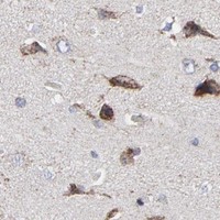
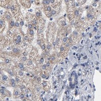
Real IHC data Anti-TXNRD2 antibody, PA1903, IHC(P) IHC(P): Human Lung Cancer Tissue
Anti-TXNRD2 Antibody ®
Cat # PA1903

PA1903 is the sole rendered SKU, with IHC(P) shown in human lung cancer tissue (PA1903 image caption; card payload). Its second caption documents paraffin-section IHC in rat intestine using 1 μg/ml antibody after citrate retrieval at pH 6 for 20 minutes (PA1903 image caption).

Which to pick: For tissue IHC, choose PA1903 because its own captions show human lung cancer and rat intestine paraffin sections; the fixative is unreported (PA1903 image captions). For mouse-inclusive IHC, M03900-1 is a rabbit monoclonal listed for human, mouse and rat, though it has no IHC image in the payload (M03900-1 catalog). Neither SKU lists IF/ICC or an IF dilution, so the payload does not support an IF/ICC antibody recommendation (PA1903 and M03900-1 catalog).

Each figure is that product's own IHC / IF validation image from its datasheet.

References

  1. UniProt Consortium. UniProt entry Q9NNW7 (TRXR2_HUMAN, Thioredoxin reductase 2, mitochondrial).
  2. Human Protein Atlas. TXNRD2 tissue IHC expression (reliability: Supported).
  3. Human Protein Atlas. TXNRD2 subcellular location (ICC-IF): Localized to the cytosol and mitochondria..
  4. Human Protein Atlas. TXNRD2 antibody validation summary (2 antibodies).
  5. Stromal response to prostate cancer: nanotechnology-based detection of thioredoxin-interacting protein partners distinguishes prostate cancer associated stroma from that of benign prostatic hyperplasia. PloS one 2013 — PMC3675098.
  6. Txnrd2 loss in skeletal muscle causes muscle atrophy and drives leanness and obesity resistance. Redox biology 2026 — PMC13099521.
  7. Loss of IDH2 Accelerates Age-related Hearing Loss in Male Mice. Scientific reports 2018 — PMC5864918.
  8. Auranofin is a potent suppressor of osteosarcoma metastasis. Oncotarget 2016 — PMC4808036.
  9. PubMed PMID:10455115 — UniProt-cited evidence.
  10. PubMed PMID:9923614 — UniProt-cited evidence.
  11. PubMed PMID:10215850 — UniProt-cited evidence.