This website uses cookies to ensure you get the best experience on our website.


- Table of Contents
Plan chromogenic FFPE staining around UBQLN4’s general nuclear tissue pattern (HPA tissue IHC). The catalog antibody A08887 has an IHC dilution of 1:100–1:300 (datasheet: A08887); respiratory epithelial and rectal glandular cells show high staining (HPA tissue IHC).

Expected localisation, validated protocols, controls and antibodies — the at-a-glance facts below, then the full design guide.
| Expected localisation | Predominantly nuclear in tissue sections (HPA tissue IHC) | |
| Staining pattern | General nuclear staining across cell types (HPA tissue IHC) | |
| Antigen retrieval | Tris-EDTA pH 9.0 HIER, 95–98 °C, 20 min (rule: nuclear antigen) | |
| Positive control | Bronchus+4 more · see all | |
| Negative control | Heart muscle+1 more · see all |
| Fixation | Matched tissue-IHC evidence does not establish the fixation claim. Validate the specimen-specific method before use. (selected-SKU IHC image A08887) | |
| Caveat | Cardiomyocytes may lack staining despite reported heart expression (HPA tissue IHC; UniProt) | |
| Regulation | DNA damage drives nuclear recruitment (UniProt) | |
| Isoform / epitope | 2 isoforms; epitope coverage is unresolved (UniProt) |
The catalog antibody’s IHC-P protocol is followed by published UBQLN4 IHC conditions from two articles (PMC8637560; PMC8688525).
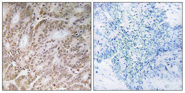
| Sample | Paraffin-embedded human colon carcinoma tissue; fixative not specified (datasheet A08887) |
| Fixation | Image fixative and duration unreported (datasheet A08887); verify before use. |
| Sectioning | 4–5 µm sections on charged slides (standard) |
| Deparaffinisation | Xylene, graded ethanol series to water (standard) |
| Antigen retrieval | Heat-induced epitope retrieval in Tris-EDTA buffer, pH 9.0, 20 min at 95–98 °C (standard rule: nuclear antigen) |
| Peroxidase block | 3% H2O2, 10 min, room temperature (standard) |
| Blocking | 10% normal serum of the secondary host, 30 min, room temperature (standard) |
| Primary antibody | Rabbit anti-UBQLN4, 1:100 - 1:300 (datasheet A08887) |
| Primary incubation | Overnight at 4 °C (standard) |
| Detection | HRP-polymer secondary, DAB chromogen 5–10 min (standard) |
| Counterstain | Hematoxylin, blue, dehydrate and mount (standard) |
| Expected result | UBQLN4-positive staining in respiratory epithelial cells of bronchus (HPA tissue IHC: High). HPA tissue profile: General nuclear expression. No signal in the no-primary control. |
UBQLN4 staining is expected mainly in nuclei across tissues (HPA tissue IHC: general nuclear expression), with High staining reported in respiratory epithelial, rectal glandular and urothelial cells (HPA tissue IHC). Nucleoplasmic localization is supported by ICC-IF (HPA ICC-IF). UniProt also lists cytoplasmic and other intracellular locations; UBQLN4 has no transmembrane segment (UniProt Q9NRR5 topology). HPA rates the tissue IHC pattern Approved, with medium consistency against RNA data (HPA tissue IHC).
| Distinct nuclear chromogen in respiratory epithelial cells of bronchus or nasopharynx, rectal glandular cells, or urothelial cells. | This matches the reported High staining in those cell populations and the general nuclear pattern (HPA tissue IHC). Assess signal within the named cells, rather than treating every cell in the section as an equally strong positive; tissue architecture helps distinguish nuclear staining from pigment or deposits (general IHC practice). |
| Predominantly extracellular, luminal or sharply cell-surface staining, with little nuclear signal. | That distribution conflicts with the HPA nuclear IHC pattern and the absence of a UBQLN4 transmembrane segment (HPA tissue IHC; UniProt Q9NRR5 topology). Review morphology and controls for nonspecific staining or detection deposits (general IHC practice). Cytoplasmic signal alone needs more careful interpretation because UniProt also lists cytoplasmic locations (UniProt Q9NRR5). |
| Strong staining in cardiomyocytes or cholangiocytes, especially when expected positive cells are unstained. | HPA reports UBQLN4 as Not detected in those specific cell populations (HPA tissue IHC). Check cell identification and reagent controls before attributing the signal to UBQLN4: cross-reactivity or endogenous detection activity can produce staining in unexpected cells (general IHC practice). A single discrepant section does not establish a new tissue pattern. |
| Uniform chromogen across nuclei, cytoplasm, stroma and empty spaces, obscuring cell boundaries. | This is diffuse background rather than an interpretable cell-specific nuclear pattern (HPA tissue IHC; general IHC practice). Check the reagent-free and primary-omission controls, detection chemistry, washing and chromogen development (general IHC practice). Do not score broadly colored tissue as UBQLN4-positive merely because some nuclei are included. |
| No nuclear signal in a bronchus, nasopharynx, rectum or urinary bladder section selected as a positive control. | The relevant epithelial, glandular or urothelial cells are reported High by HPA, so absent staining warrants a workflow check (HPA tissue IHC). First confirm that those cells are present and preserved, then inspect control performance, antibody application and detection (general IHC practice). HPA's Approved rating still carries only medium staining-to-RNA consistency (HPA tissue IHC). |
| Tissue and cell selection | HPA reports High staining in respiratory epithelial cells of bronchus and nasopharynx, rectal glandular cells, and urothelial cells; cardiomyocytes and cholangiocytes are Not detected (HPA tissue IHC). Compare identified cell populations, not whole-organ labels. UniProt describes high expression in heart and skeletal muscle, while HPA reports cardiomyocytes as undetected and myocytes as low by IHC; these observations use different readouts (UniProt Q9NRR5; HPA tissue IHC). |
| Antibody evidence | The supplied HPA antibody, HPA061797, has IHC status Approved and ICC status Supported (HPA antibodies). HPA describes tissue IHC as having medium consistency with RNA expression (HPA tissue IHC). Use that evidence to set confidence in the reported pattern; it does not turn an unexpected compartment or cell type into a validated positive. |
| Intracellular distribution | The predominant tissue interpretation is nuclear, and ICC-IF supports nucleoplasmic localization (HPA tissue IHC; HPA ICC-IF). UniProt additionally lists cytoplasm, chromosome, endoplasmic reticulum, perinuclear region and autophagosomes; it reports nuclear recruitment after DNA damage (UniProt Q9NRR5). Those annotations allow context-dependent intracellular patterns, but they do not establish the frequency or intensity of each pattern in routine tissue IHC. |
| Protein form and epitope limits | UniProt lists 2 isoforms, a single 1–601 chain, no signal peptide, no propeptide and no transmembrane segment (UniProt Q9NRR5). Without an antibody epitope or isoform-specific validation in the supplied evidence, the staining cannot be assigned to one isoform; the record also gives no basis for interpreting extracellular staining as normal UBQLN4 shedding. |
| Phosphorylation and damage response | UniProt records phosphorylation at Ser-318 by ATM and recruitment to nuclear DNA-damage sites after damage (UniProt Q9NRR5). This supports a possible change in localization under that condition. The supplied evidence does not show whether the IHC antibody recognizes phosphorylated and unphosphorylated UBQLN4 equally, so staining intensity cannot be read as a phosphorylation assay. |
| IF/ICC Q&A: what should a separate IF result show? | A mainly nucleoplasmic signal is the supported ICC-IF expectation; HPA lists images from MCF-7 and U2OS (HPA ICC-IF). UniProt's additional intracellular locations may be context-dependent (UniProt Q9NRR5). This IHC interpretation does not specify an IF/ICC protocol. |
| Situation | Likely cause | Next action |
|---|---|---|
| Expected positive cells are present but their nuclei are unstained. | The observed result conflicts with HPA's High calls for the specified epithelial, glandular or urothelial cells (HPA tissue IHC); a staining or detection failure is possible (general IHC practice). | Verify the cell identity and positive control, then check that the primary antibody, antigen-retrieval step and detection reagents were applied as intended (general IHC practice). The supplied sources give no UBQLN4-specific retrieval condition or fixation-sensitivity claim. |
| Chromogen spreads across stroma or empty spaces. | Diffuse color lacks the cell-associated nuclear distribution reported by HPA and can reflect background or detection deposits (HPA tissue IHC; general IHC practice). | Compare primary-omission and reagent controls; review washing, blocking and chromogen development using standard IHC controls (general IHC practice). Score only interpretable staining in identified cells. |
| Unexpected strong signal appears in cardiomyocytes or cholangiocytes. | Both are Not detected in the supplied HPA IHC observations (HPA tissue IHC); misidentified cells, cross-reactivity or endogenous detection activity are possible explanations (general IHC practice). | Recheck morphology and control sections. If using an enzyme-based detection system, assess its relevant endogenous activity with an appropriate control before assigning UBQLN4 positivity (general IHC practice). |
| The slide shows only cytoplasmic or perinuclear staining. | UniProt includes those locations, but HPA describes general nuclear tissue staining and supported nucleoplasmic ICC-IF staining (UniProt Q9NRR5; HPA tissue IHC; HPA ICC-IF). | Confirm that nuclei are identifiable by counterstain and compare positive and negative controls (general IHC practice). Record the compartment explicitly; do not treat an exclusively cytoplasmic pattern as matching the dominant HPA IHC pattern. |
| Nearly every cell looks equally strong, including HPA low or undetected populations. | Uniform intensity conflicts with HPA's cell-specific range, from High in selected epithelia to low in myocytes and undetected in cardiomyocytes (HPA tissue IHC). | Review exposure to chromogen, background controls and cell-level scoring (general IHC practice). Compare nuclei in the named HPA populations before concluding that the entire tissue is uniformly positive. |
| A nuclear result differs between specimens or from an IF image. | HPA's tissue IHC has medium consistency with RNA data, while ICC-IF supports nucleoplasmic localization; UniProt also documents context-dependent intracellular localization (HPA tissue IHC; HPA ICC-IF; UniProt Q9NRR5). | Compare the same cell type and compartment across specimens, document tissue condition and controls, and avoid inferring a fixation effect or phosphorylation state from the discrepancy alone (general IHC practice; UniProt Q9NRR5). |


Comprehensive Human Protein Atlas IHC scoring per tissue (reliability: Approved — Medium consistency between antibody staining and RNA expression data.). Rows are taken directly from the HPA tissue chart — click any row's HPA link to view the source.
| Tissue | Cell type | Level | Evidence | Source |
|---|---|---|---|---|
| Bronchus | Respiratory epithelial cells | High | Protein (IHC) | HPA → |
| Nasopharynx | Respiratory epithelial cells | High | Protein (IHC) | HPA → |
| Rectum | Glandular cells | High | Protein (IHC) | HPA → |
| Urinary bladder | Urothelial cells | High | Protein (IHC) | HPA → |
| Adrenal gland | Glandular cells | Medium | Protein (IHC) | HPA → |
Troubleshooting for chromogenic UBQLN4 IHC in paraffin sections, with one IF/ICC question for the separate fluorescence workflow.
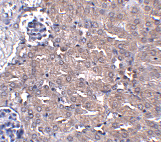
Catalog IHC images cover human colon carcinoma paraffin sections and mouse kidney tissue (image captions); IF/ICC is listed for A08887, with no IF image supplied (catalog payload).
A08887 has an IHC image of human colon carcinoma paraffin sections with a peptide-blocked comparison; its applications include IF and ICC, and its listed reactivity is human and mouse (A08887 image caption; catalog). A08887-1 has an IHC image of mouse kidney tissue at 5 μg/mL; its applications include IHC-P, and its listed reactivity is human, mouse and rat (A08887-1 image caption; catalog).
Which to pick: For human tissue IHC on paraffin sections, choose A08887 based on its own colon carcinoma image; the fixative is unreported (A08887 image caption). For IF/ICC, choose A08887 because those applications are listed, although no IF image is supplied (A08887 catalog payload). For mouse kidney IHC, A08887-1 has direct image evidence; it also lists rat reactivity, but its supplied IHC validation is in mouse tissue (A08887-1 image caption; catalog).