This website uses cookies to ensure you get the best experience on our website.


- Table of Contents
Plan WDR83 chromogenic IHC in paraffin sections using its reported cytoplasmic tissue staining as a starting point (HPA tissue IHC). The guide covers controls, staining assessment, and the uncertainty caused by low antibody–RNA concordance (HPA tissue IHC).

Expected localisation, validated protocols, controls and antibodies — the at-a-glance facts below, then the full design guide.
| Expected localisation | Cytoplasmic in tissue (HPA tissue IHC); partial nuclear localisation possible (UniProt) | |
| Staining pattern | Widespread cytoplasmic staining (HPA tissue IHC) | |
| Antigen retrieval | Tris-EDTA pH 9.0 HIER, 95–98 °C, 20 min (rule: nuclear antigen) | |
| Positive control | Adrenal gland+4 more · see all | |
| Negative control | Bone marrow+3 more · see all |
| Fixation | Keep fixation consistent across paraffin sections. (standard IHC practice; not target-specific) | |
| Caveat | Antibody staining has low concordance with RNA data (HPA tissue IHC) | |
| Regulation | Expression regulation unreported (UniProt) | |
| Isoform / epitope | No isoforms or processing annotated; epitope impact unknown (UniProt) |
The catalog antibody’s IHC-P protocol is paired with a published chromogenic WDR83 protocol for mouse brain (PMC12816048).
| Sample | Tissue sections; selected-image fixative not specified (standard IHC workflow) |
| Fixation | Image fixative and duration unreported (datasheet PA1053); verify before use. |
| Sectioning | 4–5 µm sections on charged slides (standard) |
| Deparaffinisation | Xylene, graded ethanol series to water (standard) |
| Antigen retrieval | Heat-induced epitope retrieval in Tris-EDTA buffer, pH 9.0, 20 min at 95–98 °C (standard rule: nuclear antigen) |
| Peroxidase block | 3% H2O2, 10 min, room temperature (standard) |
| Blocking | 10% normal serum of the secondary host, 30 min, room temperature (standard) |
| Primary antibody | Rabbit anti-WDR83, 0.5-1μg/ml (datasheet PA1053) |
| Primary incubation | Overnight at 4 °C (standard) |
| Detection | HRP-polymer secondary, DAB chromogen 5–10 min (standard) |
| Counterstain | Hematoxylin, blue, dehydrate and mount (standard) |
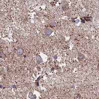
| Expected result | WDR83-positive staining in glandular cells of adrenal gland (HPA tissue IHC: Medium). HPA tissue profile: Ubiquitous cytoplasmic expression. No signal in the no-primary control. |
WDR83 is predominantly cytoplasmic and partly nuclear, with lysosomal localization; it has no transmembrane segment (UniProt Q9BRX9 localization and topology). In paraffin sections, look for cytoplasmic staining in the cell types where HPA reports medium signal, such as kidney tubular cells and liver hepatocytes (HPA tissue IHC). Treat the pattern as provisional: HPA rates tissue IHC reliability Uncertain because antibody staining and RNA expression have low consistency (HPA tissue IHC).
| Cytoplasmic staining in kidney tubular cells or liver hepatocytes, with possible nuclear signal. | This is compatible with the reported medium IHC staining in those cells (HPA tissue IHC) and predominantly cytoplasmic, partly nuclear localization (UniProt Q9BRX9). Judge whether the cytoplasmic signal is distinguishable from background on the same section; this pattern alone cannot establish specificity because tissue IHC reliability is Uncertain (HPA tissue IHC). |
| Signal appears only at cell borders, in extracellular material, or as a uniform surface outline. | That distribution does not match the reported intracellular localization (UniProt Q9BRX9; HPA subcellular ICC-IF). WDR83 has no transmembrane segment (UniProt Q9BRX9 topology). Check the counterstain and tissue structure before assigning a compartment; a boundary pattern warrants investigation as possible artifact rather than a WDR83-positive call. |
| Strong staining appears in bone marrow hematopoietic cells while expected positive cells are weak. | HPA reports WDR83 as not detected in bone marrow hematopoietic cells (HPA tissue IHC). Consider cross-reactivity or endogenous chromogenic detection activity (general IHC practice). Because HPA rates its tissue IHC evidence Uncertain, this comparison flags a result for control checks; it does not prove the stained cells lack WDR83. |
| Brown signal spreads across the section without clear cell boundaries or compartment contrast. | Treat this as diffuse background until staining can be assigned to cells and intracellular compartments (general IHC interpretation). It cannot be matched confidently to the cytoplasmic tissue pattern (HPA tissue IHC) or predominant cytoplasmic localization (UniProt Q9BRX9). Review a no-primary control and the detection and blocking steps (general IHC practice). |
| No staining is visible in kidney tubular cells or liver hepatocytes. | HPA reports medium IHC staining in these cell types (HPA tissue IHC), so an entirely blank result calls for a technical check. Confirm tissue identity and examine a positive-control section processed in the same run (general IHC practice). An absent result alone cannot resolve biological absence versus assay failure, especially with Uncertain HPA tissue IHC reliability. |
| Tissue evidence and confidence (HPA tissue IHC) | HPA describes a ubiquitous cytoplasmic profile and low tissue RNA specificity, yet rates tissue IHC reliability Uncertain because staining and RNA have low consistency (HPA tissue IHC). Use reported medium-staining cells as practical comparison sites, not proof that every section or antibody run must stain at the same intensity. |
| Compartment evidence across assays (UniProt Q9BRX9; HPA subcellular ICC-IF) | UniProt calls WDR83 predominantly cytoplasmic and partly nuclear, with lysosomal localization (UniProt Q9BRX9). HPA ICC-IF instead lists nucleoplasm and vesicles as main approved locations, with cytosol among additional locations (HPA subcellular ICC-IF). Read those as assay-specific observations; the ICC-IF pattern does not set an IHC nuclear-intensity threshold. |
| Comparator cell types (HPA tissue IHC) | HPA reports medium staining in bronchial respiratory epithelial cells, cerebellar Purkinje cells, colon and gallbladder glandular cells, adrenal glandular cells, cardiomyocytes, kidney tubular cells, and hepatocytes (HPA tissue IHC). It reports no detection in bone marrow hematopoietic cells, ovarian stroma cells, salivary gland glandular cells, and vaginal squamous epithelial cells (HPA tissue IHC). Compare named cells, not whole-organ averages. |
| Topology and processing (UniProt Q9BRX9) | The annotated protein is a single 1–315 chain without a signal peptide, propeptide, or transmembrane segment; no glycosylation sites or modified residues are annotated (UniProt Q9BRX9). This supports checking an intracellular pattern. These annotations provide no basis for predicting epitope exposure after fixation or choosing an antigen-retrieval condition. |
| Situation | Likely cause | Next action |
|---|---|---|
| No signal in a reported medium-staining cell type (HPA tissue IHC). | Possible run failure, weak detection, or tissue mismatch (general IHC practice); HPA reliability is Uncertain (HPA tissue IHC). | Verify cell identity, inspect a same-run positive control, and review antibody dilution, retrieval, and detection settings against the assay instructions (general IHC practice). Do not infer a WDR83-specific fixation effect. |
| Diffuse chromogen obscures cytoplasmic detail. | Background from detection or incomplete blocking may prevent compartment scoring (general IHC practice). | Examine a no-primary control; review blocking, wash, and chromogen development steps (general IHC practice). Score WDR83 only where intracellular signal remains distinguishable from background. |
| Stain appears exclusively extracellular or at cell boundaries. | The pattern conflicts with reported intracellular locations and absence of a transmembrane segment (UniProt Q9BRX9; HPA subcellular ICC-IF). | Recheck tissue structure and counterstain, then compare with a no-primary control (general IHC practice). Withhold a positive call until the apparent compartment is resolved. |
| Bone marrow hematopoietic cells stain strongly. | HPA reports them as not detected (HPA tissue IHC); cross-reactivity or endogenous detection activity is possible (general IHC practice). | Compare a no-primary control and reported medium-staining cells processed in the same run (general IHC practice; HPA tissue IHC). Treat the discrepancy as unresolved because HPA tissue IHC reliability is Uncertain. |
| Sections vary in the number of stained cells. | HPA levels refer to named cell types, and tissue IHC reliability is Uncertain (HPA tissue IHC); section composition can also vary (general histology practice). | Identify and score the relevant cells within each section before comparing runs (general IHC practice). Record intensity and compartment separately instead of treating every unstained cell as assay failure. |
| IF/ICC Q&A: where should WDR83 appear? | HPA ICC-IF lists nucleoplasm and vesicles as approved main locations; cytosol, centriolar satellites, and basal body are additional approved locations (HPA subcellular ICC-IF). | Interpret IF/ICC localization using its own guide and controls (general IF practice). Do not transfer IF/ICC compartment prominence into an IHC staining-intensity rule (HPA subcellular ICC-IF; HPA tissue IHC). |



Comprehensive Human Protein Atlas IHC scoring per tissue (reliability: Uncertain — Low consistency between antibody staining and RNA expression data. Pending external verification.). Rows are taken directly from the HPA tissue chart — click any row's HPA link to view the source.
| Tissue | Cell type | Level | Evidence | Source |
|---|---|---|---|---|
| Adrenal gland | Glandular cells | Medium | Protein (IHC) | HPA → |
| Bronchus | Respiratory epithelial cells | Medium | Protein (IHC) | HPA → |
| Cerebellum | Purkinje cells | Medium | Protein (IHC) | HPA → |
| Colon | Glandular cells | Medium | Protein (IHC) | HPA → |
| Gallbladder | Glandular cells | Medium | Protein (IHC) | HPA → |
| Tissue | Cell type | Level | Evidence | Source |
|---|---|---|---|---|
| Bone marrow | Hematopoietic cells | Not detected | Protein (IHC) | HPA → |
| Ovary | Ovarian stroma cells | Not detected | Protein (IHC) | HPA → |
| Salivary gland | Glandular cells | Not detected | Protein (IHC) | HPA → |
| Vagina | Squamous epithelial cells | Not detected | Protein (IHC) | HPA → |
Troubleshoot WDR83 staining in paraffin sections by checking retrieval, compartment pattern, controls, and scoring before interpreting chromogenic signal.
PA1053 provides IHC data from rat lung paraffin sections (PA1053 image caption) and lists human, mouse and rat reactivity (PA1053 catalog). No IF data are supplied (PA1053 catalog).
PA1053 is listed for IHC and WB, with human, mouse and rat reactivity (PA1053 catalog). Its IHC(P) image shows rat lung tissue; no IF image is supplied (PA1053 image caption; PA1053 catalog).
Which to pick: Choose PA1053 for tissue IHC in paraffin sections: its own image shows rat lung IHC(P), and its listed IHC concentration is 0.5–1 μg/ml (PA1053 image caption; PA1053 datasheet). Its fixative is unreported (PA1053 image caption). For cross-species work, PA1053 lists human, mouse and rat reactivity, but the supplied IHC image documents rat tissue only; no IF/ICC option is documented because IF/ICC is absent from its application list and no IF image is supplied (PA1053 catalog; PA1053 image caption).