This website uses cookies to ensure you get the best experience on our website.


- Table of Contents
Plan YES1 staining in paraffin sections using the widespread cytoplasmic pattern reported in tissue IHC (HPA tissue IHC). The guide covers controls and interpretation using the catalog antibody at 0.5–1 μg/ml (datasheet: PB9466).

Expected localisation, validated protocols, controls and antibodies — the at-a-glance facts below, then the full design guide.
| Expected localisation | Cytoplasmic staining (HPA tissue IHC) | |
| Staining pattern | Widespread cytoplasmic staining (HPA tissue IHC) | |
| Antigen retrieval | EDTA pH 8.0 HIER, heat-mediated (datasheet PB9466) | |
| Positive control | Adipose tissue+4 more · see all | |
| Negative control | Spleen |
| Fixation | Keep fixation conditions consistent across sections. (standard IHC practice; not target-specific) | |
| Caveat | Antibody staining has low consistency with RNA data (HPA tissue IHC) | |
| Regulation | Broad expression; inducer unknown (HPA tissue IHC; UniProt) | |
| Isoform / epitope | No listed isoforms or transmembrane segment (UniProt) |
The catalog antibody’s IHC-P protocol (datasheet: PB9466) is accompanied by published YES1 staining protocols for mouse tumor sections (PMC5561618) and tissue microarrays (PMC7931761).
| Sample | Paraffin-embedded human mammary cancer tissue; fixative not specified (datasheet PB9466) |
| Fixation | Image fixative and duration unreported (datasheet PB9466); verify before use. |
| Sectioning | 4–5 µm sections on charged slides (standard) |
| Deparaffinisation | Xylene, graded ethanol series to water (standard) |
| Antigen retrieval | Heat retrieval: EDTA pH 8.0 (datasheet PB9466); 20 min, 95–100 °C (standard) |
| Peroxidase block | 3% H2O2, 10 min, room temperature (standard) |
| Blocking | 10% goat serum (datasheet PB9466) |
| Primary antibody | Rabbit anti-YES1, 0.5-1μg/ml (datasheet PB9466) |
| Primary incubation | Overnight at 4 °C (datasheet PB9466) |
| Detection | Streptavidin-biotin complex (SABC), DAB chromogen (datasheet PB9466) |
| Counterstain | Hematoxylin, blue, dehydrate and mount (standard) |
| Expected result | YES1-positive staining in adipocytes of adipose tissue (HPA tissue IHC: Medium). HPA tissue profile: Ubiquitous cytoplasmic expression. No signal in the no-primary control. |
YES1 is a nontransmembrane kinase found in cytosol, at the cell membrane and at cell junctions (UniProt P07947: topology and subcellular location). In tissue IHC, expect predominantly cytoplasmic staining across many cell types, including adipocytes and respiratory epithelial cells (HPA: ubiquitous cytoplasmic expression; Medium in both). Interpret intensity cautiously: the HPA IHC profile is Approved but has low consistency with RNA expression (HPA: reliability description).
| Cytoplasmic chromogen in adipocytes or bronchial respiratory epithelium, around Medium intensity. | This matches observed YES1 staining in those cells (HPA: Medium in adipocytes and bronchial respiratory epithelial cells). Membrane enrichment can be plausible given YES1 localisation (UniProt P07947: cell membrane), but HPA tissue IHC describes a broadly cytoplasmic pattern; a crisp membrane rim is not required for a positive call (HPA: tissue profile). |
| Strong, predominantly nuclear staining with little cytoplasmic signal. | A nuclear-only pattern conflicts with the supplied localisation evidence (UniProt P07947: cytosol, membrane and junction; HPA ICC-IF: supported plasma membrane and cytosol). Treat it as a possible artefact. Review the negative control and staining distribution, then reassess antibody specificity before assigning the signal to YES1 (general IHC practice). |
| Prominent signal in splenic red-pulp cells despite a clean positive-tissue control. | HPA reports YES1 as Not detected in red-pulp cells (HPA: spleen IHC). Investigate cross-reactivity or endogenous chromogenic activity, especially if the signal persists in a no-primary control (general IHC practice). This is a caution, not proof of an absent target: HPA notes low staining–RNA consistency, and UniProt describes fetal splenic hematopoietic expression (HPA: reliability description; UniProt P07947: tissue specificity). |
| Uniform haze across tissue, blank areas or several unrelated cell types. | A widespread haze that ignores tissue boundaries is hard to reconcile with a cell-associated cytoplasmic pattern (HPA: tissue profile). Check the no-primary control for endogenous detection activity; review blocking, washing and antibody concentration if the haze requires primary antibody (general IHC practice). Broad YES1 staining alone is not abnormal, because HPA describes ubiquitous expression (HPA: tissue profile). |
| No signal in adipocytes and bronchial respiratory epithelium processed alongside the study sample. | Both are observed at Medium intensity in HPA tissue IHC (HPA: adipose tissue and bronchus). If both controls fail, first investigate the run: section integrity, retrieval conditions, antibody dilution and chromogen development (general IHC practice). A blank study sample cannot be interpreted confidently until a known-positive control stains; HPA's Approved rating does not guarantee every run (HPA: IHC reliability). |
| Compartment and topology | YES1 has no transmembrane segment, yet localises to the cell membrane, cytosol and cell junctions (UniProt P07947: topology and subcellular location). Evaluate membrane-associated staining in the context of the dominant cytoplasmic tissue pattern; absence of a sharp membrane outline does not by itself invalidate IHC (HPA: tissue profile). |
| Tissue reference and confidence | HPA calls tissue staining ubiquitous and cytoplasmic, with low RNA tissue specificity (HPA: tissue profile; RNA specificity). Its IHC reliability is Approved, with low consistency between staining and RNA expression (HPA: reliability description). Use the listed cell-level observations as practical references, not an absolute expression map. |
| Antibody validation | HPA026480 and CAB004370 each have Approved IHC status; HPA026480 also has Supported ICC status (HPA: antibody validation). These labels apply to the listed antibodies and applications. They do not validate an unlisted antibody, establish an IHC dilution or upgrade the tissue profile to an Enhanced rating. |
| Processing and target forms | UniProt lists one chain spanning residues 2–543, no signal peptide or propeptide, no annotated glycosylation sites and no isoforms in this payload (UniProt P07947: processing, glycosylation and isoforms). Those entries provide no basis for predicting tissue shedding, an isoform-specific stain or a retrieval condition. |
| IF/ICC Q: What localisation should be expected? | A: Plasma-membrane and cytosolic fluorescence is supported in the HPA ICC-IF record (HPA: supported main locations). That record lists images from A-431, U-251MG, U2OS, RPTEC/TERT1 and serum-starved hTERT-RPE1 (HPA: ICC-IF image lines). This answers localisation only; IF/ICC workflow belongs in its own guide. |
| Situation | Likely cause | Next action |
|---|---|---|
| Positive tissue and study sections are both blank. | A shared staining-run failure is possible; HPA reports Medium staining in adipocytes and bronchial respiratory epithelium (HPA: tissue IHC). | Check section integrity, retrieval, primary dilution, detection reagents and chromogen with a known-positive section (general IHC practice). Reassess the study sample only after that control works. |
| Signal is nuclear while cytoplasm is clear. | The pattern conflicts with supplied YES1 localisation (UniProt P07947: subcellular location; HPA: supported ICC-IF locations). | Inspect no-primary and positive controls, confirm staining is cell-associated, and compare a separately validated IHC antibody if available (general IHC practice; HPA: two Approved IHC antibodies). |
| Brown signal appears in splenic red pulp. | HPA lists these cells as Not detected; endogenous enzyme activity or antibody cross-reactivity could account for unexpected chromogen (HPA: spleen IHC; general IHC practice). | Run a no-primary control and appropriate endogenous-enzyme blocking control, then compare the cell-level pattern with a known-positive tissue (general IHC practice). Do not use spleen as an absolute negative reference across ages (UniProt P07947: fetal spleen expression). |
| All tissue compartments look equally hazy. | Background from detection, insufficient washing or excess antibody is possible (general IHC practice); broad YES1 expression can also produce genuine cell-associated signal (HPA: ubiquitous cytoplasmic profile). | Compare tissue-free areas and no-primary controls; adjust blocking, washing or primary concentration only when controls identify background (general IHC practice). Preserve discrete cytoplasmic staining during optimisation. |
| Only a weak signal appears in oral mucosa, liver or esophagus. | HPA records Low staining in squamous oral or esophageal epithelium and in hepatocytes (HPA: low tissue-IHC observations). | Judge these slides beside a Medium reference such as bronchial respiratory epithelium, and verify control performance before increasing signal amplification (HPA: bronchus Medium; general IHC practice). |
| A sharp membrane rim is absent, but cytoplasmic signal is present. | Membrane localisation is supported, while HPA's tissue IHC summary emphasises cytoplasmic staining (UniProt P07947: cell membrane; HPA: tissue profile). | Score the cell-associated cytoplasmic signal against an HPA-observed positive cell type and its control; avoid rejecting a section solely for lacking a continuous membrane rim (HPA: tissue IHC profile; general IHC practice). |


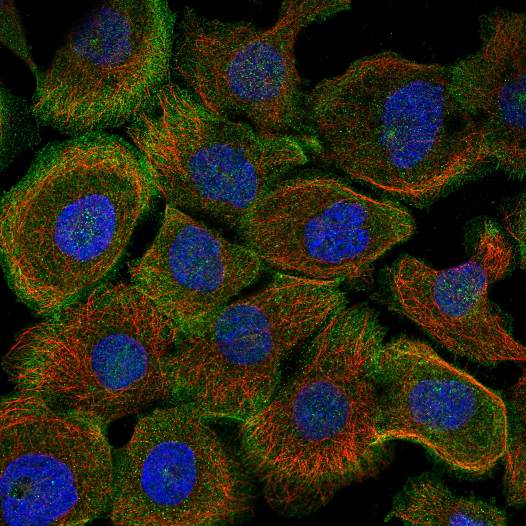
Comprehensive Human Protein Atlas IHC scoring per tissue (reliability: Approved — Low consistency between antibody staining and RNA expression data.). Rows are taken directly from the HPA tissue chart — click any row's HPA link to view the source.
| Tissue | Cell type | Level | Evidence | Source |
|---|---|---|---|---|
| Adipose tissue | Adipocytes | Medium | Protein (IHC) | HPA → |
| Adrenal gland | Glandular cells | Medium | Protein (IHC) | HPA → |
| Appendix | Glandular cells | Medium | Protein (IHC) | HPA → |
| Breast | Adipocytes | Medium | Protein (IHC) | HPA → |
| Bronchus | Respiratory epithelial cells | Medium | Protein (IHC) | HPA → |
| Tissue | Cell type | Level | Evidence | Source |
|---|---|---|---|---|
| Spleen | Cells in red pulp | Not detected | Protein (IHC) | HPA → |
Troubleshoot YES1 chromogenic IHC in paraffin sections by checking retrieval, staining compartment, controls, and scoring against the supplied tissue evidence.
The catalog lists human-reactive YES1 antibodies for IHC and IF/ICC (catalog applications/reactivity). The supplied image documents IHC in a human mammary cancer paraffin section (PB9466 IHC caption).
PB9466 is the sole rendering card, with IHC shown in a paraffin-embedded human mammary cancer section (cards payload; PB9466 IHC caption). It is listed for human IHC at 0.5–1 μg/ml; no IF image is supplied (catalog applications/reactivity/IHC dilution/IF image alts).
Which to pick: For human tissue IHC, choose PB9466: its own image documents a paraffin section with EDTA pH 8.0 retrieval and DAB detection; the fixative is unreported (PB9466 IHC caption). For IF/ICC, choose M02640-2 because those applications are listed for it, though no IF image is supplied (catalog applications/IF image alts). For mouse or rat samples, M02640-2 is the listed cross-species option: it is a rabbit monoclonal, clone 19Y44, with human, mouse and rat reactivity (catalog host/clone/reactivity).