This website uses cookies to ensure you get the best experience on our website.


- Table of Contents
Plan ZBTB4 chromogenic IHC around its predominantly nuclear tissue staining (HPA tissue IHC). Start antibody A08155 at 2.5 μg/mL (datasheet), and compare decidual cells with appendix glandular cells, where staining was high and undetected, respectively (HPA tissue IHC).

Expected localisation, validated protocols, controls and antibodies — the at-a-glance facts below, then the full design guide.
| Expected localisation | Predominantly nuclear; lesser cytoplasmic staining (HPA tissue IHC) | |
| Staining pattern | Most tissues show nuclear and lesser cytoplasmic staining (HPA tissue IHC) | |
| Antigen retrieval | Tris-EDTA pH 9.0 HIER, 95–98 °C, 20 min (rule: nuclear antigen) | |
| Positive control | Placenta+4 more · see all | |
| Negative control | Appendix+4 more · see all |
| Fixation | Keep paraffin-section fixation consistent across samples. (standard IHC practice; not target-specific) | |
| Caveat | Staining–RNA consistency is medium; verify specificity (HPA tissue IHC) | |
| Regulation | Low tissue specificity; no regulator annotated (HPA RNA; UniProt) | |
| Isoform / epitope | No isoforms annotated; one chain spans residues 1–1013 (UniProt) |
Compare the catalog antibody’s IHC-P protocol with four published ZBTB4 IHC protocols (PMC10035130; PMC7547138; PMC9941281; PMC10875348).
| Sample | Tissue sections; selected-image fixative not specified (standard IHC workflow) |
| Fixation | Image fixative and duration unreported (datasheet A08155); verify before use. |
| Sectioning | 4–5 µm sections on charged slides (standard) |
| Deparaffinisation | Xylene, graded ethanol series to water (standard) |
| Antigen retrieval | Heat-induced epitope retrieval in Tris-EDTA buffer, pH 9.0, 20 min at 95–98 °C (standard rule: nuclear antigen) |
| Peroxidase block | 3% H2O2, 10 min, room temperature (standard) |
| Blocking | 10% normal serum of the secondary host, 30 min, room temperature (standard) |
| Primary antibody | Rabbit anti-ZBTB4, 2.5 μg/mL (datasheet A08155) |
| Primary incubation | Overnight at 4 °C (standard) |
| Detection | HRP-polymer secondary, DAB chromogen 5–10 min (standard) |
| Counterstain | Hematoxylin, blue, dehydrate and mount (standard) |
| Expected result | ZBTB4-positive staining in decidual cells of placenta (HPA tissue IHC: High). HPA tissue profile: Nuclear and to a lesser extent cytoplasmic expression in most tissues. No signal in the no-primary control. |
ZBTB4 should stain mainly nuclei in paraffin sections, with weaker cytoplasmic staining possible across many tissues (HPA tissue IHC: Supported). Strong staining is reported in placental decidual cells; several other listed cell types show medium staining (HPA tissue IHC). Its nuclear and chromocenter localization, with no transmembrane segment, supports a nuclear interpretation (UniProt Q9P1Z0). HPA reports medium agreement with RNA data and says external verification is pending (HPA tissue IHC).
| Decidual cell nuclei stain strongly; adipocyte, adrenal glandular, or breast glandular cell nuclei stain moderately. | This fits the reported high placental decidual signal and medium signal in the listed cell types (HPA tissue IHC). Judge staining in the named cells and their nuclei, since the tissue label alone does not identify which cells should be positive (HPA tissue IHC). |
| Signal is predominantly membranous, or cytoplasm stains much more strongly than nuclei across the section. | That distribution conflicts with the reported mainly nuclear IHC pattern and the lack of a transmembrane segment (HPA tissue IHC; UniProt Q9P1Z0 topology). Limited cytoplasmic signal can occur, so assess whether a convincing nuclear pattern remains before calling the stain discordant (HPA tissue IHC). |
| Appendix or duodenal glandular cells, or liver cholangiocytes, stain strongly. | Those specific cell populations were reported as not detected (HPA tissue IHC). Check cell identity and controls, then consider nonspecific antibody binding or endogenous chromogen activity as possible explanations (standard IHC practice). A negative designation for these cells does not classify every cell in the section as negative (HPA tissue IHC). |
| Color is spread across tissue or present in blank regions without recognizable nuclear boundaries. | This is difficult to score as ZBTB4 because the expected pattern is mainly nuclear (HPA tissue IHC). Uneven blocking, excessive antibody concentration, or detection background are general IHC possibilities; they are not established ZBTB4-specific effects (standard IHC practice). |
| Placental decidual cells show no detectable nuclear staining. | The result disagrees with the reported high staining in those cells (HPA tissue IHC). Confirm that decidual cells are represented and examine the IHC run controls and catalog antibody conditions before interpreting the absence as biological (standard IHC practice). HPA's Supported rating still awaits external verification (HPA tissue IHC). |
| Cell population chosen for assessment | Placental decidual cells offer a reported high-signal reference; adipocytes and several listed glandular or neural cells have medium signal (HPA tissue IHC). Appendix and duodenal glandular cells, liver cholangiocytes, and splenic red-pulp cells are reported not detected; evaluate the specified cells rather than labeling whole organs positive or negative (HPA tissue IHC). |
| Expected compartment | UniProt places ZBTB4 in the nucleus and at chromocenters; tissue IHC reports mainly nuclear with lesser cytoplasmic staining (UniProt Q9P1Z0; HPA tissue IHC). Use nuclear predominance to interpret chromogenic sections. Nuclear bodies are an ICC-IF observation and should not be required as a resolvable chromogenic IHC feature (HPA subcellular ICC-IF). |
| Strength of the tissue evidence | The tissue profile is rated Supported, with medium agreement between staining and RNA expression and external verification pending (HPA tissue IHC). Two listed antibodies have Supported IHC status, but the payload does not assign Enhanced IHC validation (HPA antibodies). Treat departures from the pattern as findings to check, especially when based on a single section (standard IHC practice). |
| Topology and processing | ZBTB4 has no transmembrane segment or signal peptide, and the annotated chain spans residues 1–1013 (UniProt Q9P1Z0). These annotations support interpreting a mainly nuclear result rather than expecting a membrane rim; they do not predict IHC sensitivity or explain a failed stain (UniProt Q9P1Z0; HPA tissue IHC). |
| Fixation and epitope uncertainty | The supplied sources give no ZBTB4-specific fixation response or antibody epitope position (UniProt Q9P1Z0; HPA tissue IHC). Follow the catalog antibody's validated IHC-P conditions when available and record retrieval conditions for comparison; do not infer a ZBTB4-specific retrieval requirement from tissue staining levels (standard IHC practice). |
| Situation | Likely cause | Next action |
|---|---|---|
| No nuclear signal in placental decidual cells. | The observed result conflicts with their reported high staining; missed cell identification or an IHC run issue is possible (HPA tissue IHC; standard IHC practice). | Confirm decidual cells on morphology, inspect run controls, and check that the IHC-validated antibody and its stated IHC-P conditions were used (standard IHC practice). |
| Signal is faint in adipocytes or listed medium-staining glandular cells. | Their reported level is medium, so a weaker result than placental decidual cells can fit the profile (HPA tissue IHC). | Score nuclear signal in the named cells against a concurrent positive reference before changing conditions; document section-to-section variation (HPA tissue IHC; standard IHC practice). |
| Strong signal appears in appendix or duodenal glandular cells. | Those cells were reported not detected; nonspecific staining or endogenous detection activity is possible (HPA tissue IHC; standard IHC practice). | Verify the cell population, inspect no-primary and detection controls, and review blocking and detection steps if background persists (standard IHC practice). |
| Most staining outlines membranes or fills cytoplasm, with little nuclear signal. | This disagrees with mainly nuclear tissue IHC and nuclear UniProt localization; lesser cytoplasmic staining alone remains compatible (HPA tissue IHC; UniProt Q9P1Z0). | Compare nuclear and cytoplasmic signal in the same named cells, then inspect antibody concentration and detection controls for nonspecific signal (standard IHC practice). |
| Diffuse chromogen obscures nuclei across positive and negative regions. | General IHC background can prevent compartment scoring; the supplied sources do not establish a ZBTB4-specific fixation or retrieval cause (standard IHC practice; HPA tissue IHC). | Review no-primary controls, blocking, antibody dilution, washes, and detection timing under the validated IHC-P workflow; reassess identifiable nuclei afterward (standard IHC practice). |
| An IF image shows nuclear bodies or some cytosolic signal that the IHC slide does not resolve. | Nuclear bodies and nucleoplasm are supported ICC-IF locations; cytosol is uncertain, while tissue IHC describes mainly nuclear staining (HPA subcellular ICC-IF; HPA tissue IHC). | Interpret the chromogenic IHC section by its reported tissue and cell pattern. Use the separate IF/ICC guide for fluorescence-specific assessment (HPA tissue IHC; HPA subcellular ICC-IF). |
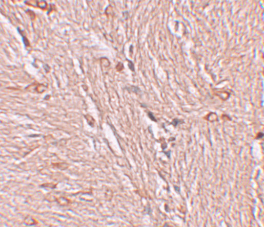
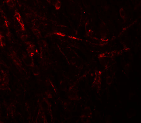


Comprehensive Human Protein Atlas IHC scoring per tissue (reliability: Supported — Medium consistency between antibody staining and RNA expression data. Pending external verification.). Rows are taken directly from the HPA tissue chart — click any row's HPA link to view the source.
| Tissue | Cell type | Level | Evidence | Source |
|---|---|---|---|---|
| Placenta | Decidual cells | High | Protein (IHC) | HPA → |
| Adipose tissue | Adipocytes | Medium | Protein (IHC) | HPA → |
| Adrenal gland | Glandular cells | Medium | Protein (IHC) | HPA → |
| Breast | Glandular cells | Medium | Protein (IHC) | HPA → |
| Bronchus | Respiratory epithelial cells | Medium | Protein (IHC) | HPA → |
| Tissue | Cell type | Level | Evidence | Source |
|---|---|---|---|---|
| Appendix | Glandular cells | Not detected | Protein (IHC) | HPA → |
| Duodenum | Glandular cells | Not detected | Protein (IHC) | HPA → |
| Liver | Cholangiocytes | Not detected | Protein (IHC) | HPA → |
| Small intestine | Glandular cells | Not detected | Protein (IHC) | HPA → |
| Spleen | Cells in red pulp | Not detected | Protein (IHC) | HPA → |
Troubleshoot ZBTB4 staining in chromogenic paraffin section IHC, using nuclear localisation and tissue patterns to guide controls and interpretation.
A08155 has human brain IHC and IF images (catalog image captions), with Human and Mouse reactivity listed (catalog: reactivity).
A08155 is listed for IHC-P and IF (catalog: applications). Its images show ZBTB4 staining in human brain tissue at 2.5 μg/mL for IHC and 20 μg/mL for IF (catalog: image captions).
Which to pick: For paraffin-section IHC, choose A08155 because IHC-P is listed (catalog: applications) and its own IHC image shows human brain tissue at 2.5 μg/mL (catalog: IHC image caption); the fixative is unreported (catalog: IHC image caption). For IF, choose A08155 based on its human brain IF image at 20 μg/mL (catalog: IF image caption); ICC validation is unreported (catalog: applications and image captions). For mouse samples, A08155 lists Mouse reactivity (catalog: reactivity), while the supplied IHC and IF images show human tissue (catalog: image captions).