ZNF521 / Zinc finger protein 521 · IHC design guide

Design Immunohistochemistry for ZNF521

Plan ZNF521 chromogenic IHC around its nuclear tissue pattern (HPA tissue IHC). Use the IHC-validated antibody at 5 μg/mL (datasheet: A06292), with Purkinje cells as a positive reference and liver cholangiocytes as a negative reference (HPA tissue IHC).

Evidence assembled Oct 2026 · For research use; verify linked source records and product datasheet before use
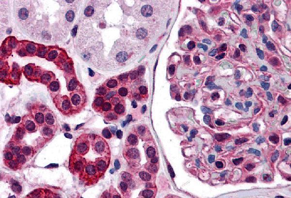
Immunohistochemistry protocol sheet for ZNF521 (IHC for ZNF521): expected localisation Nuclear staining in most tissues (HPA tissue IHC), antibody A06292, validated IHC image, and IHC protocol steps
Printable ZNF521 IHC protocol sheet — expected localisation Nuclear staining in most tissues (HPA tissue IHC), antibody A06292, controls and protocol steps. Open the full ZNF521 IHC guide →

ZNF521 Immunohistochemistry Experimental Design Guide

Expected localisation, validated protocols, controls and antibodies — the at-a-glance facts below, then the full design guide.

Must know before staining
Expected localisation Nuclear staining in most tissues (HPA tissue IHC)
Staining pattern Nuclear staining across most tissues (HPA tissue IHC)
Antigen retrieval Tris-EDTA pH 9.0 HIER, 95–98 °C, 20 min (rule: nuclear antigen)
Positive control ⓘ Caudate+4 more · see all
Negative control ⓘ Liver
Important caveats
Reasons your staining may differ from the expected pattern.
Fixation Keep paraffin-section fixation consistent (standard IHC practice; not target-specific)
Caveat Staining shows low consistency with RNA (HPA tissue IHC)
Regulation Falls as CD34(+) cells differentiate (UniProt)
Isoform / epitope No annotated isoforms or processing (UniProt)
Section 1

Recommended ZNF521 IHC & IF Protocols

The catalog antibody’s IHC-P protocol accompanies published chromogenic IHC protocols for gastric cancer tissue (PMC9606838; PMC6749787).

Recommended immunohistochemistry (IHC-P) protocol parameters
SampleTissue sections; selected-image fixative not specified (standard IHC workflow)
FixationImage fixative and duration unreported (datasheet A06292); verify before use.
Sectioning4–5 µm sections on charged slides (standard)
DeparaffinisationXylene, graded ethanol series to water (standard)
Antigen retrievalHeat-induced epitope retrieval in Tris-EDTA buffer, pH 9.0, 20 min at 95–98 °C (standard rule: nuclear antigen)
Peroxidase block3% H2O2, 10 min, room temperature (standard)
Blocking10% normal serum of the secondary host, 30 min, room temperature (standard)
Primary antibodyRabbit anti-ZNF521, 5 μg/mL (datasheet A06292)
Primary incubationOvernight at 4 °C (standard)
DetectionHRP-polymer secondary, DAB chromogen 5–10 min (standard)
CounterstainHematoxylin, blue, dehydrate and mount (standard)
Expected resultZNF521-positive staining in neuronal cells of caudate (HPA tissue IHC: High). HPA tissue profile: Nuclear expression in most tissues. No signal in the no-primary control.
💡Decision noteStart with Tris-EDTA pH 9.0 HIER at 95–98 °C for 20 min (page retrieval); compare citrate pressure-cooker retrieval (PMC9606838).
Section 2

What Is the Expected ZNF521 Staining Pattern?

ZNF521 should be read chiefly as a nuclear stain: UniProt places it in the nucleus, and its recorded topology has no transmembrane segment (UniProt Q96K83). HPA reports nuclear expression in most tissues, with High staining in selected neuronal, epithelial, glandular and decidual cells (HPA tissue IHC). HPA rates its tissue profile Enhanced while also reporting low consistency between antibody staining and RNA expression (HPA tissue IHC).

What am I looking at on my slide?
Distinct nuclear staining in Purkinje cells, cortical or caudate neurons, or placental decidual cells (HPA tissue IHC).This matches HPA's High staining in those cell populations and its predominantly nuclear profile (HPA tissue IHC). Judge the signal in the named cells; a strongly stained section alone does not show which cells produced it.
Predominantly cytoplasmic or membrane staining, with nuclei comparatively clear.That conflicts with the nuclear location reported by UniProt and HPA, and with the lack of a transmembrane segment (UniProt Q96K83; HPA tissue IHC). Treat it as suspect until the compartment is confirmed with a nuclear counterstain and an appropriate control; the image alone cannot identify the artefact.
Prominent staining in liver cholangiocytes instead of the expected negative reference cells (HPA tissue IHC).HPA reports cholangiocytes as Not detected (HPA tissue IHC). Consider cross-reactivity or endogenous chromogenic detection activity, both general IHC possibilities, and compare with a no-primary control. This reference concerns cholangiocytes, not every cell in liver.
Diffuse colour across tissue or outside cell boundaries obscures nuclear detail.This is not the resolved nuclear pattern reported for ZNF521 (HPA tissue IHC; UniProt Q96K83). In general IHC practice, background can arise from nonspecific binding, residual detection activity or inadequate washing; inspect control sections before assigning any of it to ZNF521.
No nuclear signal in an HPA-listed High cell population, such as cerebellar Purkinje cells (HPA tissue IHC).Target-specific fixation effects are not established by the supplied assay evidence. Verify with a matched IHC source before attributing a result to fixation.
💡Expected ZNF521 appearanceCall a positive result when the named HPA-positive cells show resolved, chiefly nuclear staining at the reported High level; diffuse colour or dominant extranuclear signal is suspect (HPA tissue IHC; UniProt Q96K83).
How each factor affects the staining
Tissue and cell selectionHPA reports High staining in caudate and cortical neurons, Purkinje cells, cervical and esophageal squamous cells, rectal glandular cells and placental decidual cells; cholangiocytes are Not detected (HPA tissue IHC). Use the specified cells when comparing sections, since HPA levels do not describe every cell in an organ.
Hematopoietic differentiationUniProt describes abundant expression in CD34(+) cells, a rapid decline during their differentiation, and undetectable levels in mature peripheral blood leukocytes (UniProt Q96K83). Interpret a negative mature leukocyte separately from a negative progenitor-rich sample; these are biological expectations, not guarantees of IHC assay performance.
Antibody evidence and scopeBoth HPA023056 and HPA023849 have Enhanced IHC validation, meaning a pattern reproduced by independent antibodies or orthogonal data (HPA antibodies). HPA nevertheless flags low consistency between tissue antibody staining and RNA expression (HPA tissue IHC). Keep that caveat when interpreting an unexpected positive cell population.
IF/ICC Q: Where should ZNF521 appear?A: HPA reports an enhanced nucleoplasmic location in ICC-IF images from A-431, U-251MG, U2OS, AF22 and THP-1 (HPA subcellular). This supports a nuclear localisation check; the IF/ICC workflow belongs to its separate guide and does not supply an IHC protocol option.
Why is my staining missing, weak or wrong?
SituationLikely causeNext action
Known HPA-positive cells are present, but the IHC section has no nuclear signal (HPA tissue IHC).The run may have failed, or the antibody and detection conditions may be unsuitable; the result alone does not isolate a cause.Review run controls and the catalog antibody's IHC-P instructions, including its stated retrieval and dilution conditions. Do not infer a ZNF521-specific fixation effect from HPA staining levels.
Signal is mostly cytoplasmic or outlines cells.The compartment disagrees with the nuclear assignment (UniProt Q96K83; HPA tissue IHC); nonspecific staining or interpretation error is possible.Check a nuclear counterstain, compare the no-primary control, and score signal within nuclei separately from adjacent colour (general IHC practice).
Cholangiocytes show strong colour despite the HPA negative reference (HPA tissue IHC).Cross-reactivity or endogenous detection activity may produce colour in general chromogenic IHC practice.Compare a no-primary control and appropriate detection controls, then reassess the cell identity before calling ZNF521 positive.
Background is widespread and nuclear boundaries are hard to see.Nonspecific reagent binding, residual detection activity or insufficient washing are general IHC possibilities.Check blocking and washes against the validated workflow, inspect controls, and score only resolved cellular staining.
A mature peripheral blood leukocyte sample is negative.Undetectable ZNF521 in mature peripheral blood leukocytes is the UniProt expression expectation (UniProt Q96K83).Do not use that result alone to declare assay failure; evaluate an appropriate positive cell population and the run controls (UniProt Q96K83; HPA tissue IHC).
A cell type outside the named HPA examples appears positive.The HPA profile describes nuclear expression in most tissues but flags low antibody–RNA consistency; an unlisted cell cannot be classified from the examples alone (HPA tissue IHC).Record its identity, compartment and control staining, and interpret the finding cautiously alongside the HPA tissue profile and antibody validation (HPA tissue IHC; HPA antibodies).

Sample controls for ZNF521 IHC & IF

🧪Run cerebellum first and require nuclear staining in Purkinje cells (HPA: High in cerebellar Purkinje cells; UniProt Q96K83: nucleus). Run liver cholangiocytes as the negative tissue (HPA: Not detected in liver cholangiocytes); on the cerebellum slide, inspect adjacent non-Purkinje cells for background, without assuming they are biologically ZNF521-negative from the supplied HPA rows.
Positive control tissue: Caudate (Neuronal cells, HPA High)
Negative control tissue: Liver (HPA Not detected)
ICC-IF cell lines (HPA subcellular resource): HPA ICC-IF images show ZNF521 in A-431, U-251MG, U2OS, AF22, THP-1, with annotated localisation: Nucleoplasm (enhanced) (HPA subcellular).
Technical controls: Include a no-primary, secondary-only control, an isotype-matched control appropriate to the primary antibody’s host species and clonality, and ZNF521-knockout material as a biological negative if available (standard IHC practice). Block endogenous peroxidase for chromogenic IHC; if examining cerebellum by IF, check an unstained section for neuronal lipofuscin autofluorescence (standard IHC/IF practice).
⚠️Feasibility: A target-specific fixation window and retrieval dependency are unreported in the supplied evidence; optimize antigen retrieval on paraffin sections empirically (standard IHC practice). The selected A06292 kidney tissue-IHC caption reports 5 μg/mL but leaves the fixative unreported (selected-SKU tissue-IHC caption). The evidence does not establish whether frozen sections or IF/ICC are easier for this target; HPA reports nucleoplasmic ICC-IF localization, and neuronal lipofuscin may complicate cerebellar IF interpretation (HPA: nucleoplasm; standard IF practice).

HPA tissue IHC evidence for ZNF521

Comprehensive Human Protein Atlas IHC scoring per tissue (reliability: Enhanced — Low consistency between antibody staining and RNA expression data.). Rows are taken directly from the HPA tissue chart — click any row's HPA link to view the source.

Positive expression · recommended positive controls

TissueCell typeLevelEvidenceSource
Caudate Neuronal cells High Protein (IHC) HPA →
Cerebellum Purkinje cells High Protein (IHC) HPA →
Cerebral cortex Neuronal cells High Protein (IHC) HPA →
Cervix Squamous epithelial cells High Protein (IHC) HPA →
Esophagus Squamous epithelial cells High Protein (IHC) HPA →

Undetected expression · recommended negative controls

TissueCell typeLevelEvidenceSource
Liver Cholangiocytes Not detected Protein (IHC) HPA →
Section 3

Advanced ZNF521 IHC Tips

Troubleshoot ZNF521 chromogenic IHC by checking nuclear staining, cell identity, and controls before comparing staining intensity across sections.

Which retrieval conditions should I try first for weak ZNF521 nuclear staining?
Start with Tris-EDTA pH 9.0 heat-induced retrieval at 95–98 °C for 20 min (page retrieval setting: nuclear antigen). Let sections cool in retrieval buffer, then compare a dilution series of the catalog antibody on matched sections while holding detection conditions constant (standard IHC practice). ZNF521 is nuclear, so judge recovery by crisp nuclear staining rather than diffuse cytoplasmic color (UniProt Q96K83: nucleus; HPA: nucleoplasm). If signal remains weak, test a lower-pH retrieval buffer as a fallback on adjacent sections and include the same positive tissue and no-primary control in each run (standard IHC practice).
Could fixation explain inconsistent ZNF521 staining across paraffin blocks?
Target-specific sensitivity of ZNF521 to fixation is unknown from the supplied evidence; the selected kidney IHC caption gives 5 μg/mL antibody but does not state a fixative (A06292 tissue-IHC caption). Compare blocks with documented fixation histories using the same Tris-EDTA pH 9.0 retrieval for 20 min and identical detection settings (page retrieval setting; standard IHC practice). Include a section from the same reference block in each run to distinguish a block effect from run variation (standard IHC practice). Do not infer fixation tolerance from nuclear localisation, phosphorylation sites, or tissue staining patterns (UniProt Q96K83; HPA: tissue IHC).
How should I assess cytoplasmic staining when evaluating ZNF521 IHC?
Prioritise staining within counterstained nuclei: ZNF521 is assigned to the nucleus and, more specifically, the nucleoplasm (UniProt Q96K83: nucleus; HPA: nucleoplasm). Its record lists no transmembrane segment, so a membrane outline alone does not fit the supplied localisation evidence (UniProt Q96K83: topology). Review the same field at 20× and 40×, comparing nuclear color with adjacent cytoplasm and the no-primary control (standard IHC practice). If cytoplasmic color rises with antibody concentration while nuclear contrast does not, adjust dilution and blocking, then verify the pattern in a second section (standard IHC practice).
What can epitope uncertainty tell me about discrepant ZNF521 IHC results?
The supplied record lists 0 isoforms and no annotated domain, but it does not identify this antibody’s binding epitope (UniProt Q96K83: isoforms and domains; A06292 tissue-IHC caption). ZNF521 has reported phosphoserines at residues 546, 605, and 608; their presence does not establish an effect on this antibody’s staining (UniProt Q96K83: modified residues). Compare adjacent sections across the same retrieval and antibody-dilution series before assigning a discrepancy to epitope masking (standard IHC practice). Request epitope information for the exact antibody if available, and interpret divergent staining only alongside nuclear localisation and appropriate controls (UniProt Q96K83: nucleus; standard IHC practice).
How can IF help assess a questionable ZNF521 IHC pattern?
Use IF as a separate cross-check for nuclear localisation, with a nuclear counterstain and a marker for the cell population under study; ZNF521 is assigned to the nucleoplasm (HPA: nucleoplasm; standard IF practice). Select a fluorophore channel with low background in that tissue, and inspect an unstained or secondary-only section for autofluorescence before interpreting colocalisation (standard IF practice). Because the expected epitope is intracellular and no transmembrane segment is annotated, permeabilisation may be needed; a brief 0.1% Triton X-100 trial is a general IF starting point, not antibody validation (UniProt Q96K83: topology; standard IF practice). Compare cell identity and nuclear distribution with chromogenic IHC, without treating the separate IF result as proof of IHC specificity (standard IHC/IF practice).
What should I change when ZNF521 IHC shows widespread brown background?
First inspect a no-primary control and a matched section stained with the catalog antibody; diffuse color in both suggests detection background (standard IHC practice). For peroxidase-based DAB detection, check the peroxidase block, wash steps, and detection reagent exposure before changing the ZNF521 antibody concentration (standard IHC practice). The selected kidney image reports 5 μg/mL antibody, but its caption does not establish a working concentration for other specimens (A06292 tissue-IHC caption). Titrate around the chosen concentration on adjacent sections and retain the setting that improves nuclear contrast without raising cytoplasmic or edge staining (UniProt Q96K83: nucleus; standard IHC practice).
How should I quantify ZNF521 staining across heterogeneous tissue sections? ⚠ ANSWER MARKED FOR VERIFICATION
Define the cell compartment and region before scoring, and count ZNF521-positive nuclei rather than total brown area because its expected location is nuclear (UniProt Q96K83: nucleus; HPA: nucleoplasm). Report either an H-score on a 0–300 scale, the percentage of positive nuclei, or positive-cell density per mm², with a fixed positivity threshold (standard IHC practice). Normalise percentages to eligible nuclei in the same annotated cell population and density to evaluable tissue area; exclude folds, necrosis, and section edges by preset rules (standard IHC practice). Keep retrieval, antibody concentration, imaging, and scoring thresholds constant across groups, and report how many sections and fields contributed (standard IHC practice).
Which findings distinguish credible ZNF521 staining from artefact?
A credible signal has clear nuclear distribution in identifiable cells and exceeds the no-primary control; nucleoplasmic localisation is supported independently (HPA: nucleoplasm; standard IHC practice). Consider cell identity: the record describes abundant expression in CD34(+) hematopoietic cells and loss during their differentiation, while HPA reports nuclear expression across many tissues (UniProt Q96K83: tissue specificity; HPA: tissue IHC). Treat edge-only staining, necrotic areas, diffuse cytoplasm, and residual peroxidase color as potential artefacts, then check adjacent sections and controls (standard IHC practice). Interpret small intensity differences cautiously because HPA reports low consistency between antibody staining and RNA expression data, despite its enhanced reliability designation (HPA: reliability description).
Boster reagents

Best ZNF521 / Zinc finger protein 521 IHC Antibodies

Anti-ZNF521 antibodies have IHC data from human kidney tissue (A06292 image caption) and IF/ICC data from HeLa cells (A06292-1 image captions); both list human reactivity (catalog).

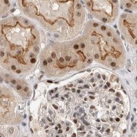
Real IHC data Immunohistochemistry of ZNF521 in human kidney tissue with ZNF521 antibody at 5 μg/mL.
Anti-Zinc finger protein 521 ZNF521 Antibody
Cat # A06292
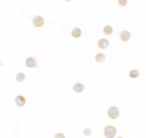
Real IF data Immunocytochemistry of ZNF521 in HeLa cells with ZNF521 antibody at 2.5 μg/mL.
Anti-Zinc finger protein 521 ZNF521 Antibody
Cat # A06292-1

A06292 will render with an IHC image of human kidney tissue at 5 μg/mL (A06292 image caption). A06292-1 will render with an ICC image of HeLa cells at 2.5 μg/mL and lists IF and ICC applications (A06292-1 image caption and applications).

Which to pick: Choose A06292 for human paraffin-section IHC: it is polyclonal and lists IHC-P, with a human kidney IHC image at 5 μg/mL (A06292 catalog and image caption); the fixative is unreported (A06292 image caption). Choose A06292-1 for human IF/ICC: it lists both applications and has HeLa cell images, while its clonality is unreported (A06292-1 catalog and image captions). Neither SKU has documented cross-species reactivity; both list Human only (catalog reactivity).

Each figure is that product's own IHC / IF validation image from its datasheet.

References

  1. UniProt Consortium. UniProt entry Q96K83 (ZN521_HUMAN, Zinc finger protein 521).
  2. Human Protein Atlas. ZNF521 tissue IHC expression (reliability: Enhanced).
  3. Human Protein Atlas. ZNF521 subcellular location (ICC-IF): Localized to the nucleoplasm..
  4. Human Protein Atlas. ZNF521 antibody validation summary (2 antibodies).
  5. ZNF521 Is Correlated with Tumor Immune Cell Infiltration and Act as a Valuable Prognostic Biomarker in Gastric Cancer. Gastroenterology research and practice 2022 — PMC9606838.
  6. Critical role of zinc finger protein 521 in the control of growth, clonogenicity and tumorigenic potential of medulloblastoma cells. Oncotarget 2013 — PMC3787157.
  7. Zinc Finger Protein 521, Negatively Regulated by MicroRNA-204-5p, Promotes Proliferation, Motility and Invasion of Gastric Cancer Cells. Technology in cancer research & treatment 2019 — PMC6749787.
  8. ZNF521 which is downregulated by miR-802 suppresses malignant progression of Hepatocellular Carcinoma through regulating Runx2 expression. Journal of Cancer 2020 — PMC7477442.
  9. PubMed PMID:14630787 — UniProt-cited evidence.
  10. PubMed PMID:14702039 — UniProt-cited evidence.
  11. PubMed PMID:15489334 — UniProt-cited evidence.