Click image to see more details
-

-
-
-
-
+19
Product Info Summary
| SKU: | M00397 |
|---|---|
| Size: | 100 μl |
| Reactive Species: | Human, Mouse, Rat |
| Host: | Rabbit |
| Application: | Flow Cytometry, IF, IHC, ICC, WB |
Customers Who Bought This Also Bought


Product info
Product Name
Anti-53BP1 TP53BP1 Rabbit Monoclonal Antibody
SKU/Catalog Number
M00397
Size
100 μl
Form
Liquid
Description
Boster Bio Anti-53BP1 TP53BP1 Rabbit Monoclonal Antibody catalog # M00397. Tested in WB, IHC, ICC/IF, Flow Cytometry applications. This antibody reacts with Human, Mouse, Rat.
Storage & Handling
Store at -20°C for one year. For short term storage and frequent use, store at 4°C for up to one month. Avoid repeated freeze-thaw cycles.
Cite This Product
Anti-53BP1 TP53BP1 Rabbit Monoclonal Antibody (Boster Biological Technology, Pleasanton CA, USA, Catalog # M00397)
Host
Rabbit
Contents
Rabbit IgG in phosphate buffered saline, pH 7.4, 150mM NaCl, 0.02% sodium azide and 50% glycerol, 0.4-0.5mg/ml BSA.
Clonality
Monoclonal
Clone Number
ABAI-20
Isotype
Rabbit IgG
Immunogen
A synthesized peptide derived from human 53BP1
*Blocking peptide can be purchased. Costs vary based on immunogen length. Contact us for pricing.
Reactive Species
M00397 is reactive to TP53BP1 in Human, Mouse, Rat
Applications
M00397 is guaranteed for Flow Cytometry, IF, IHC, ICC, WB Boster Guarantee
Observed Molecular Weight
450 kDa
Calculated molecular weight
213.574kDa
Background of 53BP1
C3 plays a central role in the activation of the complement system. Its processing by C3 convertase is the central reaction in both classical and alternative complement pathways. After activation C3b can bind covalently, via its reactive thioester, to cell surface carbohydrates or immune aggregates.
Antibody Validation
Boster validates all antibodies on WB, IHC, ICC, Immunofluorescence, and ELISA with known positive control and negative samples to ensure specificity and high affinity, including thorough antibody incubations.
Innovating Scientists Reward
If you are the first to review this product, or if you have results for a special sample, species or application this product is not validated in, share your results with us and receive product credits you can use towards any Boster products! Applicable to all scientists worldwide.
Submit A Review
Assay dilution & Images
Reconsitution
Restore with deionized water (or equivalent) for reconstitution volume of 1.0 mL
Assay Dilutions Recommendation
The recommendations below provide a starting point for assay optimization. The actual working concentration varies and should be decided by the user.
WB 1:500-1:2000
IHC 1:50-1:200
ICC/IF 1:50-1:200
FC 1:50
Validation Images & Assay Conditions
Click image to see more details
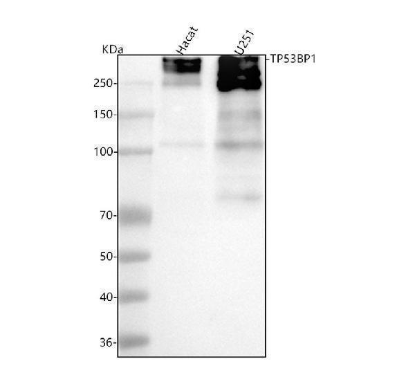
Figure 1. Western blot analysis of 53BP1 using anti-53BP1 antibody (M00397).
Electrophoresis was performed on a 5-20% SDS-PAGE gel at 70V (Stacking gel) / 90V (Resolving gel) for 2-3 hours. The sample well of each lane was loaded with 30 ug of sample under reducing conditions.
Lane 1: human Hacat whole cell lysates,
Lane 2: human U251 whole cell lysates.
After electrophoresis, proteins were transferred to a nitrocellulose membrane at 150 mA for 50-90 minutes. Blocked the membrane with 5% non-fat milk/TBS for 1.5 hour at RT. The membrane was incubated with rabbit anti-53BP1 antigen affinity purified polyclonal antibody (Catalog # M00397) at 1:500 overnight at 4°C, then washed with TBS-0.1%Tween 3 times with 5 minutes each and probed with a goat anti-rabbit IgG-HRP secondary antibody at a dilution of 1:5000 for 1.5 hour at RT. The signal is developed using an Enhanced Chemiluminescent detection (ECL) kit (Catalog # EK1002) with Tanon 5200 system. A specific band was detected for 53BP1 at approximately 450 kDa. The expected band size for 53BP1 is at 214 kDa.

Click image to see more details
Figure 2. IHC analysis of 53BP1 using anti-53BP1 antibody (M00397).
53BP1 was detected in a paraffin-embedded section of human acinic cell carcinoma of parotid tissue. Heat mediated antigen retrieval was performed in EDTA buffer (pH 8.0, epitope retrieval solution). The tissue section was blocked with 10% goat serum. The tissue section was then incubated with 1:50 rabbit anti-53BP1 Antibody (M00397) overnight at 4°C. Peroxidase Conjugated Goat Anti-rabbit IgG was used as secondary antibody and incubated for 30 minutes at 37°C. The tissue section was developed using HRP Conjugated Rabbit IgG Super Vision Assay Kit (Catalog # SV0002) with DAB as the chromogen.

Click image to see more details
Figure 3. IHC analysis of 53BP1 using anti-53BP1 antibody (M00397).
53BP1 was detected in a paraffin-embedded section of human acinic cell carcinoma of parotid tissue. Heat mediated antigen retrieval was performed in EDTA buffer (pH 8.0, epitope retrieval solution). The tissue section was blocked with 10% goat serum. The tissue section was then incubated with 1:50 rabbit anti-53BP1 Antibody (M00397) overnight at 4°C. Peroxidase Conjugated Goat Anti-rabbit IgG was used as secondary antibody and incubated for 30 minutes at 37°C. The tissue section was developed using HRP Conjugated Rabbit IgG Super Vision Assay Kit (Catalog # SV0002) with DAB as the chromogen.

Click image to see more details
Figure 4. IHC analysis of 53BP1 using anti-53BP1 antibody (M00397).
53BP1 was detected in a paraffin-embedded section of human cervical cancer tissue. Heat mediated antigen retrieval was performed in EDTA buffer (pH 8.0, epitope retrieval solution). The tissue section was blocked with 10% goat serum. The tissue section was then incubated with 1:50 rabbit anti-53BP1 Antibody (M00397) overnight at 4°C. Peroxidase Conjugated Goat Anti-rabbit IgG was used as secondary antibody and incubated for 30 minutes at 37°C. The tissue section was developed using HRP Conjugated Rabbit IgG Super Vision Assay Kit (Catalog # SV0002) with DAB as the chromogen.

Click image to see more details
Figure 5. IHC analysis of 53BP1 using anti-53BP1 antibody (M00397).
53BP1 was detected in a paraffin-embedded section of human cervical cancer tissue. Heat mediated antigen retrieval was performed in EDTA buffer (pH 8.0, epitope retrieval solution). The tissue section was blocked with 10% goat serum. The tissue section was then incubated with 1:50 rabbit anti-53BP1 Antibody (M00397) overnight at 4°C. Peroxidase Conjugated Goat Anti-rabbit IgG was used as secondary antibody and incubated for 30 minutes at 37°C. The tissue section was developed using HRP Conjugated Rabbit IgG Super Vision Assay Kit (Catalog # SV0002) with DAB as the chromogen.

Click image to see more details
Figure 6. IHC analysis of 53BP1 using anti-53BP1 antibody (M00397).
53BP1 was detected in a paraffin-embedded section of human clear cell renal cell carcinoma tissue. Heat mediated antigen retrieval was performed in EDTA buffer (pH 8.0, epitope retrieval solution). The tissue section was blocked with 10% goat serum. The tissue section was then incubated with 1:50 rabbit anti-53BP1 Antibody (M00397) overnight at 4°C. Peroxidase Conjugated Goat Anti-rabbit IgG was used as secondary antibody and incubated for 30 minutes at 37°C. The tissue section was developed using HRP Conjugated Rabbit IgG Super Vision Assay Kit (Catalog # SV0002) with DAB as the chromogen.

Click image to see more details
Figure 7. IHC analysis of 53BP1 using anti-53BP1 antibody (M00397).
53BP1 was detected in a paraffin-embedded section of human clear cell renal cell carcinoma tissue. Heat mediated antigen retrieval was performed in EDTA buffer (pH 8.0, epitope retrieval solution). The tissue section was blocked with 10% goat serum. The tissue section was then incubated with 1:50 rabbit anti-53BP1 Antibody (M00397) overnight at 4°C. Peroxidase Conjugated Goat Anti-rabbit IgG was used as secondary antibody and incubated for 30 minutes at 37°C. The tissue section was developed using HRP Conjugated Rabbit IgG Super Vision Assay Kit (Catalog # SV0002) with DAB as the chromogen.

Click image to see more details
Figure 8. IHC analysis of 53BP1 using anti-53BP1 antibody (M00397).
53BP1 was detected in a paraffin-embedded section of human colon adenocarcinoma tissue. Heat mediated antigen retrieval was performed in EDTA buffer (pH 8.0, epitope retrieval solution). The tissue section was blocked with 10% goat serum. The tissue section was then incubated with 1:50 rabbit anti-53BP1 Antibody (M00397) overnight at 4°C. Peroxidase Conjugated Goat Anti-rabbit IgG was used as secondary antibody and incubated for 30 minutes at 37°C. The tissue section was developed using HRP Conjugated Rabbit IgG Super Vision Assay Kit (Catalog # SV0002) with DAB as the chromogen.

Click image to see more details
Figure 9. IHC analysis of 53BP1 using anti-53BP1 antibody (M00397).
53BP1 was detected in a paraffin-embedded section of human colon adenocarcinoma tissue. Heat mediated antigen retrieval was performed in EDTA buffer (pH 8.0, epitope retrieval solution). The tissue section was blocked with 10% goat serum. The tissue section was then incubated with 1:50 rabbit anti-53BP1 Antibody (M00397) overnight at 4°C. Peroxidase Conjugated Goat Anti-rabbit IgG was used as secondary antibody and incubated for 30 minutes at 37°C. The tissue section was developed using HRP Conjugated Rabbit IgG Super Vision Assay Kit (Catalog # SV0002) with DAB as the chromogen.

Click image to see more details
Figure 10. IHC analysis of 53BP1 using anti-53BP1 antibody (M00397).
53BP1 was detected in a paraffin-embedded section of human liver cancer tissue. Heat mediated antigen retrieval was performed in EDTA buffer (pH 8.0, epitope retrieval solution). The tissue section was blocked with 10% goat serum. The tissue section was then incubated with 1:50 rabbit anti-53BP1 Antibody (M00397) overnight at 4°C. Peroxidase Conjugated Goat Anti-rabbit IgG was used as secondary antibody and incubated for 30 minutes at 37°C. The tissue section was developed using HRP Conjugated Rabbit IgG Super Vision Assay Kit (Catalog # SV0002) with DAB as the chromogen.

Click image to see more details
Figure 11. IHC analysis of 53BP1 using anti-53BP1 antibody (M00397).
53BP1 was detected in a paraffin-embedded section of human liver cancer tissue. Heat mediated antigen retrieval was performed in EDTA buffer (pH 8.0, epitope retrieval solution). The tissue section was blocked with 10% goat serum. The tissue section was then incubated with 1:50 rabbit anti-53BP1 Antibody (M00397) overnight at 4°C. Peroxidase Conjugated Goat Anti-rabbit IgG was used as secondary antibody and incubated for 30 minutes at 37°C. The tissue section was developed using HRP Conjugated Rabbit IgG Super Vision Assay Kit (Catalog # SV0002) with DAB as the chromogen.

Click image to see more details
Figure 12. IHC analysis of 53BP1 using anti-53BP1 antibody (M00397).
53BP1 was detected in a paraffin-embedded section of human ovarian cancer tissue. Heat mediated antigen retrieval was performed in EDTA buffer (pH 8.0, epitope retrieval solution). The tissue section was blocked with 10% goat serum. The tissue section was then incubated with 1:50 rabbit anti-53BP1 Antibody (M00397) overnight at 4°C. Peroxidase Conjugated Goat Anti-rabbit IgG was used as secondary antibody and incubated for 30 minutes at 37°C. The tissue section was developed using HRP Conjugated Rabbit IgG Super Vision Assay Kit (Catalog # SV0002) with DAB as the chromogen.

Click image to see more details
Figure 13. IHC analysis of 53BP1 using anti-53BP1 antibody (M00397).
53BP1 was detected in a paraffin-embedded section of human ovarian cancer tissue. Heat mediated antigen retrieval was performed in EDTA buffer (pH 8.0, epitope retrieval solution). The tissue section was blocked with 10% goat serum. The tissue section was then incubated with 1:50 rabbit anti-53BP1 Antibody (M00397) overnight at 4°C. Peroxidase Conjugated Goat Anti-rabbit IgG was used as secondary antibody and incubated for 30 minutes at 37°C. The tissue section was developed using HRP Conjugated Rabbit IgG Super Vision Assay Kit (Catalog # SV0002) with DAB as the chromogen.

Click image to see more details
Figure 14. IHC analysis of 53BP1 using anti-53BP1 antibody (M00397).
53BP1 was detected in a paraffin-embedded section of human placenta tissue. Heat mediated antigen retrieval was performed in EDTA buffer (pH 8.0, epitope retrieval solution). The tissue section was blocked with 10% goat serum. The tissue section was then incubated with 1:50 rabbit anti-53BP1 Antibody (M00397) overnight at 4°C. Peroxidase Conjugated Goat Anti-rabbit IgG was used as secondary antibody and incubated for 30 minutes at 37°C. The tissue section was developed using HRP Conjugated Rabbit IgG Super Vision Assay Kit (Catalog # SV0002) with DAB as the chromogen.

Click image to see more details
Figure 15. IHC analysis of 53BP1 using anti-53BP1 antibody (M00397).
53BP1 was detected in a paraffin-embedded section of human placenta tissue. Heat mediated antigen retrieval was performed in EDTA buffer (pH 8.0, epitope retrieval solution). The tissue section was blocked with 10% goat serum. The tissue section was then incubated with 1:50 rabbit anti-53BP1 Antibody (M00397) overnight at 4°C. Peroxidase Conjugated Goat Anti-rabbit IgG was used as secondary antibody and incubated for 30 minutes at 37°C. The tissue section was developed using HRP Conjugated Rabbit IgG Super Vision Assay Kit (Catalog # SV0002) with DAB as the chromogen.

Click image to see more details
Figure 16. IHC analysis of 53BP1 using anti-53BP1 antibody (M00397).
53BP1 was detected in a paraffin-embedded section of human tonsil tissue. Heat mediated antigen retrieval was performed in EDTA buffer (pH 8.0, epitope retrieval solution). The tissue section was blocked with 10% goat serum. The tissue section was then incubated with 1:50 rabbit anti-53BP1 Antibody (M00397) overnight at 4°C. Peroxidase Conjugated Goat Anti-rabbit IgG was used as secondary antibody and incubated for 30 minutes at 37°C. The tissue section was developed using HRP Conjugated Rabbit IgG Super Vision Assay Kit (Catalog # SV0002) with DAB as the chromogen.

Click image to see more details
Figure 17. IHC analysis of 53BP1 using anti-53BP1 antibody (M00397).
53BP1 was detected in a paraffin-embedded section of human tonsil tissue. Heat mediated antigen retrieval was performed in EDTA buffer (pH 8.0, epitope retrieval solution). The tissue section was blocked with 10% goat serum. The tissue section was then incubated with 1:50 rabbit anti-53BP1 Antibody (M00397) overnight at 4°C. Peroxidase Conjugated Goat Anti-rabbit IgG was used as secondary antibody and incubated for 30 minutes at 37°C. The tissue section was developed using HRP Conjugated Rabbit IgG Super Vision Assay Kit (Catalog # SV0002) with DAB as the chromogen.

Click image to see more details
Figure 18. IHC analysis of 53BP1 using anti-53BP1 antibody (M00397).
53BP1 was detected in a paraffin-embedded section of mouse liver tissue. Heat mediated antigen retrieval was performed in EDTA buffer (pH 8.0, epitope retrieval solution). The tissue section was blocked with 10% goat serum. The tissue section was then incubated with 1:50 rabbit anti-53BP1 Antibody (M00397) overnight at 4°C. Peroxidase Conjugated Goat Anti-rabbit IgG was used as secondary antibody and incubated for 30 minutes at 37°C. The tissue section was developed using HRP Conjugated Rabbit IgG Super Vision Assay Kit (Catalog # SV0002) with DAB as the chromogen.

Click image to see more details
Figure 19. IHC analysis of 53BP1 using anti-53BP1 antibody (M00397).
53BP1 was detected in a paraffin-embedded section of mouse liver tissue. Heat mediated antigen retrieval was performed in EDTA buffer (pH 8.0, epitope retrieval solution). The tissue section was blocked with 10% goat serum. The tissue section was then incubated with 1:50 rabbit anti-53BP1 Antibody (M00397) overnight at 4°C. Peroxidase Conjugated Goat Anti-rabbit IgG was used as secondary antibody and incubated for 30 minutes at 37°C. The tissue section was developed using HRP Conjugated Rabbit IgG Super Vision Assay Kit (Catalog # SV0002) with DAB as the chromogen.

Click image to see more details
Figure 20. IHC analysis of 53BP1 using anti-53BP1 antibody (M00397).
53BP1 was detected in a paraffin-embedded section of rat liver tissue. Heat mediated antigen retrieval was performed in EDTA buffer (pH 8.0, epitope retrieval solution). The tissue section was blocked with 10% goat serum. The tissue section was then incubated with 1:50 rabbit anti-53BP1 Antibody (M00397) overnight at 4°C. Peroxidase Conjugated Goat Anti-rabbit IgG was used as secondary antibody and incubated for 30 minutes at 37°C. The tissue section was developed using HRP Conjugated Rabbit IgG Super Vision Assay Kit (Catalog # SV0002) with DAB as the chromogen.

Click image to see more details
Figure 21. IHC analysis of 53BP1 using anti-53BP1 antibody (M00397).
53BP1 was detected in a paraffin-embedded section of rat liver tissue. Heat mediated antigen retrieval was performed in EDTA buffer (pH 8.0, epitope retrieval solution). The tissue section was blocked with 10% goat serum. The tissue section was then incubated with 1:50 rabbit anti-53BP1 Antibody (M00397) overnight at 4°C. Peroxidase Conjugated Goat Anti-rabbit IgG was used as secondary antibody and incubated for 30 minutes at 37°C. The tissue section was developed using HRP Conjugated Rabbit IgG Super Vision Assay Kit (Catalog # SV0002) with DAB as the chromogen.

Click image to see more details
Immunofluorescent analysis of HepG2 cells, using 53BP1 Antibody .

Click image to see more details
Immunofluorescent analysis using the Antibody at 1:50 dilution.
Protein Target Info & Infographic
Gene/Protein Information For TP53BP1 (Source: Uniprot.org, NCBI)
Gene Name
TP53BP1
Full Name
TP53-binding protein 1
Weight
213.574kDa
Alternative Names
53BP1; FLJ41424; MGC138366; p202; p53-binding protein 1; p53bp1; TP53BP1; tumor protein 53-binding protein, 1; tumor protein p53 binding protein 1; tumor protein p53-binding protein, 1; tumor suppressor p53-binding protein 1 TP53BP1 53BP1, TDRD30, p202, p53BP1 tumor protein p53 binding protein 1 TP53-binding protein 1|p53-binding protein 1|tumor protein 53-binding protein, 1|tumor suppressor p53-binding protein 1
*If product is indicated to react with multiple species, protein info is based on the gene entry specified above in "Species".For more info on TP53BP1, check out the TP53BP1 Infographic

We have 30,000+ of these available, one for each gene! Check them out.
In this infographic, you will see the following information for TP53BP1: database IDs, superfamily, protein function, synonyms, molecular weight, chromosomal locations, tissues of expression, subcellular locations, post-translational modifications, and related diseases, research areas & pathways. If you want to see more information included, or would like to contribute to it and be acknowledged, please contact [email protected].
Specific Publications For Anti-53BP1 TP53BP1 Rabbit Monoclonal Antibody (M00397)
Hello CJ!
No publications found for M00397
*Do you have publications using this product? Share with us and receive a reward. Ask us for more details.
Recommended Resources
Here are featured tools and databases that you might find useful.
- Boster's Pathways Library
- Protein Databases
- Bioscience Research Protocol Resources
- Data Processing & Analysis Software
- Photo Editing Software
- Scientific Literature Resources
- Research Paper Management Tools
- Molecular Biology Software
- Primer Design Tools
- Bioinformatics Tools
- Phylogenetic Tree Analysis
Customer Reviews
Have you used Anti-53BP1 TP53BP1 Rabbit Monoclonal Antibody?
Submit a review and receive an Amazon gift card.
- $30 for a review with an image
Be the first to review Anti-53BP1 TP53BP1 Rabbit Monoclonal Antibody
*The first user to submit a review for a product is eligible for Boster's Innovating Scientists Reward, which gives product credits. This is in addition to the gift card reward.
Customer Q&As
Have a question?
Find answers in Q&As, reviews.
Can't find your answer?
Submit your question
18 Customer Q&As for Anti-53BP1 TP53BP1 Rabbit Monoclonal Antibody
Question
Is M00397 a recombinant antibody?
Verified customer
Asked: 2020-12-21
Answer
Yes, the Anti-53BP1 TP53BP1 Rabbit Monoclonal Antibody (M00397) is a recombinant antibody.
Boster Scientific Support
Answered: 2020-12-21
Question
What is the expression system and the transfection method (virus vector, transfection reagent) of M00397?
Verified customer
Asked: 2020-12-21
Answer
For the Anti-53BP1 TP53BP1 Rabbit Monoclonal Antibody (M00397), expression system is plasmid transfection with 293F without using any viral particles.
Boster Scientific Support
Answered: 2020-12-21
Question
I was wanting to use your anti-53BP1 Rabbit Monoclonal antibody for IF for mouse cerebellum on frozen tissues, but I want to know if it has been tested for this particular application. Has this antibody been tested and is this antibody a good choice for mouse cerebellum identification?
Verified Customer
Verified customer
Asked: 2020-04-22
Answer
You can see on the product datasheet, M00397 anti-53BP1 Rabbit Monoclonal antibody has been tested for IF, WB on human, mouse, rat tissues. We have an innovator award program that if you test this antibody and show it works in mouse cerebellum in IHC-frozen, you can get your next antibody for free.
Boster Scientific Support
Answered: 2020-04-22
Question
Do you have a BSA free version of anti-53BP1 Rabbit Monoclonal antibody M00397 available?
R. Miller
Verified customer
Asked: 2020-03-04
Answer
Thanks for your recent telephone inquiry. I can confirm that some lots of this anti-53BP1 Rabbit Monoclonal antibody M00397 are BSA free. For now, these lots are available and we can make a BSA free formula for you free of charge. It will take 3 extra days to prepare. If you require this antibody BSA free again in future, please do not hesitate to contact me and I will be pleased to check which lots we have in stock that are BSA free.
Boster Scientific Support
Answered: 2020-03-04
Question
Can you help my question with product M00397, anti-53BP1 Rabbit Monoclonal antibody. I was wondering if it would be possible to conjugate this antibody with biotin. I would need it to be without BSA or sodium azide. I am planning on using a buffer exchange of sodium azide with PBS only. Would there be problems for me to conjugate the antibody and store it in -20 degrees in small aliquots?
Verified Customer
Verified customer
Asked: 2020-02-25
Answer
We do not advise storing this antibody with PBS buffer only in -20 degrees. If you want to store it in -20 degrees it is best to add some cryoprotectant like glycerol. If you want carrier free M00397 anti-53BP1 Rabbit Monoclonal antibody, we can provide it to you in a special formula with trehalose and/or glycerol. These molecules will not interfere with conjugation chemistry and provide a good level of protection for the antibody from degradation. Please be sure to specify this in your purchase order.
Boster Scientific Support
Answered: 2020-02-25
Question
you antibody to test anti-53BP1 Rabbit Monoclonal antibody M00397 on mouse cerebellum for research purposes, then I may be interested in using anti-53BP1 Rabbit Monoclonal antibody M00397 for diagnostic purposes as well. Is the antibody suitable for diagnostic purposes?
D. Taylor
Verified customer
Asked: 2020-02-25
Answer
The products we sell, including anti-53BP1 Rabbit Monoclonal antibody M00397, are only intended for research use. They would not be suitable for use in diagnostic work. If you have the means to develop a product into diagnostic use, and are interested in collaborating with us and develop our product into an IVD product, please contact us for more discussions.
Boster Scientific Support
Answered: 2020-02-25
Question
See below the WB image, lot number and protocol we used for cerebellum using anti-53BP1 Rabbit Monoclonal antibody M00397. Please let me know if you require anything else.
Verified Customer
Verified customer
Asked: 2020-01-15
Answer
Thank you very much for the data. Our lab team are working to resolve this as quickly as possible, and we appreciate your patience and understanding! You have provided everything we needed. Please let me know if there is anything you need in the meantime.
Boster Scientific Support
Answered: 2020-01-15
Question
Would M00397 anti-53BP1 Rabbit Monoclonal antibody work on parafin embedded sections? If so, which fixation method do you recommend we use (PFA, paraformaldehyde, other)?
Verified Customer
Verified customer
Asked: 2019-07-02
Answer
As indicated on the product datasheet, M00397 anti-53BP1 Rabbit Monoclonal antibody as been validated on IF. It is best to use PFA for fixation because it has better tissue penetration ability. PFA needs to be prepared fresh before use. Long term stored PFA turns into formalin, as the PFA molecules congregate and become formalin.
Boster Scientific Support
Answered: 2019-07-02
Question
We purchased anti-53BP1 Rabbit Monoclonal antibody for IF on embryonic kidney last year. I am using human, and We intend to use the antibody for WB next. I am interested in examining embryonic kidney as well as liver in our next experiment. Could you please give me some suggestion on which antibody would work the best for WB?
Verified Customer
Verified customer
Asked: 2019-06-21
Answer
I looked at the website and datasheets of our anti-53BP1 Rabbit Monoclonal antibody and I see that M00397 has been tested on human in both IF and WB. Thus M00397 should work for your application. Our Boster satisfaction guarantee will cover this product for WB in human even if the specific tissue type has not been validated. We do have a comprehensive range of products for WB detection and you can check out our website bosterbio.com to find out more information about them.
Boster Scientific Support
Answered: 2019-06-21
Question
Is this M00397 anti-53BP1 Rabbit Monoclonal antibody reactive to the isotypes of TP53BP1?
Verified Customer
Verified customer
Asked: 2019-06-03
Answer
The immunogen of M00397 anti-53BP1 Rabbit Monoclonal antibody is A synthesized peptide derived from human 53BP1. Could you tell me which isotype you are interested in so I can help see if the immunogen is part of this isotype?
Boster Scientific Support
Answered: 2019-06-03
Question
Thanks for helping with my inquiry over the phone. Here are the WB image, lot number and protocol we used for cerebellum using anti-53BP1 Rabbit Monoclonal antibody M00397. Let me know if you need anything else.
Verified Customer
Verified customer
Asked: 2018-03-23
Answer
I appreciate the data. You have provided everything we needed. Our lab team are working to resolve your inquiry as quickly as possible, and we appreciate your patience and understanding! Please let me know if there is anything you need in the meantime.
Boster Scientific Support
Answered: 2018-03-23
Question
My boss were satisfied with the WB result of your anti-53BP1 Rabbit Monoclonal antibody. However we have observed positive staining in cervix nucleus using this antibody. Is that expected? Could you tell me where is TP53BP1 supposed to be expressed?
Verified Customer
Verified customer
Asked: 2017-11-09
Answer
From what I have seen in literature, cervix does express TP53BP1. Generally TP53BP1 expresses in nucleus. Regarding which tissues have TP53BP1 expression, here are a few articles citing expression in various tissues:
Cerebellum, Pubmed ID: 15489334
Cervix, Pubmed ID: 17974005
Cervix carcinoma, Pubmed ID: 16964243, 17081983, 18220336, 18669648, 20068231
Cervix carcinoma, and Erythroleukemia, Pubmed ID: 23186163
Embryonic kidney, Pubmed ID: 17525332
Leukemic T-cell, Pubmed ID: 19690332
Liver, Pubmed ID: 24275569
Skeletal muscle, Pubmed ID: 9748285
Boster Scientific Support
Answered: 2017-11-09
Question
I see that the anti-53BP1 Rabbit Monoclonal antibody M00397 works with IF, what is the protocol used to produce the result images on the product page?
A. Wu
Verified customer
Asked: 2017-02-22
Answer
You can find protocols for IF on the "support/technical resources" section of our navigation menu. If you have any further questions, please send an email to [email protected]
Boster Scientific Support
Answered: 2017-02-22
Question
We have been able to see staining in human liver. Any tips? Is anti-53BP1 Rabbit Monoclonal antibody supposed to stain liver positively?
T. Kulkarni
Verified customer
Asked: 2017-02-20
Answer
Based on literature liver does express TP53BP1. Based on Uniprot.org, TP53BP1 is expressed in lung, skeletal muscle, myeloid leukemia cell, cervix, cerebellum, cervix carcinoma, embryonic kidney, leukemic t-cell, cervix carcinoma erythroleukemia, liver, among other tissues. Regarding which tissues have TP53BP1 expression, here are a few articles citing expression in various tissues:
Cerebellum, Pubmed ID: 15489334
Cervix, Pubmed ID: 17974005
Cervix carcinoma, Pubmed ID: 16964243, 17081983, 18220336, 18669648, 20068231
Cervix carcinoma, and Erythroleukemia, Pubmed ID: 23186163
Embryonic kidney, Pubmed ID: 17525332
Leukemic T-cell, Pubmed ID: 19690332
Liver, Pubmed ID: 24275569
Skeletal muscle, Pubmed ID: 9748285
Boster Scientific Support
Answered: 2017-02-20
Question
Will anti-53BP1 Rabbit Monoclonal antibody M00397 work for IF with cerebellum?
V. Evans
Verified customer
Asked: 2016-05-04
Answer
According to the expression profile of cerebellum, TP53BP1 is highly expressed in cerebellum. So, it is likely that anti-53BP1 Rabbit Monoclonal antibody M00397 will work for IF with cerebellum.
Boster Scientific Support
Answered: 2016-05-04
Question
We are currently using anti-53BP1 Rabbit Monoclonal antibody M00397 for rat tissue, and we are content with the IF results. The species of reactivity given in the datasheet says human, mouse, rat. Is it true that the antibody can work on zebrafish tissues as well?
L. Krishna
Verified customer
Asked: 2015-09-18
Answer
The anti-53BP1 Rabbit Monoclonal antibody (M00397) has not been tested for cross reactivity specifically with zebrafish tissues, though there is a good chance of cross reactivity. We have an innovator award program that if you test this antibody and show it works in zebrafish you can get your next antibody for free. Please contact me if I can help you with anything.
Boster Scientific Support
Answered: 2015-09-18
Question
We are interested in using your anti-53BP1 Rabbit Monoclonal antibody for nonhomologous end-joining (nhej) studies. Has this antibody been tested with western blotting on hela cell lysate? We would like to see some validation images before ordering.
R. Singh
Verified customer
Asked: 2013-05-14
Answer
I appreciate your inquiry. This M00397 anti-53BP1 Rabbit Monoclonal antibody is tested on hela cell lysate. It is guaranteed to work for IF, WB in human, mouse, rat. Our Boster guarantee will cover your intended experiment even if the sample type has not been be directly tested.
Boster Scientific Support
Answered: 2013-05-14
Question
Is a blocking peptide available for product anti-53BP1 Rabbit Monoclonal antibody (M00397)?
A. Brown
Verified customer
Asked: 2013-02-22
Answer
We do provide the blocking peptide for product anti-53BP1 Rabbit Monoclonal antibody (M00397). If you would like to place an order for it please contact [email protected] and make a special request.
Boster Scientific Support
Answered: 2013-02-22
