Click image to see more details
-

-
-
-
-
+4
Product Info Summary
| SKU: | PB10032 |
|---|---|
| Size: | 100 μg/vial |
| Reactive Species: | Human, Mouse, Rat |
| Host: | Rabbit |
| Application: | Flow Cytometry, IF, IHC, ICC, WB |
Customers Who Bought This Also Bought

Product info
Product Name
Anti-ADO Antibody Picoband™
SKU/Catalog Number
PB10032
Size
100 μg/vial
Form
Lyophilized
Description
Boster Bio Anti-ADO Antibody Picoband™ catalog # PB10032. Tested in Flow Cytometry, IF, IHC, ICC, WB applications. This antibody reacts with Human, Mouse, Rat.
Storage & Handling
Store at -20˚C for one year from date of receipt. After reconstitution, at 4˚C for one month. It can also be aliquotted and stored frozen at -20˚C for six months. Avoid repeated freeze-thaw cycles.
Cite This Product
Anti-ADO Antibody Picoband™ (Boster Biological Technology, Pleasanton CA, USA, Catalog # PB10032)
Host
Rabbit
Contents
Each vial contains 5mg BSA, 0.9mg NaCl, 0.2mg Na2HPO4, 0.05mg NaN3.
Clonality
Polyclonal
Isotype
Rabbit IgG
Immunogen
E. coli-derived human ADO recombinant protein (Position: E49-E261). Human ADO shares 90.1% amino acid (aa) sequence identity with mouse ADO.
*Blocking peptide can be purchased. Costs vary based on immunogen length. Contact us for pricing.
Cross-reactivity
No cross-reactivity with other proteins.
Reactive Species
PB10032 is reactive to ADO in Human, Mouse, Rat
Applications
PB10032 is guaranteed for Flow Cytometry, IF, IHC, ICC, WB Boster Guarantee
Observed Molecular Weight
28 kDa, 30 kDa
Calculated molecular weight
29.751kDa
Background of ADO
Human thiol dioxygenases include cysteine dioxygenase (CDO) and cysteamine (2-aminoethanethiol) dioxygenase (ADO). CDO adds 2 oxygen atoms to free cysteine, whereas ADO adds 2 oxygen atoms to free cysteamine to form hypotaurine. It is demonstrated that mouse Ado has strong and specific dioxygenase activity in vitro towards cysteamine but not cysteine. Recombinant Ado was shown to bind iron. Overexpression of Ado in HepG2/C3A cells increased the production of hypotaurine from cysteamine. Similar results were found with human ADO. When endogenous expression of ADO was reduced by RNA-mediated interference, hypotaurine production decreased. It is also noted that the demonstration of high levels of ADO in brain challenges the previous assumption that most of the taurine in the brain is a consequence of CDO activity.
Antibody Validation
Boster validates all antibodies on WB, IHC, ICC, Immunofluorescence, and ELISA with known positive control and negative samples to ensure specificity and high affinity, including thorough antibody incubations.
Innovating Scientists Reward
If you are the first to review this product, or if you have results for a special sample, species or application this product is not validated in, share your results with us and receive product credits you can use towards any Boster products! Applicable to all scientists worldwide.
Submit A Review
Assay dilution & Images
Reconsitution
Add 0.2ml of distilled water will yield a concentration of 500ug/ml.
Assay Dilutions Recommendation
The recommendations below provide a starting point for assay optimization. The actual working concentration varies and should be decided by the user.
Western blot, 0.1-0.5μg/ml, Human, Mouse, Rat
Immunohistochemistry (Paraffin-embedded Section), 2-5μg/ml, Mouse, Rat
Immunocytochemistry/Immunofluorescence, 2μg/ml, Human
Flow Cytometry, 1-3μg/1x106 cells, Human
Validation Images & Assay Conditions
Click image to see more details
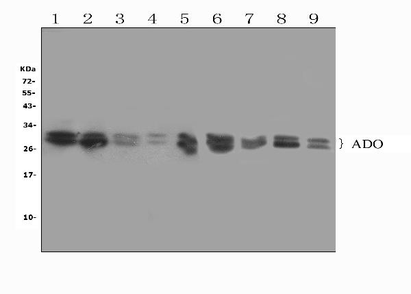
Figure 1. Western blot analysis of ADO using anti-ADO antibody (PB10032).
Electrophoresis was performed on a 5-20% SDS-PAGE gel at 70V (Stacking gel) / 90V (Resolving gel) for 2-3 hours. The sample well of each lane was loaded with 50ug of sample under reducing conditions.
Lane 1: rat brain tissue lysates,
Lane 2: rat testicular tissue lysates,
Lane 3: rat spleen tissue lysates,
Lane 4: human MCF-7 whole cell lysates,
Lane 5: human SW620 whole cell lysates,
Lane 6: mouse spleen tissue lysates,
Lane 7: mouse heart tissue lysates,
Lane 8: mouse HEPA1-6 whole cell lysates,
Lane 9: mouse SP2/0 whole cell lysates,
After Electrophoresis, proteins were transferred to a Nitrocellulose membrane at 150mA for 50-90 minutes. Blocked the membrane with 5% Non-fat Milk/ TBS for 1.5 hour at RT. The membrane was incubated with rabbit anti-ADO antigen affinity purified polyclonal antibody (Catalog # PB10032) at 0.5 μg/mL overnight at 4°C, then washed with TBS-0.1%Tween 3 times with 5 minutes each and probed with a goat anti-rabbit IgG-HRP secondary antibody at a dilution of 1:10000 for 1.5 hour at RT. The signal is developed using an Enhanced Chemiluminescent detection (ECL) kit (Catalog # EK1002) with Tanon 5200 system. A specific band was detected for ADO at approximately 28,30KD. The expected band size for ADO is at 30KD.

Click image to see more details
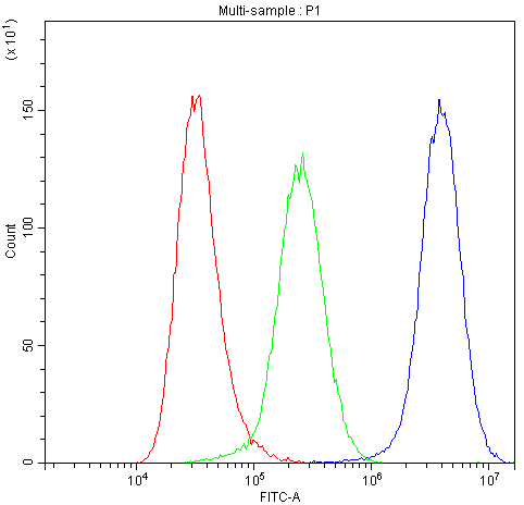
Figure 2. Flow Cytometry analysis of A549 cells using anti-ADO antibody (PB10032).
Overlay histogram showing A549 cells stained with PB10032 (Blue line).The cells were blocked with 10% normal goat serum. And then incubated with rabbit anti-ADO Antibody (PB10032,1μg/1x106 cells) for 30 min at 20°C. DyLight®488 conjugated goat anti-rabbit IgG (BA1127, 5-10μg/1x106 cells) was used as secondary antibody for 30 minutes at 20°C. Isotype control antibody (Green line) was rabbit IgG (1μg/1x106) used under the same conditions. Unlabelled sample (Red line) was also used as a control.

Click image to see more details
Figure 3. Flow Cytometry analysis of U251 cells using anti-ADO antibody (PB10032).
Overlay histogram showing U251 cells stained with PB10032 (Blue line).The cells were blocked with 10% normal goat serum. And then incubated with rabbit anti-ADO Antibody (PB10032,1μg/1x106 cells) for 30 min at 20°C. DyLight®488 conjugated goat anti-rabbit IgG (BA1127, 5-10μg/1x106 cells) was used as secondary antibody for 30 minutes at 20°C. Isotype control antibody (Green line) was rabbit IgG (1μg/1x106) used under the same conditions. Unlabelled sample (Red line) was also used as a control.

Click image to see more details
Figure 4. Flow Cytometry analysis of Hela cells using anti-ADO antibody (PB10032).
Overlay histogram showing Hela cells stained with PB10032 (Blue line).The cells were blocked with 10% normal goat serum. And then incubated with rabbit anti-ADO Antibody (PB10032,1μg/1x106 cells) for 30 min at 20°C. DyLight®488 conjugated goat anti-rabbit IgG (BA1127, 5-10μg/1x106 cells) was used as secondary antibody for 30 minutes at 20°C. Isotype control antibody (Green line) was rabbit IgG (1μg/1x106) used under the same conditions. Unlabelled sample (Red line) was also used as a control.

Click image to see more details
Figure 5. Flow Cytometry analysis of PC-3 cells using anti-ADO antibody (PB10032).
Overlay histogram showing PC-3 cells stained with PB10032 (Blue line).The cells were blocked with 10% normal goat serum. And then incubated with rabbit anti-ADO Antibody (PB10032,1μg/1x106 cells) for 30 min at 20°C. DyLight®488 conjugated goat anti-rabbit IgG (BA1127, 5-10μg/1x106 cells) was used as secondary antibody for 30 minutes at 20°C. Isotype control antibody (Green line) was rabbit IgG (1μg/1x106) used under the same conditions. Unlabelled sample (Red line) was also used as a control.

Click image to see more details
Figure 6. IF analysis of ADO using anti-ADO antibody (PB10032).
ADO was detected in immunocytochemical section of A549 cells. Enzyme antigen retrieval was performed using IHC enzyme antigen retrieval reagent (AR0022) for 15 mins. The cells were blocked with 10% goat serum. And then incubated with 2μg/mL rabbit anti-ADO Antibody (PB10032) overnight at 4°C. DyLight®488 Conjugated Goat Anti-Rabbit IgG (BA1127) was used as secondary antibody at 1:100 dilution and incubated for 30 minutes at 37°C. The section was counterstained with DAPI. Visualize using a fluorescence microscope and filter sets appropriate for the label used.

Click image to see more details
Figure 7. IHC analysis of ADO using anti-ADO antibody (PB10032).
ADO was detected in paraffin-embedded section of rat brain tissue. Heat mediated antigen retrieval was performed in EDTA buffer (pH8.0, epitope retrieval solution). The tissue section was blocked with 10% goat serum. The tissue section was then incubated with 2μg/ml rabbit anti-ADO Antibody (PB10032) overnight at 4°C. Biotinylated goat anti-rabbit IgG was used as secondary antibody and incubated for 30 minutes at 37°C. The tissue section was developed using Strepavidin-Biotin-Complex (SABC) (Catalog # SA1022) with DAB as the chromogen.

Click image to see more details
Figure 8. IHC analysis of ADO using anti-ADO antibody (PB10032).
ADO was detected in paraffin-embedded section of mouse brain tissue. Heat mediated antigen retrieval was performed in EDTA buffer (pH8.0, epitope retrieval solution). The tissue section was blocked with 10% goat serum. The tissue section was then incubated with 2μg/ml rabbit anti-ADO Antibody (PB10032) overnight at 4°C. Biotinylated goat anti-rabbit IgG was used as secondary antibody and incubated for 30 minutes at 37°C. The tissue section was developed using Strepavidin-Biotin-Complex (SABC) (Catalog # SA1022) with DAB as the chromogen.
Protein Target Info & Infographic
Gene/Protein Information For ADO (Source: Uniprot.org, NCBI)
Gene Name
ADO
Full Name
2-aminoethanethiol dioxygenase
Weight
29.751kDa
Alternative Names
2-aminoethanethiol (cysteamine) dioxygenase; 2-aminoethanethiol dioxygenase; C10orf22; chromosome 10 open reading frame 22; cysteamine (2-aminoethanethiol) dioxygenase (ADO); Cysteamine dioxygenase; DKFZp564C046; EC 1.13.11.19; FLJ14547 ADO C10orf22 2-aminoethanethiol dioxygenase 2-aminoethanethiol dioxygenase|2-aminoethanethiol (cysteamine) dioxygenase|cysteamine (2-aminoethanethiol) dioxygenase (ADO)|cysteamine dioxygenase
*If product is indicated to react with multiple species, protein info is based on the gene entry specified above in "Species".For more info on ADO, check out the ADO Infographic

We have 30,000+ of these available, one for each gene! Check them out.
In this infographic, you will see the following information for ADO: database IDs, superfamily, protein function, synonyms, molecular weight, chromosomal locations, tissues of expression, subcellular locations, post-translational modifications, and related diseases, research areas & pathways. If you want to see more information included, or would like to contribute to it and be acknowledged, please contact [email protected].
Specific Publications For Anti-ADO Antibody Picoband™ (PB10032)
Hello CJ!
No publications found for PB10032
*Do you have publications using this product? Share with us and receive a reward. Ask us for more details.
Recommended Resources
Here are featured tools and databases that you might find useful.
- Boster's Pathways Library
- Protein Databases
- Bioscience Research Protocol Resources
- Data Processing & Analysis Software
- Photo Editing Software
- Scientific Literature Resources
- Research Paper Management Tools
- Molecular Biology Software
- Primer Design Tools
- Bioinformatics Tools
- Phylogenetic Tree Analysis
Customer Reviews
Have you used Anti-ADO Antibody Picoband™?
Submit a review and receive an Amazon gift card.
- $30 for a review with an image
1 Reviews For Anti-ADO Antibody Picoband™
0
ADO Antibody Works in Western Blot--Hua Jiang, Pediatrics, University of Colorado Denver, Research Associate
Excellent

Source: Biocompare.com
| Applications | Western Blot |
|---|---|
| Sample | Human liver, rat liver, mouse liver, mouse kidney, and mouse brain |
| Detection | Typhoon 940 |
"We are studying the metabolism of amino acid and we are interested in knowing if the pathway involved this protein has changed under the background of a knockout mouse. The western blot image for this antibody looks clean and the signal looks strong. The band shown on western blot is at the right size and the intensity is strong enough. Equally important, the price is generally lower than other companies. This antibody works ok in western blot experiments with human, rat, and mouse tissues."
Hua Jiang
Verified customer
Submitted 2017-11-29
Customer Q&As
Have a question?
Find answers in Q&As, reviews.
Can't find your answer?
Submit your question
1 Customer Q&As for Anti-ADO Antibody Picoband™
Question
We are currently using anti-ADO antibody PB10032 for mouse tissue, and we are content with the IHC results. The species of reactivity given in the datasheet says human, mouse, rat. Is it likely that the antibody can work on zebrafish tissues as well?
S. Thomas
Verified customer
Asked: 2013-02-25
Answer
The anti-ADO antibody (PB10032) has not been validated for cross reactivity specifically with zebrafish tissues, but there is a good chance of cross reactivity. We have an innovator award program that if you test this antibody and show it works in zebrafish you can get your next antibody for free. Please contact me if I can help you with anything.
Boster Scientific Support
Answered: 2013-02-25
