
Click image to see more details
-

-
-
-
-
+7
Product Info Summary
| SKU: | PB9473 |
|---|---|
| Size: | 100 μg/vial |
| Reactive Species: | Human, Mouse, Rat |
| Host: | Rabbit |
| Application: | Flow Cytometry, IF, IHC, WB |
Customers Who Bought This Also Bought

Product info
Product Name
Anti-Aquaporin 1/AQP1 Antibody Picoband™
View all Aquaporin 1/AQP1 Antibodies
SKU/Catalog Number
PB9473
Size
100 μg/vial
Form
Lyophilized
Description
Boster Bio Anti-Aquaporin 1/AQP1 Antibody Picoband™ catalog # PB9473. Tested in Flow Cytometry, IF, IHC, WB applications. This antibody reacts with Human, Mouse, Rat.
Storage & Handling
Store at -20˚C for one year from date of receipt. After reconstitution, at 4˚C for one month. It can also be aliquotted and stored frozen at -20˚C for six months. Avoid repeated freeze-thaw cycles.
Cite This Product
Anti-Aquaporin 1/AQP1 Antibody Picoband™ (Boster Biological Technology, Pleasanton CA, USA, Catalog # PB9473)
Host
Rabbit
Contents
Each vial contains 4 mg Trehalose, 0.9 mg NaCl and 0.2 mg Na2HPO4.
Clonality
Polyclonal
Isotype
Rabbit IgG
Immunogen
A synthetic peptide corresponding to a sequence at the C-terminus of human Aquaporin 1, different from the related mouse and rat sequences by one amino acid.
*Blocking peptide can be purchased. Costs vary based on immunogen length. Contact us for pricing.
Cross-reactivity
No cross-reactivity with other proteins
Reactive Species
PB9473 is reactive to AQP1 in Human, Mouse, Rat
Applications
PB9473 is guaranteed for Flow Cytometry, IF, IHC, WB Boster Guarantee
Observed Molecular Weight
25 kDa
Calculated molecular weight
28.526kDa
Background of Aquaporin 1/AQP1
Aquaporin 1 is a 28-kD integral protein thought at first to be a breakdown product of the Rh polypeptide but was later shown to be a unique molecule that is abundant in erythrocytes and renal tubules. AQP1 is also expressed by the choroid plexus and various other tissues. It forms a water-specific channel that provides the plasma membranes of red cells and kidney proximal tubules with high permeability to water, thereby permitting water to move in the direction of an osmotic gradient.
Antibody Validation
Boster validates all antibodies on WB, IHC, ICC, Immunofluorescence, and ELISA with known positive control and negative samples to ensure specificity and high affinity, including thorough antibody incubations.
Innovating Scientists Reward
If you are the first to review this product, or if you have results for a special sample, species or application this product is not validated in, share your results with us and receive product credits you can use towards any Boster products! Applicable to all scientists worldwide.
Submit A Review
Assay dilution & Images
Reconsitution
Add 0.2ml of distilled water will yield a concentration of 500ug/ml.
Assay Dilutions Recommendation
The recommendations below provide a starting point for assay optimization. The actual working concentration varies and should be decided by the user.
Western blot, 0.1-0.5μg/ml, Mouse, Rat
Immunohistochemistry (Paraffin-embedded Section), 2-5μg/ml, Human, Mouse, Rat, By Heat
Immunofluorescence, 5μg/ml, Human, Rat
Flow Cytometry(Fixed), 1-3μg/1x106 cells, Human
Validation Images & Assay Conditions
Click image to see more details
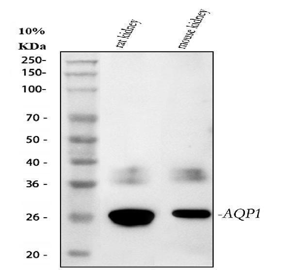
Figure 1. Western blot analysis of AQP1 using anti-AQP1 antibody (PB9473).
Electrophoresis was performed on a 5-20% SDS-PAGE gel at 70V (Stacking gel) / 90V (Resolving gel) for 2-3 hours. The sample well of each lane was loaded with 30 ug of sample under reducing conditions.
Lane 1: rat kidney tissue lysates,
Lane 2: mouse kidney tissue lysates,
Lane 3: mouse lung tissue lysates.
After electrophoresis, proteins were transferred to a nitrocellulose membrane at 150 mA for 50-90 minutes. Blocked the membrane with 5% non-fat milk/TBS for 1.5 hour at RT. The membrane was incubated with rabbit anti-AQP1 antigen affinity purified polyclonal antibody (Catalog # PB9473) at 0.5 μg/mL overnight at 4°C, then washed with TBS-0.1%Tween 3 times with 5 minutes each and probed with a goat anti-rabbit IgG-HRP secondary antibody at a dilution of 1:5000 for 1.5 hour at RT. The signal is developed using an Enhanced Chemiluminescent detection (ECL) kit (Catalog # EK1002) with Tanon 5200 system. A specific band was detected for AQP1 at approximately 25 kDa. The expected band size for AQP1 is at 29 kDa.

Click image to see more details
Figure 2. IHC analysis of AQP1 using anti-AQP1 antibody (PB9473).
AQP1 was detected in a paraffin-embedded section of human bladder urothelial carcinoma tissue. Heat mediated antigen retrieval was performed in EDTA buffer (pH 8.0, epitope retrieval solution). The tissue section was blocked with 10% goat serum. The tissue section was then incubated with 2 μg/ml rabbit anti-AQP1 Antibody (PB9473) overnight at 4°C. Peroxidase Conjugated Goat Anti-rabbit IgG was used as secondary antibody and incubated for 30 minutes at 37°C. The tissue section was developed using HRP Conjugated Rabbit IgG Super Vision Assay Kit (Catalog # SV0002) with DAB as the chromogen.

Click image to see more details
Figure 3. IHC analysis of AQP1 using anti-AQP1 antibody (PB9473).
AQP1 was detected in a paraffin-embedded section of human liver cancer tissue. Heat mediated antigen retrieval was performed in EDTA buffer (pH 8.0, epitope retrieval solution). The tissue section was blocked with 10% goat serum. The tissue section was then incubated with 2 μg/ml rabbit anti-AQP1 Antibody (PB9473) overnight at 4°C. Peroxidase Conjugated Goat Anti-rabbit IgG was used as secondary antibody and incubated for 30 minutes at 37°C. The tissue section was developed using HRP Conjugated Rabbit IgG Super Vision Assay Kit (Catalog # SV0002) with DAB as the chromogen.

Click image to see more details
Figure 4. IHC analysis of AQP1 using anti-AQP1 antibody (PB9473).
AQP1 was detected in a paraffin-embedded section of human lung cancer tissue. Heat mediated antigen retrieval was performed in EDTA buffer (pH 8.0, epitope retrieval solution). The tissue section was blocked with 10% goat serum. The tissue section was then incubated with 2 μg/ml rabbit anti-AQP1 Antibody (PB9473) overnight at 4°C. Peroxidase Conjugated Goat Anti-rabbit IgG was used as secondary antibody and incubated for 30 minutes at 37°C. The tissue section was developed using HRP Conjugated Rabbit IgG Super Vision Assay Kit (Catalog # SV0002) with DAB as the chromogen.

Click image to see more details
Figure 5. IHC analysis of AQP1 using anti-AQP1 antibody (PB9473).
AQP1 was detected in a paraffin-embedded section of human rectal cancer tissue. Heat mediated antigen retrieval was performed in EDTA buffer (pH 8.0, epitope retrieval solution). The tissue section was blocked with 10% goat serum. The tissue section was then incubated with 2 μg/ml rabbit anti-AQP1 Antibody (PB9473) overnight at 4°C. Peroxidase Conjugated Goat Anti-rabbit IgG was used as secondary antibody and incubated for 30 minutes at 37°C. The tissue section was developed using HRP Conjugated Rabbit IgG Super Vision Assay Kit (Catalog # SV0002) with DAB as the chromogen.

Click image to see more details
Figure 6. IHC analysis of AQP1 using anti-AQP1 antibody (PB9473).
AQP1 was detected in a paraffin-embedded section of human tonsil tissue. Heat mediated antigen retrieval was performed in EDTA buffer (pH 8.0, epitope retrieval solution). The tissue section was blocked with 10% goat serum. The tissue section was then incubated with 2 μg/ml rabbit anti-AQP1 Antibody (PB9473) overnight at 4°C. Peroxidase Conjugated Goat Anti-rabbit IgG was used as secondary antibody and incubated for 30 minutes at 37°C. The tissue section was developed using HRP Conjugated Rabbit IgG Super Vision Assay Kit (Catalog # SV0002) with DAB as the chromogen.

Click image to see more details
Figure 7. IHC analysis of AQP1 using anti-AQP1 antibody (PB9473).
AQP1 was detected in a paraffin-embedded section of mouse kidney tissue. Heat mediated antigen retrieval was performed in EDTA buffer (pH 8.0, epitope retrieval solution). The tissue section was blocked with 10% goat serum. The tissue section was then incubated with 2 μg/ml rabbit anti-AQP1 Antibody (PB9473) overnight at 4°C. Peroxidase Conjugated Goat Anti-rabbit IgG was used as secondary antibody and incubated for 30 minutes at 37°C. The tissue section was developed using HRP Conjugated Rabbit IgG Super Vision Assay Kit (Catalog # SV0002) with DAB as the chromogen.

Click image to see more details
Figure 8. IHC analysis of AQP1 using anti-AQP1 antibody (PB9473).
AQP1 was detected in a paraffin-embedded section of rat kidney tissue. Heat mediated antigen retrieval was performed in EDTA buffer (pH 8.0, epitope retrieval solution). The tissue section was blocked with 10% goat serum. The tissue section was then incubated with 2 μg/ml rabbit anti-AQP1 Antibody (PB9473) overnight at 4°C. Peroxidase Conjugated Goat Anti-rabbit IgG was used as secondary antibody and incubated for 30 minutes at 37°C. The tissue section was developed using HRP Conjugated Rabbit IgG Super Vision Assay Kit (Catalog # SV0002) with DAB as the chromogen.

Click image to see more details
Figure 9. IF analysis of AQP1 using anti-AQP1 antibody (PB9473).
AQP1 was detected in a paraffin-embedded section of human lung cancer tissue. Heat mediated antigen retrieval was performed in EDTA buffer (pH 8.0, epitope retrieval solution). The tissue section was blocked with 10% goat serum. The tissue section was then incubated with 2 μg/mL rabbit anti-AQP1 Antibody (PB9473) overnight at 4°C. Cy3 Conjugated Goat Anti-Rabbit IgG (BA1032) was used as secondary antibody at 1:500 dilution and incubated for 30 minutes at 37°C. The section was counterstained with DAPI. Visualize using a fluorescence microscope and filter sets appropriate for the label used.

Click image to see more details
Figure 10. IF analysis of AQP1 using anti-AQP1 antibody (PB9473).
AQP1 was detected in a paraffin-embedded section of rat kidney tissue. Heat mediated antigen retrieval was performed in EDTA buffer (pH 8.0, epitope retrieval solution). The tissue section was blocked with 10% goat serum. The tissue section was then incubated with 2 μg/mL rabbit anti-AQP1 Antibody (PB9473) overnight at 4°C. Cy3 Conjugated Goat Anti-Rabbit IgG (BA1032) was used as secondary antibody at 1:500 dilution and incubated for 30 minutes at 37°C. The section was counterstained with DAPI. Visualize using a fluorescence microscope and filter sets appropriate for the label used.

Click image to see more details
Figure 11. Flow Cytometry analysis of SH-SY5Y cells using anti-AQP1 antibody (PB9473).
Overlay histogram showing SH-SY5Y cells stained with PB9473 (Blue line). The cells were fixed with 4% paraformaldehyde and blocked with 10% normal goat serum. And then incubated with rabbit anti-AQP1 Antibody (PB9473, 1 μg/1x106 cells) for 30 min at 20°C. DyLight®488 conjugated goat anti-rabbit IgG (BA1127, 5-10 μg/1x106 cells) was used as secondary antibody for 30 minutes at 20°C. Isotype control antibody (Green line) was rabbit IgG (1 μg/1x106) used under the same conditions. Unlabelled sample (Red line) was also used as a control.
Protein Target Info & Infographic
Gene/Protein Information For AQP1 (Source: Uniprot.org, NCBI)
Gene Name
AQP1
Full Name
Aquaporin-1
Weight
28.526kDa
Superfamily
MIP/aquaporin (TC 1.A.8) family
Alternative Names
28-kDa; AQP1; AQP-1; AQP-CHIP; aquaporin 1 (channel-forming integral protein, 28kDa); aquaporin 1 (Colton blood group); Aquaporin 1; Aquaporin-CHIP; CHIP28; CHIP2828kDa, CO blood group); CO; MGC26324 AQP1 AQP-CHIP, CHIP28, CO aquaporin 1 (Colton blood group) aquaporin-1|Colton blood group antigen|aquaporin 1 (channel-forming integral protein, 28kDa, CO blood group)|aquaporin 1, Colton blood group antigen|aquaporin-CHIP|channel-like integral membrane protein, 28-kDa|urine water channel|water channel protein for red blood cells and kidney proximal tubule
*If product is indicated to react with multiple species, protein info is based on the gene entry specified above in "Species".For more info on AQP1, check out the AQP1 Infographic

We have 30,000+ of these available, one for each gene! Check them out.
In this infographic, you will see the following information for AQP1: database IDs, superfamily, protein function, synonyms, molecular weight, chromosomal locations, tissues of expression, subcellular locations, post-translational modifications, and related diseases, research areas & pathways. If you want to see more information included, or would like to contribute to it and be acknowledged, please contact [email protected].
Specific Publications For Anti-Aquaporin 1/AQP1 Antibody Picoband™ (PB9473)
Hello CJ!
PB9473 has been cited in 9 publications:
*The publications in this section are manually curated by our staff scientists. They may differ from Bioz's machine gathered results. Both are accurate. If you find a publication citing this product but is missing from this list, please let us know we will issue you a thank-you coupon.
Expression of AQP1, AQP3, AQP4 and AQP5 in upper respiratory tract of buffaloes during different seasons
Seasonal effect in expression of AQP1, AQP3 and AQP5 in skin of Murrah buffaloes
Lead Exposure in Developmental Ages Promotes Aβ Accumulation by Disturbing Aβ Transportation in Blood-Cerebrospinal Fluid Barrier/Blood–Brain Barriers and Impairing Aβ Clearance in the Liver
Sunitinib treatment enhances metastasis of innately drug-resistant breast tumors
Effect of selective inhibition of aquaporin 1 on chemotherapy sensitivity of J82 human bladder cancer cells
Effects of dexmedetomidine on the protection of hyperoxia-induced lung injury in newborn rats
Expression pattern of aquaporins in patients with primary nephrotic syndrome with edema
Huo J, Liu J, Wang J, Zhang Y, Wang C, Yang Y, Sun W, Xu S. Neural Regen Res. 2012 Aug 5;7(22):1729-35. Doi: 10.3969/J.Issn.1673-5374.2012.22.008. Early Hyperbaric Oxygen Therapy Inhibits Aquaporin 4 And Adrenocorticotropic Hormone Expression In T...
Ran X, Wang H, Chen Y, Zeng Z, Zhou Q, Zheng R, Sun J, Wang B, Lv X, Liang Y, Zhang K, Liu W. Heart Vessels. 2010 May;25(3):237-47. Doi: 10.1007/S00380-009-1179-5. Epub 2010 May 29. Aquaporin-1 Expression And Angiogenesis In Rabbit Chronic Myocard...
Recommended Resources
Here are featured tools and databases that you might find useful.
- Boster's Pathways Library
- Protein Databases
- Bioscience Research Protocol Resources
- Data Processing & Analysis Software
- Photo Editing Software
- Scientific Literature Resources
- Research Paper Management Tools
- Molecular Biology Software
- Primer Design Tools
- Bioinformatics Tools
- Phylogenetic Tree Analysis
Customer Reviews
Have you used Anti-Aquaporin 1/AQP1 Antibody Picoband™?
Submit a review and receive an Amazon gift card.
- $30 for a review with an image
Be the first to review Anti-Aquaporin 1/AQP1 Antibody Picoband™
*The first user to submit a review for a product is eligible for Boster's Innovating Scientists Reward, which gives product credits. This is in addition to the gift card reward.
Customer Q&As
Have a question?
Find answers in Q&As, reviews.
Can't find your answer?
Submit your question
6 Customer Q&As for Anti-Aquaporin 1/AQP1 Antibody Picoband™
Question
We are currently using anti-Aquaporin 1/AQP1 antibody PB9473 for mouse tissue, and we are content with the IHC results. The species of reactivity given in the datasheet says human, mouse, rat. Is it true that the antibody can work on horse tissues as well?
Verified Customer
Verified customer
Asked: 2019-08-26
Answer
The anti-Aquaporin 1/AQP1 antibody (PB9473) has not been validated for cross reactivity specifically with horse tissues, but there is a good chance of cross reactivity. We have an innovator award program that if you test this antibody and show it works in horse you can get your next antibody for free. Please contact me if I can help you with anything.
Boster Scientific Support
Answered: 2019-08-26
Question
My colleagues were satisfied with the WB result of your anti-Aquaporin 1/AQP1 antibody. However we have seen positive staining in retinal pigment epithelium cell membrane using this antibody. Is that expected? Could you tell me where is AQP1 supposed to be expressed?
Verified Customer
Verified customer
Asked: 2019-08-21
Answer
From what I have seen in literature, retinal pigment epithelium does express AQP1. Generally AQP1 expresses in cell membrane. Regarding which tissues have AQP1 expression, here are a few articles citing expression in various tissues:
Articular cartilage, Pubmed ID: 2007592, 1373524
Brain, Pubmed ID: 15489334
Mesangial cell, Pubmed ID: 14702039
Retinal pigment epithelium, Pubmed ID: 8703970
Uterus, Pubmed ID: 7517253
Boster Scientific Support
Answered: 2019-08-21
Question
We have observed staining in rat retinal pigment epithelium. What should we do? Is anti-Aquaporin 1/AQP1 antibody supposed to stain retinal pigment epithelium positively?
Verified Customer
Verified customer
Asked: 2019-06-10
Answer
From what I have seen in literature retinal pigment epithelium does express AQP1. From what I have seen in Uniprot.org, AQP1 is expressed in right lung, retinal pigment epithelium, uterus, mesangial cell, brain, articular cartilage, among other tissues. Regarding which tissues have AQP1 expression, here are a few articles citing expression in various tissues:
Articular cartilage, Pubmed ID: 2007592, 1373524
Brain, Pubmed ID: 15489334
Mesangial cell, Pubmed ID: 14702039
Retinal pigment epithelium, Pubmed ID: 8703970
Uterus, Pubmed ID: 7517253
Boster Scientific Support
Answered: 2019-06-10
Question
We purchased anti-Aquaporin 1/AQP1 antibody for IHC on retinal pigment epithelium in the past. I am using mouse, and We want to use the antibody for WB next. Our lab want to know about examining retinal pigment epithelium as well as uterus in our next experiment. Could give a recommendation on which antibody would work the best for WB?
Verified Customer
Verified customer
Asked: 2019-05-22
Answer
I have checked the website and datasheets of our anti-Aquaporin 1/AQP1 antibody and it seems that PB9473 has been tested on mouse in both IHC and WB. Thus PB9473 should work for your application. Our Boster satisfaction guarantee will cover this product for WB in mouse even if the specific tissue type has not been validated. We do have a comprehensive range of products for WB detection and you can check out our website bosterbio.com to find out more information about them.
Boster Scientific Support
Answered: 2019-05-22
Question
Has this antibody been used to image erythrocytes by fluorescence confocal microscopy?
Verified Customer
Verified customer
Asked: 2019-01-02
Answer
No. This antibody has not been validated with erythrocytes.
Boster Scientific Support
Answered: 2019-01-02
Question
I am looking for using your anti-Aquaporin 1/AQP1 antibody for vasopressin regulates renal water homeostasis via aquaporins studies. Has this antibody been tested with western blotting on intestinal cancer tissue? We would like to see some validation images before ordering.
W. Kulkarni
Verified customer
Asked: 2016-04-04
Answer
Thank you for your inquiry. This PB9473 anti-Aquaporin 1/AQP1 antibody is tested on rat lung tissue, kidney tissue, tissue lysate, cardiac muscle tissue, hepa whole cell lysate, mouse kidney tissue, intestinal cancer tissue. It is guaranteed to work for IHC, WB in human, mouse, rat. Our Boster guarantee will cover your intended experiment even if the sample type has not been be directly tested.
Boster Scientific Support
Answered: 2016-04-04
