Click image to see more details
Product Info Summary
| SKU: | M09735 |
|---|---|
| Size: | 100 μg/vial |
| Reactive Species: | Human, Monkey, Mouse, Rat |
| Host: | Mouse |
| Application: | Flow Cytometry, WB |
Customers Who Bought This Also Bought


Product info
Product Name
Anti-ATP5F1,2,3/ATP5MC1,2,3 Picoband™ Antibody (monoclonal, 12E9)
View all ATP synthase C mature Antibodies
SKU/Catalog Number
M09735
Size
100 μg/vial
Form
Lyophilized
Description
Boster Bio Anti-ATP5F1,2,3/ATP5MC1,2,3 Picoband™ Antibody (monoclonal, 12E9) catalog # M09735. Tested in Flow Cytometry, WB applications. This antibody reacts with Human, Monkey, Mouse, Rat.
Storage & Handling
Store at -20˚C for one year from date of receipt. After reconstitution, at 4˚C for one month. It can also be aliquotted and stored frozen at -20˚C for six months. Avoid repeated freeze-thaw cycles.
Cite This Product
Anti-ATP5F1,2,3/ATP5MC1,2,3 Picoband™ Antibody (monoclonal, 12E9) (Boster Biological Technology, Pleasanton CA, USA, Catalog # M09735)
Host
Mouse
Contents
Each vial contains 4mg Trehalose, 0.9mg NaCl and 0.2mg Na2HPO4.
Clonality
Monoclonal
Clone Number
12E9
Isotype
Mouse IgG2b
Immunogen
E.coli-derived human ATP5G1,2,3/ATP5MC1,2,3 recombinant protein (Position: D62-L113).
*Blocking peptide can be purchased. Costs vary based on immunogen length. Contact us for pricing.
Cross-reactivity
No cross-reactivity with other proteins.
Reactive Species
M09735 is reactive to ATP5MC1 in Human, Monkey, Mouse, Rat
Applications
M09735 is guaranteed for Flow Cytometry, WB Boster Guarantee
Observed Molecular Weight
10-14 kDa
Calculated molecular weight
14.277kDa
Background of ATP synthase C mature
The ATP5MC1 gene is one of three human paralogs that encode membrane subunit c of the mitochondrial ATP synthase. It is mapped to 17q21.32. This gene encodes a subunit of mitochondrial ATP synthase. Mitochondrial ATP synthase catalyzes ATP synthesis, utilizing an electrochemical gradient of protons across the inner membrane during oxidative phosphorylation. ATP synthase is composed of two linked multi-subunit complexes: the soluble catalytic core, F1, and the membrane-spanning component, Fo, comprising the proton channel. The catalytic portion of mitochondrial ATP synthase consists of 5 different subunits (alpha, beta, gamma, delta, and epsilon) assembled with a stoichiometry of 3 alpha, 3 beta, and a single representative of the other 3. The proton channel seems to have nine subunits (a, b, c, d, e, f, g, F6 and 8). This gene is one of three genes that encode subunit c of the proton channel. Each of the three genes have distinct mitochondrial import sequences but encode the identical mature protein. Alternatively spliced transcript variants encoding the same protein have been identified.
Antibody Validation
Boster validates all antibodies on WB, IHC, ICC, Immunofluorescence, and ELISA with known positive control and negative samples to ensure specificity and high affinity, including thorough antibody incubations.
Innovating Scientists Reward
If you are the first to review this product, or if you have results for a special sample, species or application this product is not validated in, share your results with us and receive product credits you can use towards any Boster products! Applicable to all scientists worldwide.
Submit A Review
Assay dilution & Images
Reconsitution
Add 0.2ml of distilled water will yield a concentration of 500ug/ml.
Assay Dilutions Recommendation
The recommendations below provide a starting point for assay optimization. The actual working concentration varies and should be decided by the user.
Western blot, 0.25-0.5μg/ml, Human, Mouse, Rat, Monkey
Flow Cytometry, 1-3μg/1x106 cells, Human, Mouse, Rat
Validation Images & Assay Conditions
Click image to see more details
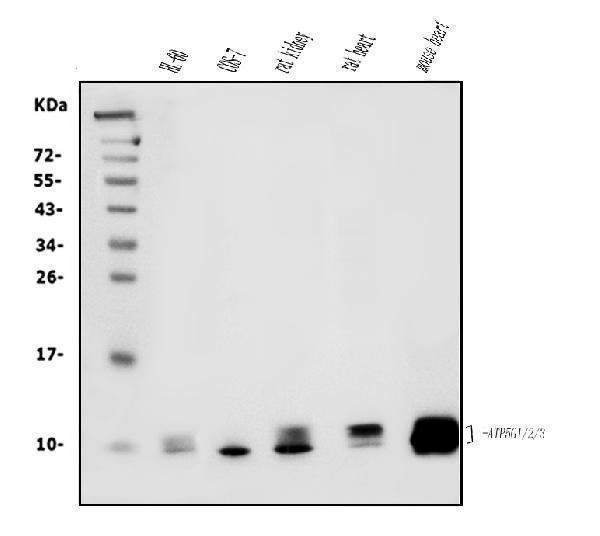
Figure 1. Western blot analysis of ATP5F1,2,3/ATP5MC1,2,3 using anti-ATP5F1,2,3/ATP5MC1,2,3 antibody (M09735).
Electrophoresis was performed on a 5-20% SDS-PAGE gel at 70V (Stacking gel) / 90V (Resolving gel) for 2-3 hours. The sample well of each lane was loaded with 50ug of sample under reducing conditions.
Lane 1: human HL-60 whole cell lysates,
Lane 2: monkey COS-7 whole cell lysates,
Lane 3: rat kidney tissue lysates,
Lane 4: rat heart tissue lysates,
Lane 5: mouse heart tissue lysates.
After Electrophoresis, proteins were transferred to a Nitrocellulose membrane at 150mA for 50-90 minutes. Blocked the membrane with 5% Non-fat Milk/ TBS for 1.5 hour at RT. The membrane was incubated with mouse anti-ATP5F1,2,3/ATP5MC1,2,3 antigen affinity purified monoclonal antibody (Catalog # M09735) at 0.5 μg/mL overnight at 4°C, then washed with TBS-0.1%Tween 3 times with 5 minutes each and probed with a goat anti-mouse IgG-HRP secondary antibody at a dilution of 1:10000 for 1.5 hour at RT. The signal is developed using an Enhanced Chemiluminescent detection (ECL) kit (Catalog # EK1001) with Tanon 5200 system. A specific band was detected for ATP5F1,2,3/ATP5MC1,2,3 at approximately 10-14KD. The expected band size for ATP5F1,2,3/ATP5MC1,2,3 is at 10-14KD.

Click image to see more details
Figure 2. Flow Cytometry analysis of HEPA1-6 cells using anti-ATP5F1,2,3/ATP5MC1,2,3 antibody (M09735).
Overlay histogram showing HEPA1-6 cells stained with M09735 (Blue line).The cells were blocked with 10% normal goat serum. And then incubated with mouse anti-ATP5F1,2,3/ATP5MC1,2,3 Antibody (M09735, 1μg/1x106 cells) for 30 min at 20°C. DyLight®488 conjugated goat anti-mouse IgG (BA1126, 5-10μg/1x106 cells) was used as secondary antibody for 30 minutes at 20°C. Isotype control antibody (Green line) was mouse IgG (1μg/1x106) used under the same conditions. Unlabelled sample (Red line) was also used as a control.

Click image to see more details
Figure 3. Flow Cytometry analysis of HEPG2 cells using anti-ATP5F1,2,3/ATP5MC1,2,3 antibody (M09735).
Overlay histogram showing HEPG2 cells stained with M09735 (Blue line).The cells were blocked with 10% normal goat serum. And then incubated with mouse anti-ATP5F1,2,3/ATP5MC1,2,3 Antibody (M09735, 1μg/1x106 cells) for 30 min at 20°C. DyLight®488 conjugated goat anti-mouse IgG (BA1126, 5-10μg/1x106 cells) was used as secondary antibody for 30 minutes at 20°C. Isotype control antibody (Green line) was mouse IgG (1μg/1x106) used under the same conditions. Unlabelled sample (Red line) was also used as a control.

Click image to see more details
Figure 4. Flow Cytometry analysis of RH35 cells using anti-ATP5F1,2,3/ATP5MC1,2,3 antibody (M09735).
Overlay histogram showing RH35 cells stained with M09735 (Blue line).The cells were blocked with 10% normal goat serum. And then incubated with mouse anti-ATP5F1,2,3/ATP5MC1,2,3 Antibody (M09735, 1μg/1x106 cells) for 30 min at 20°C. DyLight®488 conjugated goat anti-mouse IgG (BA1126, 5-10μg/1x106 cells) was used as secondary antibody for 30 minutes at 20°C. Isotype control antibody (Green line) was mouse IgG (1μg/1x106) used under the same conditions. Unlabelled sample (Red line) was also used as a control.
Protein Target Info & Infographic
Gene/Protein Information For ATP5MC1 (Source: Uniprot.org, NCBI)
Gene Name
ATP5MC1
Full Name
ATP synthase F(0) complex subunit C1, mitochondrial
Weight
14.277kDa
Superfamily
ATPase C chain family
Alternative Names
ATP synthase, H+ transporting, mitochondrial F0 complex, subunit c (subunit 9); ATP synthase, H+ transporting, mitochondrial Fo complex, subunit C1 (subunit 9); ATP5A; ATP5G; ATPase protein 9; ATPase subunit 9; H+ transporting, mitochondrial F0 complex, subunit C1 (subunit 9); isoform 1; mitochondrial ATP synthase, subunit 9; mitochondrial ATP synthase, subunit C; mitochondrial ATP5MC1 ATP5A, ATP5G, ATP5G1 ATP synthase membrane subunit c locus 1 ATP synthase F(0) complex subunit C1, mitochondrial|ATP synthase lipid-binding protein, mitochondrial|ATP synthase proteolipid P1|ATP synthase proton-transporting mitochondrial F(0) complex subunit C1|ATP synthase subunit 9|ATP synthase, H+ transporting, mitochondrial F0 complex, subunit C1 (subunit 9)|ATP synthase, H+ transporting, mitochondrial F0 complex, subunit c (subunit 9)|ATP synthase, H+ transporting, mitochondrial Fo complex subunit C1 (subunit 9)|ATPase protein 9|ATPase subunit 9|ATPase subunit C|dicyclohexylcarbodiimide (DCCD)-reactive proteolipid subunit|mitochondrial ATP synthase, subunit 9|mitochondrial ATP synthase, subunit C
*If product is indicated to react with multiple species, protein info is based on the gene entry specified above in "Species".For more info on ATP5MC1, check out the ATP5MC1 Infographic

We have 30,000+ of these available, one for each gene! Check them out.
In this infographic, you will see the following information for ATP5MC1: database IDs, superfamily, protein function, synonyms, molecular weight, chromosomal locations, tissues of expression, subcellular locations, post-translational modifications, and related diseases, research areas & pathways. If you want to see more information included, or would like to contribute to it and be acknowledged, please contact [email protected].
Specific Publications For Anti-ATP5F1,2,3/ATP5MC1,2,3 Picoband™ Antibody (monoclonal, 12E9) (M09735)
Hello CJ!
No publications found for M09735
*Do you have publications using this product? Share with us and receive a reward. Ask us for more details.
Recommended Resources
Here are featured tools and databases that you might find useful.
- Boster's Pathways Library
- Protein Databases
- Bioscience Research Protocol Resources
- Data Processing & Analysis Software
- Photo Editing Software
- Scientific Literature Resources
- Research Paper Management Tools
- Molecular Biology Software
- Primer Design Tools
- Bioinformatics Tools
- Phylogenetic Tree Analysis
Customer Reviews
Have you used Anti-ATP5F1,2,3/ATP5MC1,2,3 Picoband™ Antibody (monoclonal, 12E9)?
Submit a review and receive an Amazon gift card.
- $30 for a review with an image
Be the first to review Anti-ATP5F1,2,3/ATP5MC1,2,3 Picoband™ Antibody (monoclonal, 12E9)
*The first user to submit a review for a product is eligible for Boster's Innovating Scientists Reward, which gives product credits. This is in addition to the gift card reward.
Customer Q&As
Have a question?
Find answers in Q&As, reviews.
Can't find your answer?
Submit your question
