Click image to see more details
Product Info Summary
| SKU: | M00360 |
|---|---|
| Size: | 100 μl |
| Reactive Species: | Human, Mouse, Rat |
| Host: | Rabbit |
| Application: | Flow Cytometry, IF, ICC, WB |
Customers Who Bought This Also Bought


Product info
Product Name
Anti-CD47 Rabbit Monoclonal Antibody
SKU/Catalog Number
M00360
Size
100 μl
Form
Liquid
Description
Boster Bio Anti-CD47 Rabbit Monoclonal Antibody catalog # M00360. Tested in WB, ICC/IF, Flow Cytometry applications. This antibody reacts with Human, Mouse, Rat.
Storage & Handling
Store at -20°C for one year. For short term storage and frequent use, store at 4°C for up to one month. Avoid repeated freeze-thaw cycles.
Cite This Product
Anti-CD47 Rabbit Monoclonal Antibody (Boster Biological Technology, Pleasanton CA, USA, Catalog # M00360)
Host
Rabbit
Contents
Rabbit IgG in phosphate buffered saline, pH 7.4, 150mM NaCl, 0.02% sodium azide and 50% glycerol, 0.4-0.5mg/ml BSA.
Clonality
Monoclonal
Clone Number
D-3
Isotype
Rabbit IgG
Immunogen
A synthesized peptide derived from human CD47
*Blocking peptide can be purchased. Costs vary based on immunogen length. Contact us for pricing.
Reactive Species
M00360 is reactive to CD47 in Human, Mouse, Rat
Applications
M00360 is guaranteed for Flow Cytometry, IF, ICC, WB Boster Guarantee
Observed Molecular Weight
305 kDa
Calculated molecular weight
35.214kDa
Background of CD47
C3 plays a central role in the activation of the complement system. Its processing by C3 convertase is the central reaction in both classical and alternative complement pathways. After activation C3b can bind covalently, via its reactive thioester, to cell surface carbohydrates or immune aggregates.
Antibody Validation
Boster validates all antibodies on WB, IHC, ICC, Immunofluorescence, and ELISA with known positive control and negative samples to ensure specificity and high affinity, including thorough antibody incubations.
Innovating Scientists Reward
If you are the first to review this product, or if you have results for a special sample, species or application this product is not validated in, share your results with us and receive product credits you can use towards any Boster products! Applicable to all scientists worldwide.
Submit A Review
Assay dilution & Images
Reconsitution
Restore with deionized water (or equivalent) for reconstitution volume of 1.0 mL
Assay Dilutions Recommendation
The recommendations below provide a starting point for assay optimization. The actual working concentration varies and should be decided by the user.
WB 1:1000-1:2000
ICC/IF 1:50-1:200
FC 1:50
Validation Images & Assay Conditions
Click image to see more details
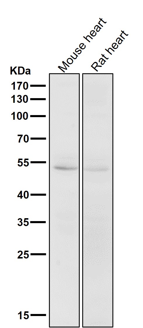
All lanes use the Antibody at 1:1K dilution for 1 hour at room temperature.

Click image to see more details
All lanes use the Antibody at 1:1K dilution for 1 hour at room temperature.

Click image to see more details
Western blot analysis of extracts of NIH/3T3 cell lysate, using CD47 antibody.
Protein Target Info & Infographic
Gene/Protein Information For CD47 (Source: Uniprot.org, NCBI)
Gene Name
CD47
Full Name
Leukocyte surface antigen CD47
Weight
35.214kDa
Alternative Names
antigen identified by monoclonal 1D8; Antigenic surface determinant protein OA3; CD47 antigen (Rh-related antigen, integrin-associated signal transducer); CD47 antigen; CD47 glycoprotein; CD47 molecule; CD47; IAP; IAPintegrin associated protein; Integrin-associated protein; leukocyte surface antigen CD47; MER6integrin-associated signal transducer; OA3; Protein MER6; Rh-related antigen CD47 IAP, MER6, OA3 CD47 molecule leukocyte surface antigen CD47|CD47 antigen (Rh-related antigen, integrin-associated signal transducer)|CD47 glycoprotein|Rh-related antigen|antigen identified by monoclonal antibody 1D8|antigenic surface determinant protein OA3|integrin associated protein|integrin-associated signal transducer
*If product is indicated to react with multiple species, protein info is based on the gene entry specified above in "Species".For more info on CD47, check out the CD47 Infographic

We have 30,000+ of these available, one for each gene! Check them out.
In this infographic, you will see the following information for CD47: database IDs, superfamily, protein function, synonyms, molecular weight, chromosomal locations, tissues of expression, subcellular locations, post-translational modifications, and related diseases, research areas & pathways. If you want to see more information included, or would like to contribute to it and be acknowledged, please contact [email protected].
Specific Publications For Anti-CD47 Rabbit Monoclonal Antibody (M00360)
Hello CJ!
No publications found for M00360
*Do you have publications using this product? Share with us and receive a reward. Ask us for more details.
Recommended Resources
Here are featured tools and databases that you might find useful.
- Boster's Pathways Library
- Protein Databases
- Bioscience Research Protocol Resources
- Data Processing & Analysis Software
- Photo Editing Software
- Scientific Literature Resources
- Research Paper Management Tools
- Molecular Biology Software
- Primer Design Tools
- Bioinformatics Tools
- Phylogenetic Tree Analysis
Customer Reviews
Have you used Anti-CD47 Rabbit Monoclonal Antibody?
Submit a review and receive an Amazon gift card.
- $30 for a review with an image
Be the first to review Anti-CD47 Rabbit Monoclonal Antibody
*The first user to submit a review for a product is eligible for Boster's Innovating Scientists Reward, which gives product credits. This is in addition to the gift card reward.
Customer Q&As
Have a question?
Find answers in Q&As, reviews.
Can't find your answer?
Submit your question
4 Customer Q&As for Anti-CD47 Rabbit Monoclonal Antibody
Question
We are currently using anti-CD47 Rabbit Monoclonal antibody M00360 for mouse tissue, and we are happy with the IF results. The species of reactivity given in the datasheet says human, mouse, rat. Is it true that the antibody can work on bovine tissues as well?
D. Banerjee
Verified customer
Asked: 2020-05-01
Answer
The anti-CD47 Rabbit Monoclonal antibody (M00360) has not been tested for cross reactivity specifically with bovine tissues, though there is a good chance of cross reactivity. We have an innovator award program that if you test this antibody and show it works in bovine you can get your next antibody for free. Please contact me if I can help you with anything.
Boster Scientific Support
Answered: 2020-05-01
Question
We have tried in the past anti-CD47 Rabbit Monoclonal antibody for IF on visceral pleura a few years ago. I am using mouse, and We are going to use the antibody for WB next. My lab would like examining visceral pleura as well as hippocampus in our next experiment. Could you please give me some suggestion on which antibody would work the best for WB?
T. Banerjee
Verified customer
Asked: 2020-01-24
Answer
I took a look at the website and datasheets of our anti-CD47 Rabbit Monoclonal antibody and I see that M00360 has been validated on mouse in both IF and WB. Thus M00360 should work for your application. Our Boster satisfaction guarantee will cover this product for WB in mouse even if the specific tissue type has not been validated. We do have a comprehensive range of products for WB detection and you can check out our website bosterbio.com to find out more information about them.
Boster Scientific Support
Answered: 2020-01-24
Question
My boss were happy with the WB result of your anti-CD47 Rabbit Monoclonal antibody. However we have observed positive staining in leukemic t-cell cell membrane using this antibody. Is that expected? Could you tell me where is CD47 supposed to be expressed?
Verified Customer
Verified customer
Asked: 2019-11-12
Answer
Based on literature, leukemic t-cell does express CD47. Generally CD47 expresses in cell membrane. Regarding which tissues have CD47 expression, here are a few articles citing expression in various tissues:
Brain, Pubmed ID: 14702039
Erythrocyte, Pubmed ID: 7998989
Hippocampus, Leiomyosarcoma, and Ovarian adenocarcinoma, Pubmed ID: 15489334
Leukemic T-cell, Pubmed ID: 19349973
Liver, Pubmed ID: 19159218
Myelomonocyte, Pubmed ID: 7691831
Ovary, Pubmed ID: 1394148
T-cell, Pubmed ID: 15383453
Boster Scientific Support
Answered: 2019-11-12
Question
We have seen staining in mouse brain. Are there any suggestions? Is anti-CD47 Rabbit Monoclonal antibody supposed to stain brain positively?
Verified Customer
Verified customer
Asked: 2019-08-12
Answer
From what I have seen in literature brain does express CD47. From what I have seen in Uniprot.org, CD47 is expressed in visceral pleura, ovary, myelomonocyte, brain, hippocampus, leiomyosarcoma ovarian adenocarcinoma, erythrocyte, t-cell, liver, leukemic t-cell, among other tissues. Regarding which tissues have CD47 expression, here are a few articles citing expression in various tissues:
Brain, Pubmed ID: 14702039
Erythrocyte, Pubmed ID: 7998989
Hippocampus, Leiomyosarcoma, and Ovarian adenocarcinoma, Pubmed ID: 15489334
Leukemic T-cell, Pubmed ID: 19349973
Liver, Pubmed ID: 19159218
Myelomonocyte, Pubmed ID: 7691831
Ovary, Pubmed ID: 1394148
T-cell, Pubmed ID: 15383453
Boster Scientific Support
Answered: 2019-08-12
