Click image to see more details
-

-
-
-
-
+1
Product Info Summary
| SKU: | M00914-2 |
|---|---|
| Size: | 100 μg/vial |
| Reactive Species: | Human |
| Host: | Mouse |
| Application: | Flow Cytometry, IF, IHC, ICC, WB |
Customers Who Bought This Also Bought

Product info
Product Name
Anti-CD59 Antibody Picoband™(monoclonal, 3C10)
SKU/Catalog Number
M00914-2
Size
100 μg/vial
Form
Lyophilized
Description
Boster Bio Anti-CD59 Antibody Picoband™ (monoclonal, 3C10) catalog # M00914-2. Tested in Flow Cytometry, IF, IHC, ICC, WB applications. This antibody reacts with Human.
Storage & Handling
Store at -20˚C for one year from date of receipt. After reconstitution, at 4˚C for one month. It can also be aliquotted and stored frozen at -20˚C for six months. Avoid repeated freeze-thaw cycles.
Cite This Product
Anti-CD59 Antibody Picoband™(monoclonal, 3C10) (Boster Biological Technology, Pleasanton CA, USA, Catalog # M00914-2)
Host
Mouse
Contents
Each vial contains 4mg Trehalose, 0.9mg NaCl, 0.2mg Na2HPO4, 0.05mg NaN3.
Clonality
Monoclonal
Clone Number
3C10
Isotype
Mouse IgG2b
Immunogen
E.coli-derived human CD59 recombinant protein (Position: L26-N102). Human CD59 shares 47.1% amino acid (aa) sequence identity with rat CD59.
*Blocking peptide can be purchased. Costs vary based on immunogen length. Contact us for pricing.
Cross-reactivity
No cross-reactivity with other proteins.
Reactive Species
M00914-2 is reactive to CD59 in Human
Applications
M00914-2 is guaranteed for Flow Cytometry, IF, IHC, ICC, WB Boster Guarantee
Observed Molecular Weight
19 kDa
Calculated molecular weight
14.177kDa
Background of CD59
This gene encodes a cell surface glycoprotein that regulates complement-mediated cell lysis, and it is involved in lymphocyte signal transduction. And this protein is a potent inhibitor of the complement membrane attack complex, whereby it binds complement C8 and/or C9 during the assembly of this complex, thereby inhibiting the incorporation of multiple copies of C9 into the complex, which is necessary for osmolytic pore formation. It also plays a role in signal transduction pathways in the activation of T cells. Mutations in this gene cause CD59 deficiency, a disease resulting in hemolytic anemia and thrombosis, and which causes cerebral infarction. Multiple alternatively spliced transcript variants, which encode the same protein, have been identified for this gene.
Antibody Validation
Boster validates all antibodies on WB, IHC, ICC, Immunofluorescence, and ELISA with known positive control and negative samples to ensure specificity and high affinity, including thorough antibody incubations.
Innovating Scientists Reward
If you are the first to review this product, or if you have results for a special sample, species or application this product is not validated in, share your results with us and receive product credits you can use towards any Boster products! Applicable to all scientists worldwide.
Submit A Review
Assay dilution & Images
Reconsitution
Add 0.2ml of distilled water will yield a concentration of 500μg/ml.
Assay Dilutions Recommendation
The recommendations below provide a starting point for assay optimization. The actual working concentration varies and should be decided by the user.
Western blot, 0.1-0.5μg/ml, Human
Immunohistochemistry (Paraffin-embedded Section), 2-5μg/ml, Human
Immunocytochemistry/Immunofluorescence, 2μg/ml, Human
Flow Cytometry, 1-3μg/1x106, Human
Validation Images & Assay Conditions
Click image to see more details
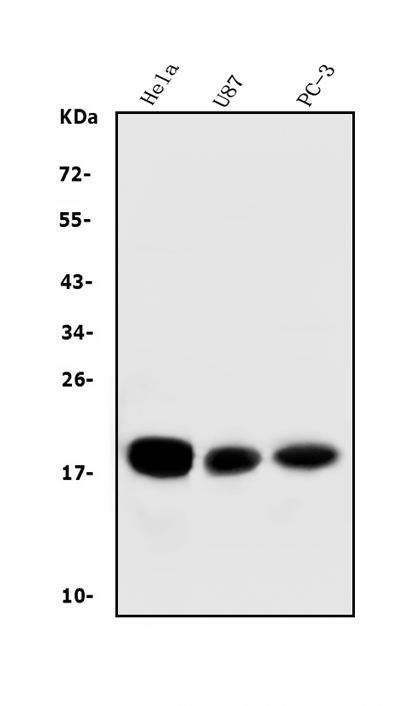
Figure 1. Western blot analysis of CD59 using anti-CD59 antibody (M00914-2).
Electrophoresis was performed on a 5-20% SDS-PAGE gel at 70V (Stacking gel) / 90V (Resolving gel) for 2-3 hours. The sample well of each lane was loaded with 50ug of sample under reducing conditions.
Lane 1: human Hela whole cell lysates;
Lane 2: human U-87MG whole cell lysates;
Lane 3: human PC-3 whole cell lysates.
After Electrophoresis, proteins were transferred to a Nitrocellulose membrane at 150mA for 50-90 minutes. Blocked the membrane with 5% Non-fat Milk/ TBS for 1.5 hour at RT. The membrane was incubated with mouse anti-CD59 antigen affinity purified monoclonal antibody (Catalog # M00914-2) at 0.5 μg/mL overnight at 4°C, then washed with TBS-0.1%Tween 3 times with 5 minutes each and probed with a goat anti-mouse IgG-HRP secondary antibody at a dilution of 1:10000 for 1.5 hour at RT. The signal is developed using an Enhanced Chemiluminescent detection (ECL) kit (Catalog # EK1001) with Tanon 5200 system. A specific band was detected for CD59 at approximately 19KD. The expected band size for CD59 is at 19KD.

Click image to see more details
Figure 2. IF analysis of CD59 using anti-CD59 antibody (M00914-2).
CD59 was detected in immunocytochemical section of A431 cells. Enzyme antigen retrieval was performed using IHC enzyme antigen retrieval reagent (AR0022) for 15 mins. The cells were blocked with 10% goat serum. And then incubated with 2μg/mL mouse anti-CD59 Antibody (M00914-2) overnight at 4°C. DyLight®488 Conjugated Goat Anti-Mouse IgG (BA1126) was used as secondary antibody at 1:100 dilution and incubated for 30 minutes at 37°C. The section was counterstained with DAPI. Visualize using a fluorescence microscope and filter sets appropriate for the label used.

Click image to see more details
Figure 3. IF analysis of CD59 using anti-CD59 antibody (M00914-2).
CD59 was detected in immunocytochemical section of Hela cells. Enzyme antigen retrieval was performed using IHC enzyme antigen retrieval reagent (AR0022) for 15 mins. The cells were blocked with 10% goat serum. And then incubated with 2μg/mL mouse anti-CD59 Antibody (M00914-2) overnight at 4°C. DyLight®488 Conjugated Goat Anti-Mouse IgG (BA1126) was used as secondary antibody at 1:100 dilution and incubated for 30 minutes at 37°C. The section was counterstained with DAPI. Visualize using a fluorescence microscope and filter sets appropriate for the label used.

Click image to see more details
Figure 4. Flow Cytometry analysis of A549 cells using anti-CD59 antibody (M00914-2).
Overlay histogram showing A549 cells stained with M00914-2 (Blue line).The cells were blocked with 10% normal goat serum. And then incubated with mouse anti-CD59 Antibody (M00914-2, 1μg/1x106 cells) for 30 min at 20°C. DyLight®488 conjugated goat anti-mouse IgG (BA1126, 5-10μg/1x106 cells) was used as secondary antibody for 30 minutes at 20°C. Isotype control antibody (Green line) was mouse IgG (1μg/1x106) used under the same conditions. Unlabelled sample (Red line) was also used as a control.

Click image to see more details
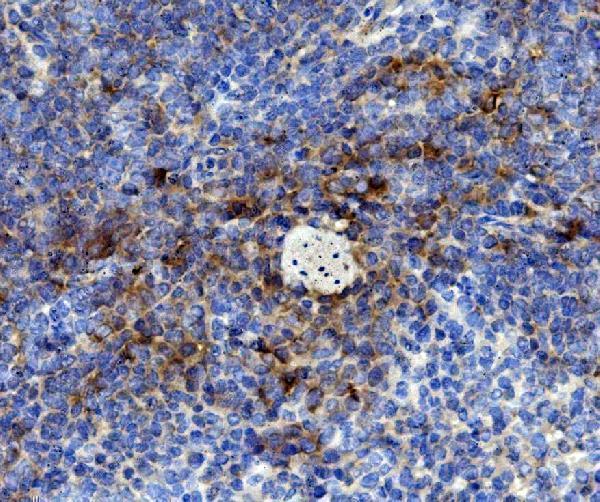
Figure 5. IHC analysis of CD59 using anti-CD59 antibody (M00914-2).
CD59 was detected in paraffin-embedded section of human colorectal cancer tissue. Heat mediated antigen retrieval was performed in EDTA buffer (pH8.0, epitope retrieval solution). The tissue section was blocked with 10% goat serum. The tissue section was then incubated with 2μg/ml mouse anti-CD59 Antibody (M00914-2) overnight at 4°C. Biotinylated goat anti-mouse IgG was used as secondary antibody and incubated for 30 minutes at 37°C. The tissue section was developed using Strepavidin-Biotin-Complex (SABC) (Catalog # SA1021) with DAB as the chromogen.
Protein Target Info & Infographic
Gene/Protein Information For CD59 (Source: Uniprot.org, NCBI)
Gene Name
CD59
Full Name
CD59 glycoprotein
Weight
14.177kDa
Alternative Names
16.3A5; 1F5 antigen; 1F5; 20 kDa homologous restriction factor; CD59 antigen p18-20 (antigen identified by monoclonal antibodies 16.3A5, EJ16; CD59 antigen; CD59 antigen, complement regulatory protein; CD59 glycoprotein; CD59 molecule, complement regulatory protein; CD59; EJ16; EJ30; EJ30, EL32 and G344); EL32; FLJ38134; FLJ92039; G344; HRF20; HRF-20; human leukocyte antigen MIC11; Ly-6-like protein; lymphocytic antigen CD59/MEM43; MACIF; MAC-inhibitory protein; MAC-IP; MEM43 antigen; MEM43; membrane attack complex (MAC) inhibition factor; Membrane attack complex inhibition factor; Membrane in CD59 16.3A5, 1F5, EJ16, EJ30, EL32, G344, HRF-20, HRF20, MAC-IP, MACIF, MEM43, MIC11, MIN1, MIN2, MIN3, MIRL, MSK21, p18-20 CD59 molecule (CD59 blood group) CD59 glycoprotein|1F5 antigen|20 kDa homologous restriction factor|CD59 antigen p18-20 (antigen identified by monoclonal antibodies 16.3A5, EJ16, EJ30, EL32 and G344)|CD59 blood group antigen|CD59 molecule, complement regulatory protein|Ly-6-like protein|MEM43 antigen|T cell-activating protein|human leukocyte antigen MIC11|lymphocytic antigen CD59/MEM43|membrane attack complex (MAC) inhibition factor|membrane attack complex inhibition factor|membrane inhibitor of reactive lysis|protectin|surface anitgen recognized by monoclonal antibody 16.3A5
*If product is indicated to react with multiple species, protein info is based on the gene entry specified above in "Species".For more info on CD59, check out the CD59 Infographic

We have 30,000+ of these available, one for each gene! Check them out.
In this infographic, you will see the following information for CD59: database IDs, superfamily, protein function, synonyms, molecular weight, chromosomal locations, tissues of expression, subcellular locations, post-translational modifications, and related diseases, research areas & pathways. If you want to see more information included, or would like to contribute to it and be acknowledged, please contact [email protected].
Specific Publications For Anti-CD59 Antibody Picoband™(monoclonal, 3C10) (M00914-2)
Hello CJ!
No publications found for M00914-2
*Do you have publications using this product? Share with us and receive a reward. Ask us for more details.
Recommended Resources
Here are featured tools and databases that you might find useful.
- Boster's Pathways Library
- Protein Databases
- Bioscience Research Protocol Resources
- Data Processing & Analysis Software
- Photo Editing Software
- Scientific Literature Resources
- Research Paper Management Tools
- Molecular Biology Software
- Primer Design Tools
- Bioinformatics Tools
- Phylogenetic Tree Analysis
Customer Reviews
Have you used Anti-CD59 Antibody Picoband™(monoclonal, 3C10)?
Submit a review and receive an Amazon gift card.
- $30 for a review with an image
Be the first to review Anti-CD59 Antibody Picoband™(monoclonal, 3C10)
*The first user to submit a review for a product is eligible for Boster's Innovating Scientists Reward, which gives product credits. This is in addition to the gift card reward.
Customer Q&As
Have a question?
Find answers in Q&As, reviews.
Can't find your answer?
Submit your question
