
Click image to see more details
-

-
-
-
-
+5
Product Info Summary
| SKU: | PA1118 |
|---|---|
| Size: | 100 μg/vial |
| Reactive Species: | Human, Monkey, Mouse, Rat |
| Host: | Rabbit |
| Application: | Flow Cytometry, IF, IHC, IHC-F, ICC, WB |
Customers Who Bought This Also Bought

Product info
Product Name
Anti-Cytochrome C/CYCS Antibody
View all Cytochrome c Antibodies
SKU/Catalog Number
PA1118
Size
100 μg/vial
Form
Lyophilized
Description
Boster Bio Anti-Cytochrome C/CYCS Antibody catalog # PA1118. Tested in Flow Cytometry, IF, IHC, IHC-F, ICC, WB applications. This antibody reacts with Human, Monkey, Mouse, Rat.
Storage & Handling
Store at -20˚C for one year from date of receipt. After reconstitution, at 4˚C for one month. It can also be aliquotted and stored frozen at -20˚C for six months. Avoid repeated freeze-thaw cycles.
Cite This Product
Anti-Cytochrome C/CYCS Antibody (Boster Biological Technology, Pleasanton CA, USA, Catalog # PA1118)
Host
Rabbit
Contents
Each vial contains 5mg BSA, 0.9mg NaCl, 0.2mg Na2HPO4, 0.05mg Thimerosal, 0.05mg NaN3.
Clonality
Polyclonal
Isotype
Rabbit IgG
Immunogen
A synthetic peptide corresponding to a sequence at the C-terminus of human Cytochrome C, identical to the related mouse and rat sequences.
*Blocking peptide can be purchased. Costs vary based on immunogen length. Contact us for pricing.
Cross-reactivity
No cross-reactivity with other proteins
Reactive Species
PA1118 is reactive to Cycs in Human, Monkey, Mouse, Rat
Applications
PA1118 is guaranteed for Flow Cytometry, IF, IHC, IHC-F, ICC, WB Boster Guarantee
Observed Molecular Weight
12 kDa
Calculated molecular weight
11.605kDa
Background of Cytochrome c
Cytochrome C is located in the mitochondria of all aerobic cells and is involved in the electron transport system. Human cytochrome c has 104 amino acid residues and a molecular weight of 11,458 and is mapped to 7p15.2. Cytochrome c released from mitochondria has been proposed to be an essential component of an apoptotic pathway responsive to DNA damage and other forms of cell stress. And it has a role in different apoptotic signaling cascades.
Antibody Validation
Boster validates all antibodies on WB, IHC, ICC, Immunofluorescence, and ELISA with known positive control and negative samples to ensure specificity and high affinity, including thorough antibody incubations.
Innovating Scientists Reward
If you are the first to review this product, or if you have results for a special sample, species or application this product is not validated in, share your results with us and receive product credits you can use towards any Boster products! Applicable to all scientists worldwide.
Submit A Review
Assay dilution & Images
Reconsitution
Add 0.2ml of distilled water will yield a concentration of 500ug/ml.
Assay Dilutions Recommendation
The recommendations below provide a starting point for assay optimization. The actual working concentration varies and should be decided by the user.
Western blot, 0.1-0.5μg/ml, Human, Mouse, Rat, Monkey
Immunohistochemistry (Paraffin-embedded Section), 0.5-1μg/ml, Human, Rat, Mouse, By Heat
Immunohistochemistry (Frozen Section), 0.5-1μg/ml, Human, Rat, Mouse
Immunocytochemistry , 0.5-1μg/ml, Human, -
Immunocytochemistry/Immunofluorescence, 5μg/ml, Human
Flow Cytometry, 1-μg/1x106 cells, Human
Validation Images & Assay Conditions
Click image to see more details
Anti-Cytochrome C antibody, PA1118, IHC(P)
IHC(P): Rat Lung Tissue

Click image to see more details
Anti-Cytochrome C antibody, PA1118, IHC(F)
IHC(F): Rat Brain Tissue

Click image to see more details
Anti-Cytochrome C antibody, PA1118, ICC
ICC: A549 Cell

Click image to see more details
Anti-Cytochrome C antibody, PA1118, Western blotting
All lanes: Anti Cytochrome C (PA1118) at 0.5ug/ml
Lane 1: Rat Liver Tissue Lysate at 50ug
Lane 2: HELA Whole Cell Lysate at 40ug
Lane 3: MCF-7 Whole Cell Lysate at 40ug
Lane 4: HEPA Whole Cell Lysate at 40ug
Predicted bind size: 12KD
Observed bind size: 12KD

Click image to see more details
Figure 5. IHC analysis of Cytochrome C using anti-Cytochrome C antibody (PA1118).
Cytochrome C was detected in paraffin-embedded section of human skeletal muscle tissues. Heat mediated antigen retrieval was performed in citrate buffer (pH6, epitope retrieval solution) for 20 mins. The tissue section was blocked with 10% goat serum. The tissue section was then incubated with 1μg/ml rabbit anti-Cytochrome C Antibody (PA1118) overnight at 4°C. Biotinylated goat anti-rabbit IgG was used as secondary antibody and incubated for 30 minutes at 37°C. The tissue section was developed using Strepavidin-Biotin-Complex (SABC)(Catalog # SA1022) with DAB as the chromogen.

Click image to see more details
Figure 6. IHC analysis of Cytochrome C using anti-Cytochrome C antibody (PA1118).
Cytochrome C was detected in paraffin-embedded section of rat kidney tissues. Heat mediated antigen retrieval was performed in citrate buffer (pH6, epitope retrieval solution) for 20 mins. The tissue section was blocked with 10% goat serum. The tissue section was then incubated with 1μg/ml rabbit anti-Cytochrome C Antibody (PA1118) overnight at 4°C. Biotinylated goat anti-rabbit IgG was used as secondary antibody and incubated for 30 minutes at 37°C. The tissue section was developed using Strepavidin-Biotin-Complex (SABC)(Catalog # SA1022) with DAB as the chromogen.

Click image to see more details
Figure 7. IF analysis of CYCS using anti-CYCS antibody (PA1118).
CYCS was detected in immunocytochemical section of A431 cell. Enzyme antigen retrieval was performed using IHC enzyme antigen retrieval reagent (AR0022) for 15 mins. The cells were blocked with 10% goat serum. And then incubated with 2μg/mL rabbit anti-CYCS Antibody (PA1118) overnight at 4°C. DyLight®488 Conjugated Goat Anti-Rabbit IgG (BA1127) was used as secondary antibody at 1:100 dilution and incubated for 30 minutes at 37°C. The section was counterstained with DAPI. Visualize using a fluorescence microscope and filter sets appropriate for the label used.

Click image to see more details
Figure 8. IF analysis of CYCS using anti-CYCS antibody (PA1118).
CYCS was detected in immunocytochemical section of A431 cells. Enzyme antigen retrieval was performed using IHC enzyme antigen retrieval reagent (AR0022) for 15 mins. The cells were blocked with 10% goat serum. And then incubated with 5μg/mL rabbit anti-CYCS Antibody (PA1118) overnight at 4°C. DyLight®488 Conjugated Goat Anti-Rabbit IgG (BA1127) was used as secondary antibody at 1:100 dilution and incubated for 30 minutes at 37°C. The section was counterstained with DAPI. Visualize using a fluorescence microscope and filter sets appropriate for the label used.

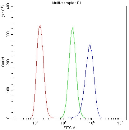
Click image to see more details
Figure 9. Flow Cytometry analysis of CACO-2 cells using anti-CYCS antibody (PA1118).
Overlay histogram showing CACO-2 cells stained with PA1118 (Blue line).The cells were blocked with 10% normal goat serum. And then incubated with rabbit anti-CYCS Antibody (PA1118, 1μg/1x106 cells) for 30 min at 20°C. DyLight®488 conjugated goat anti-rabbit IgG (BA1127, 5-10μg/1x106 cells) was used as secondary antibody for 30 minutes at 20°C. Isotype control antibody (Green line) was rabbit IgG (1μg/1x106) used under the same conditions. Unlabelled sample (Red line) was also used as a control.
Protein Target Info & Infographic
Gene/Protein Information For Cycs (Source: Uniprot.org, NCBI)
Gene Name
Cycs
Full Name
Cytochrome c, somatic
Weight
11.605kDa
Superfamily
cytochrome c family
Alternative Names
CYCHCS; CYCS; Cytochrome c; cytochrome c, somatic; THC4 Cycs|cytochrome c, somatic|cytochrome c, somatic
*If product is indicated to react with multiple species, protein info is based on the gene entry specified above in "Species".For more info on Cycs, check out the Cycs Infographic

We have 30,000+ of these available, one for each gene! Check them out.
In this infographic, you will see the following information for Cycs: database IDs, superfamily, protein function, synonyms, molecular weight, chromosomal locations, tissues of expression, subcellular locations, post-translational modifications, and related diseases, research areas & pathways. If you want to see more information included, or would like to contribute to it and be acknowledged, please contact [email protected].
Specific Publications For Anti-Cytochrome C/CYCS Antibody (PA1118)
Hello CJ!
PA1118 has been cited in 20 publications:
*The publications in this section are manually curated by our staff scientists. They may differ from Bioz's machine gathered results. Both are accurate. If you find a publication citing this product but is missing from this list, please let us know we will issue you a thank-you coupon.
A Hexokinase 2 Modulator for Field-Directed Treatment of Experimental Actinic Keratoses
Zhang T,Chen Y,Cai J,Pan M,Sun Q,Zhang J,Sun C.SOCS2 Inhibits Mitochondrial Fatty Acid Oxidation via Suppressing LepR/JAK2/AMPK Signaling Pathway in Mouse Adipocytes.Oxid Med Cell Longev.2020 Jul 13;2020:3742542.doi:10.1155/2020/3742542.PMID:32733634;PMCI
Species: Mouse
PA1118 usage in article: APP:IF, SAMPLE:ADIPOCYTES, DILUTION:1:100
Neuroprotective Effect of Tea Polyphenols on Oxyhemoglobin Induced Subarachnoid Hemorrhage in Mice
Chinese herbal medicine Yougui Pill reduces exogenous glucocorticoid-induced apoptosis in anterior pituitary cells
Catalpol inhibits apoptosis in hydrogen peroxide-induced cardiac myocytes through a mitochondrial-dependent caspase pathway
Methacryloxylethyl Cetyl Ammonium Chloride Induces DNA Damage and Apoptosis in Human Dental Pulp Cells via Generation of Oxidative Stress
N-Acetyl Cysteine Depletes Reactive Oxygen Species and Prevents Dental Monomer-Induced Intrinsic Mitochondrial Apoptosis In Vitro in Human Dental Pulp Cells
HOXB1 Is a Tumor Suppressor Gene Regulated by miR-3175 in Glioma
FABP4 reversed the regulation of leptin on mitochondrial fatty acid oxidation in mice adipocytes
Lin W, Tongyi S. Tumour Biol. 2014 Aug;35(8):8065-75. Doi: 10.1007/S13277-014-2064-0. Epub 2014 May 19. Role Of Bax/Bcl-2 Family Members In Green Tea Polyphenol Induced Necroptosis Of P53-Deficient Hep3B Cells.
Recommended Resources
Here are featured tools and databases that you might find useful.
- Boster's Pathways Library
- Protein Databases
- Bioscience Research Protocol Resources
- Data Processing & Analysis Software
- Photo Editing Software
- Scientific Literature Resources
- Research Paper Management Tools
- Molecular Biology Software
- Primer Design Tools
- Bioinformatics Tools
- Phylogenetic Tree Analysis
Customer Reviews
Have you used Anti-Cytochrome C/CYCS Antibody?
Submit a review and receive an Amazon gift card.
- $30 for a review with an image
Be the first to review Anti-Cytochrome C/CYCS Antibody
*The first user to submit a review for a product is eligible for Boster's Innovating Scientists Reward, which gives product credits. This is in addition to the gift card reward.
Customer Q&As
Have a question?
Find answers in Q&As, reviews.
Can't find your answer?
Submit your question
16 Customer Q&As for Anti-Cytochrome C/CYCS Antibody
Question
My question regarding product PA1118, anti-Cytochrome C/CYCS antibody. I was wondering if it would be possible to conjugate this antibody with biotin. I would need it to be without BSA or sodium azide. I am planning on using a buffer exchange of sodium azide with PBS only. Would there be problems for me to conjugate the antibody and store it in -20 degrees in small aliquots?
A. Carter
Verified customer
Asked: 2020-01-14
Answer
We suggest not storing this antibody with PBS buffer only in -20 degrees. If you want to store it in -20 degrees it is best to add some cryoprotectant like glycerol. If you want carrier free PA1118 anti-Cytochrome C/CYCS antibody, we can provide it to you in a special formula with trehalose and/or glycerol. These molecules will not interfere with conjugation chemistry and provide a good level of protection for the antibody from degradation. Please be sure to specify this in your purchase order.
Boster Scientific Support
Answered: 2020-01-14
Question
Our lab used your anti-Cytochrome C/CYCS antibody for IHC-P on bone marrow last year. I am using mouse, and We are going to use the antibody for WB next. you antibody examining bone marrow as well as amygdala in our next experiment. Do you have any suggestion on which antibody would work the best for WB?
Verified Customer
Verified customer
Asked: 2019-11-26
Answer
I have checked the website and datasheets of our anti-Cytochrome C/CYCS antibody and it appears that PA1118 has been validated on mouse in both IHC-P and WB. Thus PA1118 should work for your application. Our Boster satisfaction guarantee will cover this product for WB in mouse even if the specific tissue type has not been validated. We do have a comprehensive range of products for WB detection and you can check out our website bosterbio.com to find out more information about them.
Boster Scientific Support
Answered: 2019-11-26
Question
Does anti-Cytochrome C/CYCS antibody PA1118 work for ICC with kidney?
E. Dhar
Verified customer
Asked: 2019-11-25
Answer
According to the expression profile of kidney, CYCS is highly expressed in kidney. So, it is likely that anti-Cytochrome C/CYCS antibody PA1118 will work for ICC with kidney.
Boster Scientific Support
Answered: 2019-11-25
Question
Is this PA1118 anti-Cytochrome C/CYCS antibody reactive to the isotypes of CYCS?
Verified Customer
Verified customer
Asked: 2019-10-18
Answer
The immunogen of PA1118 anti-Cytochrome C/CYCS antibody is A synthetic peptide corresponding to a sequence at the C-terminus of human Cytochrome C(91-105 aa ERADLIAYLKKATNE), identical to the related mouse and rat sequences. Could you tell me which isotype you are interested in so I can help see if the immunogen is part of this isotype?
Boster Scientific Support
Answered: 2019-10-18
Question
We need using your anti-Cytochrome C/CYCS antibody for mitochondrial electron transport studies. Has this antibody been tested with western blotting on hela whole cell lysate? We would like to see some validation images before ordering.
Verified Customer
Verified customer
Asked: 2019-08-30
Answer
Thanks for your inquiry. This PA1118 anti-Cytochrome C/CYCS antibody is validated on rat lung tissue, tissue lysate, brain tissue, liver tissue, hela whole cell lysate, hepa whole cell lysate. It is guaranteed to work for IHC-P, IHC-F, ICC, WB in human, mouse, rat. Our Boster guarantee will cover your intended experiment even if the sample type has not been be directly tested.
Boster Scientific Support
Answered: 2019-08-30
Question
My team were happy with the WB result of your anti-Cytochrome C/CYCS antibody. However we have been able to see positive staining in skin, rc testis urinary bladder mitochondrion intermembrane space. using this antibody. Is that expected? Could you tell me where is CYCS supposed to be expressed?
Verified Customer
Verified customer
Asked: 2019-08-13
Answer
From what I have seen in literature, skin,
rc testis urinary bladder does express CYCS. Generally CYCS expresses in mitochondrion intermembrane space. Regarding which tissues have CYCS expression, here are a few articles citing expression in various tissues:
Amygdala, Pubmed ID: 17974005
Bone marrow, Brain, Kidney, Lung, Skeletal muscle, Skin, Testis, and Urinary bladder, Pubmed ID: 15489334
Cerebellum, Pubmed ID: 14702039
Erythroleukemia, Pubmed ID: 23186163
Heart, Pubmed ID: 13933734, 14063298, 9515723
Liver, Pubmed ID: 24275569
Boster Scientific Support
Answered: 2019-08-13
Question
Do you have a BSA free version of anti-Cytochrome C/CYCS antibody PA1118 available?
Verified Customer
Verified customer
Asked: 2018-12-03
Answer
We appreciate your recent telephone inquiry. I can confirm that some lots of this anti-Cytochrome C/CYCS antibody PA1118 are BSA free. For now, these lots are available and we can make a BSA free formula for you free of charge. It will take 3 extra days to prepare. If you require this antibody BSA free again in future, please do not hesitate to contact me and I will be pleased to check which lots we have in stock that are BSA free.
Boster Scientific Support
Answered: 2018-12-03
Question
I appreciate helping with my inquiry over the phone. Here are the WB image, lot number and protocol we used for kidney using anti-Cytochrome C/CYCS antibody PA1118. Let me know if you need anything else.
Verified Customer
Verified customer
Asked: 2018-09-14
Answer
We appreciate the data. You have provided everything we needed. Our lab team are working to resolve your inquiry as quickly as possible, and we appreciate your patience and understanding! Please let me know if there is anything you need in the meantime.
Boster Scientific Support
Answered: 2018-09-14
Question
Is a blocking peptide available for product anti-Cytochrome C/CYCS antibody (PA1118)?
T. Collins
Verified customer
Asked: 2018-09-04
Answer
We do provide the blocking peptide for product anti-Cytochrome C/CYCS antibody (PA1118). If you would like to place an order for it please contact [email protected] and make a special request.
Boster Scientific Support
Answered: 2018-09-04
Question
I was wanting to use your anti-Cytochrome C/CYCS antibody for ICC for mouse kidney on frozen tissues, but I want to know if it has been validated for this particular application. Has this antibody been validated and is this antibody a good choice for mouse kidney identification?
Verified Customer
Verified customer
Asked: 2018-06-19
Answer
It shows on the product datasheet, PA1118 anti-Cytochrome C/CYCS antibody has been tested for IHC-P, IHC-F, ICC, WB on human, mouse, rat tissues. We have an innovator award program that if you test this antibody and show it works in mouse kidney in IHC-frozen, you can get your next antibody for free.
Boster Scientific Support
Answered: 2018-06-19
Question
We have been able to see staining in mouse liver. Are there any suggestions? Is anti-Cytochrome C/CYCS antibody supposed to stain liver positively?
Verified Customer
Verified customer
Asked: 2018-01-09
Answer
Based on literature liver does express CYCS. Based on Uniprot.org, CYCS is expressed in heart, cerebellum, amygdala, bone marrow, brain, kidney, lung, skeletal muscle, skin,
rc testis urinary bladder, erythroleukemia, liver, among other tissues. Regarding which tissues have CYCS expression, here are a few articles citing expression in various tissues:
Amygdala, Pubmed ID: 17974005
Bone marrow, Brain, Kidney, Lung, Skeletal muscle, Skin, Testis, and Urinary bladder, Pubmed ID: 15489334
Cerebellum, Pubmed ID: 14702039
Erythroleukemia, Pubmed ID: 23186163
Heart, Pubmed ID: 13933734, 14063298, 9515723
Liver, Pubmed ID: 24275569
Boster Scientific Support
Answered: 2018-01-09
Question
We are currently using anti-Cytochrome C/CYCS antibody PA1118 for human tissue, and we are well pleased with the ICC results. The species of reactivity given in the datasheet says human, mouse, rat. Is it true that the antibody can work on pig tissues as well?
P. Parker
Verified customer
Asked: 2017-04-24
Answer
The anti-Cytochrome C/CYCS antibody (PA1118) has not been validated for cross reactivity specifically with pig tissues, but there is a good chance of cross reactivity. We have an innovator award program that if you test this antibody and show it works in pig you can get your next antibody for free. Please contact me if I can help you with anything.
Boster Scientific Support
Answered: 2017-04-24
Question
Please see the WB image, lot number and protocol we used for kidney using anti-Cytochrome C/CYCS antibody PA1118. Please let me know if you require anything else.
K. Carter
Verified customer
Asked: 2016-10-13
Answer
Thank you very much for the data. Our lab team are working to resolve this as quickly as possible, and we appreciate your patience and understanding! You have provided everything we needed. Please let me know if there is anything you need in the meantime.
Boster Scientific Support
Answered: 2016-10-13
Question
Would PA1118 anti-Cytochrome C/CYCS antibody work on parafin embedded sections? If so, which fixation method do you recommend we use (PFA, paraformaldehyde, other)?
N. Li
Verified customer
Asked: 2016-03-21
Answer
It shows on the product datasheet, PA1118 anti-Cytochrome C/CYCS antibody as been validated on ICC. It is best to use PFA for fixation because it has better tissue penetration ability. PFA needs to be prepared fresh before use. Long term stored PFA turns into formalin, as the PFA molecules congregate and become formalin.
Boster Scientific Support
Answered: 2016-03-21
Question
you antibody to test anti-Cytochrome C/CYCS antibody PA1118 on mouse kidney for research purposes, then I may be interested in using anti-Cytochrome C/CYCS antibody PA1118 for diagnostic purposes as well. Is the antibody suitable for diagnostic purposes?
A. Zhao
Verified customer
Asked: 2015-10-23
Answer
The products we sell, including anti-Cytochrome C/CYCS antibody PA1118, are only intended for research use. They would not be suitable for use in diagnostic work. If you have the means to develop a product into diagnostic use, and are interested in collaborating with us and develop our product into an IVD product, please contact us for more discussions.
Boster Scientific Support
Answered: 2015-10-23
Question
I see that the anti-Cytochrome C/CYCS antibody PA1118 works with ICC, what is the protocol used to produce the result images on the product page?
L. Wu
Verified customer
Asked: 2013-04-24
Answer
You can find protocols for ICC on the "support/technical resources" section of our navigation menu. If you have any further questions, please send an email to [email protected]
Boster Scientific Support
Answered: 2013-04-24
