Click image to see more details
-

-
-
-
-
+7
Product Info Summary
| SKU: | M00023-2 |
|---|---|
| Size: | 100 μl |
| Reactive Species: | Human, Mouse, Rat |
| Host: | Rabbit |
| Application: | Flow Cytometry, IP, IF, IHC, ICC, WB |
Customers Who Bought This Also Bought


Product info
Product Name
Anti-EGFR (ErbB 1) Monoclonal Antibody
SKU/Catalog Number
M00023-2
Size
100 μl
Form
Liquid
Description
Boster Bio Anti-EGFR (ErbB 1) Monoclonal Antibody catalog # M00023-2. Tested in WB, IHC, ICC/IF, IP, Flow Cytometry applications. This antibody reacts with Human, Mouse, Rat.
Storage & Handling
Store at -20°C for one year. For short term storage and frequent use, store at 4°C for up to one month. Avoid repeated freeze-thaw cycles.
Cite This Product
Anti-EGFR (ErbB 1) Monoclonal Antibody (Boster Biological Technology, Pleasanton CA, USA, Catalog # M00023-2)
Host
Rabbit
Contents
Rabbit IgG in phosphate buffered saline, pH 7.4, 150mM NaCl, 0.02% sodium azide and 50% glycerol, 0.4-0.5mg/ml BSA.
Clonality
Monoclonal
Clone Number
AEF-5
Isotype
Rabbit IgG
Immunogen
A synthesized peptide derived from human EGFR (ErbB 1)
*Blocking peptide can be purchased. Costs vary based on immunogen length. Contact us for pricing.
Reactive Species
M00023-2 is reactive to EGFR in Human, Mouse, Rat
Applications
M00023-2 is guaranteed for Flow Cytometry, IP, IF, IHC, ICC, WB Boster Guarantee
Observed Molecular Weight
175 kDa
Calculated molecular weight
134.277kDa
Background of EGFR
The ion channels activated by glutamate are typically divided into two classes. Those that are sensitive to N-methyl-D-aspartate (NMDA) are designated NMDA receptors (NMDAR) while those activated by α-amino-3-hydroxy-5-methyl-4-isoxalone propionic acid (AMPA) are known as AMPA receptors (AMPAR). The AMPAR are comprised of four distinct glutamate receptor subunits designated (GluR1-4) and they play key roles in virtually all excitatory neurotransmission in the brain (Keinänen et al., 1990; Hollmann and Heinemann, 1994). The GluR1 subunit is widely expressed throughout the nervous system. Phosphorylation of Ser-845 on GluR1 is thought to be mediated by PKA and phosphorylation of this site increases the conductance of the AMPAR (Roche et al., 1996; Banke et al., 2000). In addition, phosphorylation of this site has been linked to synaptic plasticity as well as learning and memory (Lee at al., 2003; Esteban at al., 2003).
Antibody Validation
Boster validates all antibodies on WB, IHC, ICC, Immunofluorescence, and ELISA with known positive control and negative samples to ensure specificity and high affinity, including thorough antibody incubations.
Innovating Scientists Reward
If you are the first to review this product, or if you have results for a special sample, species or application this product is not validated in, share your results with us and receive product credits you can use towards any Boster products! Applicable to all scientists worldwide.
Submit A Review
Assay dilution & Images
Reconsitution
Restore with deionized water (or equivalent) for reconstitution volume of 1.0 mL
Assay Dilutions Recommendation
The recommendations below provide a starting point for assay optimization. The actual working concentration varies and should be decided by the user.
WB 1:5000-1:10000
IHC 1:50-1:200
ICC/IF 1:50-1:200
IP 1:50
FC 1:50
Validation Images & Assay Conditions
Click image to see more details
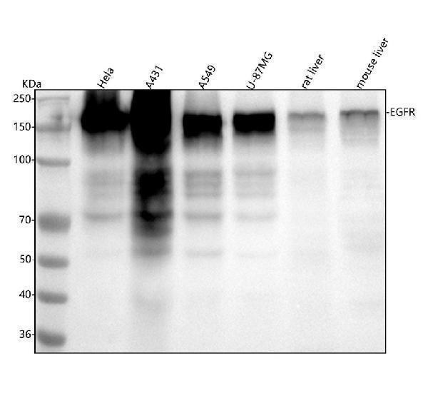
Figure 1. Western blot analysis of EGFR using anti-EGFR antibody (M00023-2).
Electrophoresis was performed on a 5-20% SDS-PAGE gel at 70V (Stacking gel) / 90V (Resolving gel) for 2-3 hours. The sample well of each lane was loaded with 30 ug of sample under reducing conditions.
Lane 1: human Hela whole cell lysates,
Lane 2: human A431 whole cell lysates,
Lane 3: human A549 whole cell lysates,
Lane 4: human U-87MG whole cell lysates,
Lane 5: rat liver tissue lysates,
Lane 6: mouse liver tissue lysates.
After electrophoresis, proteins were transferred to a nitrocellulose membrane at 150 mA for 50-90 minutes. Blocked the membrane with 5% non-fat milk/TBS for 1.5 hour at RT. The membrane was incubated with rabbit anti-EGFR antigen affinity purified monoclonal antibody (Catalog # M00023-2) at 1:5000 overnight at 4°C, then washed with TBS-0.1%Tween 3 times with 5 minutes each and probed with a goat anti-rabbit IgG-HRP secondary antibody at a dilution of 1:500 for 1.5 hour at RT. The signal is developed using an Enhanced Chemiluminescent detection (ECL) kit (Catalog # EK1002) with Tanon 5200 system. A specific band was detected for EGFR at approximately 175 kDa. The expected band size for EGFR is at 134 kDa.

Click image to see more details
Immunohistochemical analysis of paraffin-embedded Rat cerebral cortex, using the Antibody at 1:100 dilution.

Click image to see more details
Immunohistochemical analysis of paraffin-embedded Rat stomach, using the Antibody at 1:100 dilution.

Click image to see more details
Immunohistochemical analysis of paraffin-embedded Human thyroid cancer, using the Antibody at 1:100 dilution.

Click image to see more details
Immunohistochemical analysis of paraffin-embedded Human glioblastoma, using the Antibody at 1:100 dilution.

Click image to see more details
Immunohistochemical analysis of paraffin-embedded Mouse intestine, using the Antibody at 1:100 dilution.

Click image to see more details
Immunohistochemical analysis of paraffin-embedded human stomach cancer, using EGFR (ErbB 1) Antibody.

Click image to see more details
Immunofluorescent analysis using the Antibody at 1:50 dilution.

Click image to see more details
Immunofluorescent analysis using the Antibody at 1:50 dilution.

Click image to see more details
Immunofluorescent analysis using the Antibody at 1:150 dilution.

Click image to see more details
Immunofluorescent analysis using the Antibody at 1:500 dilution.
Protein Target Info & Infographic
Gene/Protein Information For EGFR (Source: Uniprot.org, NCBI)
Gene Name
EGFR
Full Name
Epidermal growth factor receptor
Weight
134.277kDa
Superfamily
protein kinase superfamily
Alternative Names
avian erythroblastic leukemia viral (v-erb-b) oncogene homolog; cell growth inhibiting protein 40; cell proliferation-inducing protein 61; EC 2.7.10; EC 2.7.10.1; EGF R; EGFR; epidermal growth factor receptor (avian erythroblastic leukemia viral (v-erb-b)oncogene homolog); epidermal growth factor receptor; ErbB; ErbB1; ERBB1PIG61; HER1; HER-1; mENA; Proto-oncogene c-ErbB-1; Receptor tyrosine-protein kinase erbB-1 EGFR ERBB, ERBB1, ERRP, HER1, NISBD2, PIG61, mENA epidermal growth factor receptor epidermal growth factor receptor|avian erythroblastic leukemia viral (v-erb-b) oncogene homolog|cell growth inhibiting protein 40|cell proliferation-inducing protein 61|epidermal growth factor receptor tyrosine kinase domain|erb-b2 receptor tyrosine kinase 1|proto-oncogene c-ErbB-1|receptor tyrosine-protein kinase erbB-1
*If product is indicated to react with multiple species, protein info is based on the gene entry specified above in "Species".For more info on EGFR, check out the EGFR Infographic

We have 30,000+ of these available, one for each gene! Check them out.
In this infographic, you will see the following information for EGFR: database IDs, superfamily, protein function, synonyms, molecular weight, chromosomal locations, tissues of expression, subcellular locations, post-translational modifications, and related diseases, research areas & pathways. If you want to see more information included, or would like to contribute to it and be acknowledged, please contact [email protected].
Specific Publications For Anti-EGFR (ErbB 1) Monoclonal Antibody (M00023-2)
Hello CJ!
M00023-2 has been cited in 1 publications:
*The publications in this section are manually curated by our staff scientists. They may differ from Bioz's machine gathered results. Both are accurate. If you find a publication citing this product but is missing from this list, please let us know we will issue you a thank-you coupon.
Study of the correlation between the expression of nuclear factor kappa B and proliferation regulatory proteins and chronic superficial gastritis,Hu Hui,Ma Zhijian,Ren Shouzhong,Xie Yiqiang, Vojnosanitetski pregled 2020 OnLine-First,00,SP - 135 EP-135
Species: Rat
M00023-2 usage in article: APP:IHC, SAMPLE:STOMACH TISSUE, DILUTION:NA
Recommended Resources
Here are featured tools and databases that you might find useful.
- Boster's Pathways Library
- Protein Databases
- Bioscience Research Protocol Resources
- Data Processing & Analysis Software
- Photo Editing Software
- Scientific Literature Resources
- Research Paper Management Tools
- Molecular Biology Software
- Primer Design Tools
- Bioinformatics Tools
- Phylogenetic Tree Analysis
Customer Reviews
Have you used Anti-EGFR (ErbB 1) Monoclonal Antibody?
Submit a review and receive an Amazon gift card.
- $30 for a review with an image
Be the first to review Anti-EGFR (ErbB 1) Monoclonal Antibody
*The first user to submit a review for a product is eligible for Boster's Innovating Scientists Reward, which gives product credits. This is in addition to the gift card reward.
Customer Q&As
Have a question?
Find answers in Q&As, reviews.
Can't find your answer?
Submit your question
