
Click image to see more details
Product Info Summary
| SKU: | M00676-4 |
|---|---|
| Size: | 200ug |
| Reactive Species: | Human |
| Host: | Mouse |
| Application: | ELISA, IP, IF, IHC, ICC, WB |
Customers Who Bought This Also Bought

Product info
Product Name
Anti-HSP27 Monoclonal Antibody
SKU/Catalog Number
M00676-4
Size
200ug
Form
liquid
Description
Boster Bio Anti-HSP27 Monoclonal Antibody catalog # M00676-4. Tested in ELISA, IP, IF, WB applications. This antibody reacts with Human.
Storage & Handling
Store at -20°C for one year. Avoid repeated freeze-thaw cycles.
Cite This Product
Anti-HSP27 Monoclonal Antibody (Boster Biological Technology, Pleasanton CA, USA, Catalog # M00676-4)
Host
Mouse
Contents
PBS pH7.4, 50% glycerol, 0.09% sodium azide
Clonality
Monoclonal
Clone Number
5D12-A12
Isotype
IgG2b Kappa
Immunogen
Full length human HSP27
*Blocking peptide can be purchased. Costs vary based on immunogen length. Contact us for pricing.
Cross-reactivity
Detects ~27kDa. Has no cross-reactivity to Alpha B crystallin. Very limited cross-reactivity to other species.
Reactive Species
M00676-4 is reactive to HSPB1 in Human
Applications
M00676-4 is guaranteed for ELISA, IP, IF, IHC, ICC, WB Boster Guarantee
Observed Molecular Weight
68 kDa
Calculated molecular weight
22.783kDa
Background of HSP27
HSP27s belong to an abundant and ubiquitous family of small heat shock proteins (sHSP). It is an important HSP found in both normal human cells and cancer cells. The basic structure of most sHSPs is a homologous and highly conserved amino acid sequence, with an α-crystallin domain at the C-terminus and the WD/EPF domain at the less conserved N-terminus. This N-terminus is essential for the development of high molecular oligomers (1, 2). HSP27-oligomers consist of stable dimers formed by as many as 8-40 HSP27 protein monomers (3). The oligomerization status is connected with the chaperone activity: aggregates of large oligomers have high chaperone activity, whereas dimers have no chaperone activity (4). HSP27 is localized to the cytoplasm of unstressed cells but can redistribute to the nucleus in response to stress, where it may function to stabilize DNA and/or the nuclear membrane. Other functions include chaperone activity (as mentioned above), thermo tolerance in vivo, inhibition of apoptosis, and signal transduction. Specifically, in vitro, it acts as an ATP-independent chaperone by inhibiting protein aggregation and by stabilizing partially denatured proteins, which ensures refolding of the HSP70 complex. HSP27 is also involved in the apoptotic signaling pathway because it interferes with the activation of cytochrome c/Apaf-1/dATP complex, thereby inhibiting the activation of procaspase-9. It is also hypothesized that HSP27 may serve some role in cross-bridge formation between actin and myosin (5). And finally, HSP27 is also thought to be involved in the process of cell differentiation. The up-regulation of HSP27 correlates with the rate of phosphorylation and with an increase of large oligomers. It is possible that HSP27 may play a crucial role in termination of growth (6). For more information visit our HSP27 Scientific Resource Guide at http://www.HSP27.com.
Antibody Validation
Boster validates all antibodies on WB, IHC, ICC, Immunofluorescence, and ELISA with known positive control and negative samples to ensure specificity and high affinity, including thorough antibody incubations.
Innovating Scientists Reward
If you are the first to review this product, or if you have results for a special sample, species or application this product is not validated in, share your results with us and receive product credits you can use towards any Boster products! Applicable to all scientists worldwide.
Submit A Review
Assay dilution & Images
Assay Dilutions Recommendation
The recommendations below provide a starting point for assay optimization. The actual working concentration varies and should be decided by the user.
WB (1:2000), ICC/IF (1:100); optimal dilutions for assays should be determined by the user.
Validation Images & Assay Conditions
Click image to see more details
Immunocytochemistry/Immunofluorescence analysis using Mouse Anti-Hsp27 Monoclonal Antibody, Clone 5D12-A3 (M00676-4) . Tissue: HaCaT cells. Species: Human. Fixation: Cold 100% methanol for 10 minutes at -20°C. Primary Antibody: Mouse Anti-Hsp27 Monoclonal Antibody (M00676-4) at 1:100 for 1 hour at RT. Secondary Antibody: FITC Goat Anti-Mouse (green) at 1:50 for 1 hour at RT. Localization: Dull heterogeneous staining, some perinuclear, some nuclear and some cytoplasmic staining .

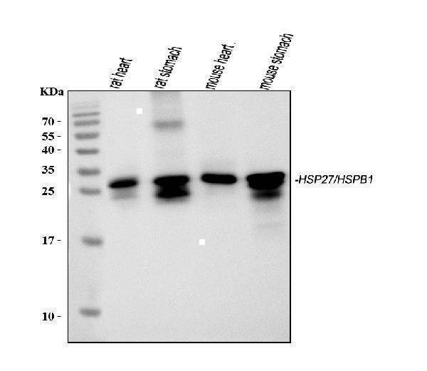
Click image to see more details
Figure 4. Western blot analysis of HSPB1 using anti-HSPB1 antibody (M00676-4).
Electrophoresis was performed on a 5-20% SDS-PAGE gel at 70V (Stacking gel) / 90V (Resolving gel) for 2-3 hours. The sample well of each lane was loaded with 50ug of sample under reducing conditions.
After Electrophoresis, proteins were transferred to a Nitrocellulose membrane at 150mA for 50-90 minutes. Blocked the membrane with 5% Non-fat Milk/ TBS for 1.5 hour at RT. The membrane was incubated with rabbit anti-HSPB1 antigen affinity purified polyclonal antibody (Catalog # M00676-4) at 0.5 ug/mL overnight at 4°C, then washed with TBS-0.1%Tween 3 times with 5 minutes each and probed with a goat anti-Mouse IgG-HRP secondary antibody at a dilution of 1:10000 for 1.5 hour at RT. The signal is developed using an Enhanced Chemiluminescent detection (ECL) kit (Catalog # SA1021) with Tanon 5200 system. A specific band was detected for HSPB1.

Click image to see more details
Immunocytochemistry/Immunofluorescence analysis using Mouse Anti-Hsp27 Monoclonal Antibody, Clone 5D12-A3 (M00676-4) . Tissue: Heat Shocked HeLa Cells. Species: Human. Fixation: 2% Formaldehyde for 20 min at RT. Primary Antibody: Mouse Anti-Hsp27 Monoclonal Antibody (M00676-4) at 1:100 for 12 hours at 4°C. Secondary Antibody: FITC Goat Anti-Mouse (green) at 1:200 for 2 hours at RT. Counterstain: DAPI (blue) nuclear stain at 1:40000 for 2 hours at RT. Localization: Cytoplasm. Nucleus. Magnification: 20x. (A) DAPI (blue) nuclear stain. (B) Anti-Hsp27 Antibody. (C) Composite. Heat Shocked at 42°C for 1h.

Click image to see more details
Immunocytochemistry/Immunofluorescence analysis using Mouse Anti-Hsp27 Monoclonal Antibody, Clone 5D12-A3 (M00676-4) . Tissue: Heat Shocked HeLa Cells. Species: Human. Fixation: 2% Formaldehyde for 20 min at RT. Primary Antibody: Mouse Anti-Hsp27 Monoclonal Antibody (M00676-4) at 1:100 for 12 hours at 4°C. Secondary Antibody: FITC Goat Anti-Mouse (green) at 1:200 for 2 hours at RT. Counterstain: DAPI (blue) nuclear stain at 1:40000 for 2 hours at RT. Localization: Cytoplasm. Nucleus. Magnification: 100x. (A) DAPI (blue) nuclear stain. (B) Anti-Hsp27 Antibody. (C) Composite. Heat Shocked at 42°C for 1h.
Protein Target Info & Infographic
Gene/Protein Information For HSPB1 (Source: Uniprot.org, NCBI)
Gene Name
HSPB1
Full Name
Heat shock protein beta-1
Weight
22.783kDa
Superfamily
small heat shock protein (HSP20) family
Alternative Names
28 kDa heat shock protein; DKFZp586P1322; Estrogen-regulated 24 kDa protein; Heat shock 27 kDa protein; heat shock 27kD protein 1; heat shock 27kDa protein 1; heat shock protein beta-1; HMN2B; HS.76067; HSP25; HSP27; HSP27HSP 27; HSP28CMT2F; HSPB1; SRP27; Stress-responsive protein 27 HSPB1 CMT2F, HEL-S-102, HMN2B, HS.76067, HSP27, HSP28, Hsp25, SRP27 heat shock protein family B (small) member 1 heat shock protein beta-1|28 kDa heat shock protein|epididymis secretory protein Li 102|estrogen-regulated 24 kDa protein|heat shock 27 kDa protein|heat shock 27kD protein 1|heat shock 27kDa protein 1|stress-responsive protein 27
*If product is indicated to react with multiple species, protein info is based on the gene entry specified above in "Species".For more info on HSPB1, check out the HSPB1 Infographic

We have 30,000+ of these available, one for each gene! Check them out.
In this infographic, you will see the following information for HSPB1: database IDs, superfamily, protein function, synonyms, molecular weight, chromosomal locations, tissues of expression, subcellular locations, post-translational modifications, and related diseases, research areas & pathways. If you want to see more information included, or would like to contribute to it and be acknowledged, please contact [email protected].
Specific Publications For Anti-HSP27 Monoclonal Antibody (M00676-4)
Hello CJ!
No publications found for M00676-4
*Do you have publications using this product? Share with us and receive a reward. Ask us for more details.
Recommended Resources
Here are featured tools and databases that you might find useful.
- Boster's Pathways Library
- Protein Databases
- Bioscience Research Protocol Resources
- Data Processing & Analysis Software
- Photo Editing Software
- Scientific Literature Resources
- Research Paper Management Tools
- Molecular Biology Software
- Primer Design Tools
- Bioinformatics Tools
- Phylogenetic Tree Analysis
Customer Reviews
Have you used Anti-HSP27 Monoclonal Antibody?
Submit a review and receive an Amazon gift card.
- $30 for a review with an image
Be the first to review Anti-HSP27 Monoclonal Antibody
*The first user to submit a review for a product is eligible for Boster's Innovating Scientists Reward, which gives product credits. This is in addition to the gift card reward.
Customer Q&As
Have a question?
Find answers in Q&As, reviews.
Can't find your answer?
Submit your question
