Click image to see more details
-

-
-
-
-
+7
Product Info Summary
| SKU: | A02304 |
|---|---|
| Size: | 100 µg/vial |
| Reactive Species: | Human, Mouse, Rat |
| Host: | Rabbit |
| Application: | ELISA, Flow Cytometry, IF, IHC, WB |
Customers Who Bought This Also Bought

Product info
Product Name
Anti-ITPA Antibody Picoband™
SKU/Catalog Number
A02304
Size
100 µg/vial
Form
Lyophilized
Description
Boster Bio Anti-ITPA Antibody Picoband™ catalog # A02304. Tested in ELISA, IF, IHC, WB, Flow Cytometry applications. This antibody reacts with Human, Mouse, Rat.
Storage & Handling
At -20°C for one year from date of receipt. After reconstitution, at 4°C for one month. It can also be aliquotted and stored frozen at -20°C for six months. Avoid repeated freezing and thawing.
Cite This Product
Anti-ITPA Antibody Picoband™ (Boster Biological Technology, Pleasanton CA, USA, Catalog # A02304)
Host
Rabbit
Contents
Each vial contains 4 mg Trehalose, 0.9 mg NaCl, 0.2 mg Na2HPO4.
Clonality
Polyclonal
Clone Number
1B9
Isotype
IgG
Immunogen
E.coli-derived human ITPA recombinant protein (Position: M1-A194). Human ITPA shares 89.6% and 92.1% amino acid (aa) sequence identity with mouse and rat ITPA, respectively.
*Blocking peptide can be purchased. Costs vary based on immunogen length. Contact us for pricing.
Cross-reactivity
No cross reactivity with other proteins.
Reactive Species
A02304 is reactive to ITPA in Human, Mouse, Rat
Applications
A02304 is guaranteed for ELISA, Flow Cytometry, IF, IHC, WB Boster Guarantee
Observed Molecular Weight
21 kDa
Calculated molecular weight
21.446kDa
Background of ITPA
Inosine triphosphate pyrophosphatase is an enzyme that in humans is encoded by the ITPA gene, by the rdgB gene in bacteria E.coli and the HAM1 gene in yeast S. cerevisiae; the protein is also encoded by some RNA viruses of the Potyviridae family. This gene encodes an inosine triphosphate pyrophosphohydrolase. The encoded protein hydrolyzes inosine triphosphate and deoxyinosine triphosphate to the monophosphate nucleotide and diphosphate. This protein, which is a member of the HAM1 NTPase protein family, is found in the cytoplasm and acts as a homodimer. Defects in the encoded protein can result in inosine triphosphate pyrophosphorylase deficiency which causes an accumulation of ITP in red blood cells. Alternate splicing results in multiple transcript variants.
Antibody Validation
Boster validates all antibodies on WB, IHC, ICC, Immunofluorescence, and ELISA with known positive control and negative samples to ensure specificity and high affinity, including thorough antibody incubations.
Innovating Scientists Reward
If you are the first to review this product, or if you have results for a special sample, species or application this product is not validated in, share your results with us and receive product credits you can use towards any Boster products! Applicable to all scientists worldwide.
Submit A Review
Assay dilution & Images
Reconsitution
Adding 0.2 ml of distilled water will yield a concentration of 500 µg/ml.
Assay Dilutions Recommendation
The recommendations below provide a starting point for assay optimization. The actual working concentration varies and should be decided by the user.
Western blot, 0.25-0.5 µg/ml, Human, Mouse, Rat
Immunohistochemistry, 2-5 µg/ml, Human, Rat
Immunofluorescence, 5 µg/ml, Human
Flow Cytometry (Fixed), 1-3 µg/1x106 cells, Human
ELISA, 0.1-0.5 µg/ml, Human
Validation Images & Assay Conditions
Click image to see more details
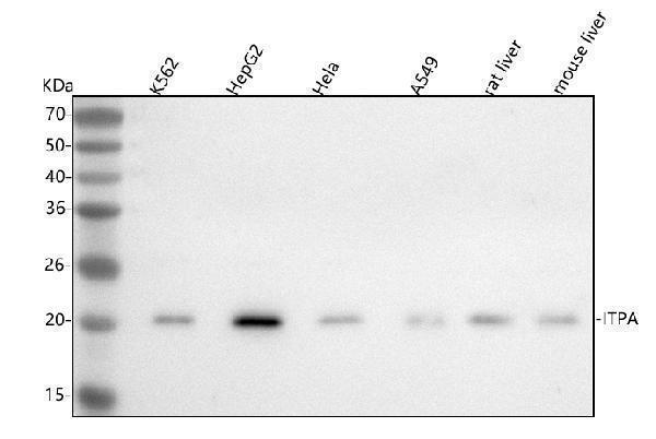
Figure 1. Western blot analysis of ITPA using anti-ITPA antibody (A02304).
Electrophoresis was performed on a 5-20% SDS-PAGE gel at 70V (Stacking gel) / 90V (Resolving gel) for 2-3 hours. The sample well of each lane was loaded with 30 ug of sample under reducing conditions.
Lane 1: human K562 whole cell lysates,
Lane 2: human HepG2 whole cell lysates,
Lane 3: human Hela whole cell lysates,
Lane 4: human A549 whole cell lysates,
Lane 5: rat liver tissue lysates,
Lane 6: mouse liver tissue lysates.
After electrophoresis, proteins were transferred to a nitrocellulose membrane at 150 mA for 50-90 minutes. Blocked the membrane with 5% non-fat milk/TBS for 1.5 hour at RT. The membrane was incubated with rabbit anti-ITPA antigen affinity purified polyclonal antibody (Catalog # A02304) at 0.5 μg/mL overnight at 4°C, then washed with TBS-0.1%Tween 3 times with 5 minutes each and probed with a goat anti-rabbit IgG-HRP secondary antibody at a dilution of 1:5000 for 1.5 hour at RT. The signal is developed using an Enhanced Chemiluminescent detection (ECL) kit (Catalog # EK1002) with Tanon 5200 system. A specific band was detected for ITPA at approximately 21 kDa. The expected band size for ITPA is at 21 kDa.

Click image to see more details
Figure 2. IHC analysis of ITPA using anti-ITPA antibody (A02304).
ITPA was detected in a paraffin-embedded section of human colon adenocarcinoma tissue. Heat mediated antigen retrieval was performed in EDTA buffer (pH 8.0, epitope retrieval solution). The tissue section was blocked with 10% goat serum. The tissue section was then incubated with 2 μg/ml rabbit anti-ITPA Antibody (A02304) overnight at 4°C. Peroxidase Conjugated Goat Anti-rabbit IgG was used as secondary antibody and incubated for 30 minutes at 37°C. The tissue section was developed using HRP Conjugated Rabbit IgG Super Vision Assay Kit (Catalog # SV0002) with DAB as the chromogen.

Click image to see more details
Figure 3. IHC analysis of ITPA using anti-ITPA antibody (A02304).
ITPA was detected in a paraffin-embedded section of human glioblastoma tissue. Heat mediated antigen retrieval was performed in EDTA buffer (pH 8.0, epitope retrieval solution). The tissue section was blocked with 10% goat serum. The tissue section was then incubated with 2 μg/ml rabbit anti-ITPA Antibody (A02304) overnight at 4°C. Peroxidase Conjugated Goat Anti-rabbit IgG was used as secondary antibody and incubated for 30 minutes at 37°C. The tissue section was developed using HRP Conjugated Rabbit IgG Super Vision Assay Kit (Catalog # SV0002) with DAB as the chromogen.

Click image to see more details
Figure 4. IHC analysis of ITPA using anti-ITPA antibody (A02304).
ITPA was detected in a paraffin-embedded section of human liver cancer tissue. Heat mediated antigen retrieval was performed in EDTA buffer (pH 8.0, epitope retrieval solution). The tissue section was blocked with 10% goat serum. The tissue section was then incubated with 2 μg/ml rabbit anti-ITPA Antibody (A02304) overnight at 4°C. Peroxidase Conjugated Goat Anti-rabbit IgG was used as secondary antibody and incubated for 30 minutes at 37°C. The tissue section was developed using HRP Conjugated Rabbit IgG Super Vision Assay Kit (Catalog # SV0002) with DAB as the chromogen.

Click image to see more details
Figure 5. IHC analysis of ITPA using anti-ITPA antibody (A02304).
ITPA was detected in a paraffin-embedded section of human lung cancer tissue. Heat mediated antigen retrieval was performed in EDTA buffer (pH 8.0, epitope retrieval solution). The tissue section was blocked with 10% goat serum. The tissue section was then incubated with 2 μg/ml rabbit anti-ITPA Antibody (A02304) overnight at 4°C. Peroxidase Conjugated Goat Anti-rabbit IgG was used as secondary antibody and incubated for 30 minutes at 37°C. The tissue section was developed using HRP Conjugated Rabbit IgG Super Vision Assay Kit (Catalog # SV0002) with DAB as the chromogen.

Click image to see more details
Figure 6. IHC analysis of ITPA using anti-ITPA antibody (A02304).
ITPA was detected in a paraffin-embedded section of human pancreas ductal adenocarcinoma tissue. Heat mediated antigen retrieval was performed in EDTA buffer (pH 8.0, epitope retrieval solution). The tissue section was blocked with 10% goat serum. The tissue section was then incubated with 2 μg/ml rabbit anti-ITPA Antibody (A02304) overnight at 4°C. Peroxidase Conjugated Goat Anti-rabbit IgG was used as secondary antibody and incubated for 30 minutes at 37°C. The tissue section was developed using HRP Conjugated Rabbit IgG Super Vision Assay Kit (Catalog # SV0002) with DAB as the chromogen.

Click image to see more details
Figure 7. IHC analysis of ITPA using anti-ITPA antibody (A02304).
ITPA was detected in a paraffin-embedded section of human testicular seminoma tissue. Heat mediated antigen retrieval was performed in EDTA buffer (pH 8.0, epitope retrieval solution). The tissue section was blocked with 10% goat serum. The tissue section was then incubated with 2 μg/ml rabbit anti-ITPA Antibody (A02304) overnight at 4°C. Peroxidase Conjugated Goat Anti-rabbit IgG was used as secondary antibody and incubated for 30 minutes at 37°C. The tissue section was developed using HRP Conjugated Rabbit IgG Super Vision Assay Kit (Catalog # SV0002) with DAB as the chromogen.

Click image to see more details
Figure 8. IHC analysis of ITPA using anti-ITPA antibody (A02304).
ITPA was detected in a paraffin-embedded section of human urothelial carcinoma tissue. Heat mediated antigen retrieval was performed in EDTA buffer (pH 8.0, epitope retrieval solution). The tissue section was blocked with 10% goat serum. The tissue section was then incubated with 2 μg/ml rabbit anti-ITPA Antibody (A02304) overnight at 4°C. Peroxidase Conjugated Goat Anti-rabbit IgG was used as secondary antibody and incubated for 30 minutes at 37°C. The tissue section was developed using HRP Conjugated Rabbit IgG Super Vision Assay Kit (Catalog # SV0002) with DAB as the chromogen.

Click image to see more details
Figure 9. IHC analysis of ITPA using anti-ITPA antibody (A02304).
ITPA was detected in a paraffin-embedded section of rat colon tissue. Heat mediated antigen retrieval was performed in EDTA buffer (pH 8.0, epitope retrieval solution). The tissue section was blocked with 10% goat serum. The tissue section was then incubated with 2 μg/ml rabbit anti-ITPA Antibody (A02304) overnight at 4°C. Peroxidase Conjugated Goat Anti-rabbit IgG was used as secondary antibody and incubated for 30 minutes at 37°C. The tissue section was developed using HRP Conjugated Rabbit IgG Super Vision Assay Kit (Catalog # SV0002) with DAB as the chromogen.

Click image to see more details
Figure 10. IF analysis of ITPA using anti-ITPA antibody (A02304).
ITPA was detected in a paraffin-embedded section of human intestinal cancer tissue. Heat mediated antigen retrieval was performed in EDTA buffer (pH 8.0, epitope retrieval solution). The tissue section was blocked with 10% goat serum. The tissue section was then incubated with 5 μg/mL rabbit anti-ITPA Antibody (A02304) overnight at 4°C. Cy3 Conjugated Goat Anti-Rabbit IgG (BA1032) was used as secondary antibody at 1:500 dilution and incubated for 30 minutes at 37°C. The section was counterstained with DAPI. Visualize using a fluorescence microscope and filter sets appropriate for the label used.

Click image to see more details
Figure 11. Flow Cytometry analysis of K562 cells using anti-ITPA antibody (A02304).
Overlay histogram showing K562 cells stained with A02304 (Blue line). To facilitate intracellular staining, cells were fixed with 4% paraformaldehyde and permeabilized with permeabilization buffer. The cells were blocked with 10% normal goat serum. And then incubated with rabbit anti-ITPA Antibody (A02304, 1 μg/1x106 cells) for 30 min at 20°C. DyLight®488 conjugated goat anti-rabbit IgG (BA1127, 5-10 μg/1x106 cells) was used as secondary antibody for 30 minutes at 20°C. Isotype control antibody (Green line) was rabbit IgG (1 μg/1x106) used under the same conditions. Unlabelled sample (Red line) was also used as a control.
Protein Target Info & Infographic
Gene/Protein Information For ITPA (Source: Uniprot.org, NCBI)
Gene Name
ITPA
Full Name
Inosine triphosphate pyrophosphatase
Weight
21.446kDa
Superfamily
HAM1 NTPase family
Alternative Names
C20orf37; dJ794I6.3; EC 3.6.1; EC 3.6.1.19; HLC14-06-P; inosine triphosphatase (nucleoside triphosphate pyrophosphatase); Inosine triphosphatase; inosine triphosphatase-A; inosine triphosphate pyrophosphatase; inosine triphosphate pyrophosphohydrolase; ITPase; My049 protein; nucleoside triphosphate diphosphatase; Putative oncogene protein hlc14-06-p ITPA C20orf37, DEE35, HLC14-06-P, ITPase, My049, NTPase, dJ794I6.3 inosine triphosphatase inosine triphosphate pyrophosphatase|epididymis secretory sperm binding protein|inosine triphosphatase (nucleoside triphosphate pyrophosphatase)|inosine triphosphate pyrophosphohydrolase|non-canonical purine NTP pyrophosphatase|non-standard purine NTP pyrophosphatase|nucleoside-triphosphate diphosphatase|putative oncogene protein HLC14-06-P
*If product is indicated to react with multiple species, protein info is based on the gene entry specified above in "Species".For more info on ITPA, check out the ITPA Infographic

We have 30,000+ of these available, one for each gene! Check them out.
In this infographic, you will see the following information for ITPA: database IDs, superfamily, protein function, synonyms, molecular weight, chromosomal locations, tissues of expression, subcellular locations, post-translational modifications, and related diseases, research areas & pathways. If you want to see more information included, or would like to contribute to it and be acknowledged, please contact [email protected].
Specific Publications For Anti-ITPA Antibody Picoband™ (A02304)
Hello CJ!
No publications found for A02304
*Do you have publications using this product? Share with us and receive a reward. Ask us for more details.
Recommended Resources
Here are featured tools and databases that you might find useful.
- Boster's Pathways Library
- Protein Databases
- Bioscience Research Protocol Resources
- Data Processing & Analysis Software
- Photo Editing Software
- Scientific Literature Resources
- Research Paper Management Tools
- Molecular Biology Software
- Primer Design Tools
- Bioinformatics Tools
- Phylogenetic Tree Analysis
Customer Reviews
Have you used Anti-ITPA Antibody Picoband™?
Submit a review and receive an Amazon gift card.
- $30 for a review with an image
Be the first to review Anti-ITPA Antibody Picoband™
*The first user to submit a review for a product is eligible for Boster's Innovating Scientists Reward, which gives product credits. This is in addition to the gift card reward.
Customer Q&As
Have a question?
Find answers in Q&As, reviews.
Can't find your answer?
Submit your question
