This website uses cookies to ensure you get the best experience on our website.


- Table of Contents

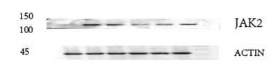
JAK2 antibody (M00027) performs well in IP, IF, IHC, ICC, WB and shows reactivity with Human, Mouse, Rat.



| Reactive Species | Host | Application |
|---|---|---|
| Human, Mouse, Rat | Rabbit | IP, IF, IHC, ICC, WB |
JAK2 antibody was positively detected by WB in:
human HepG2 whole cell, human THP-1 whole cell, rat NRK whole cell, rat PC-12 whole cell, mouse RAW2647 whole cell
Suggested blocking solution with 5% non-fat milk or BSA; (*)Recommended protein loading: 20-40 ug per lane
JAK2 antibody was positively detected by IHC in:
human prostate cancer tissue
Use TE buffer pH 9.0 for antigen retrieval; (*) citrate buffer pH 6.0 is an alternative.
JAK2 antibody was positively detected by ICC/IF in:
A549 cell
* It is recommended to optimize the amount of this reagent in each experimental system to achieve the best results.
130.7 kDa
Submit a review and receive $370 in product credits, a $185 Amazon gift card, or our carefully curated gift bundles.

| SKU | M00027 |
|---|---|
| Application | Western Blot |
| Sample | Liver tissue from an ANIT-induced rat model |
| Sample Processing Description | Cholestatic liver injury was induced in SD rats via intragastric administration of ANIT. Liver tissues were harvested 48 hours later, and proteins were extracted using RIPA lysis buffer. Following BCA quantification, the samples were mixed with loading buffer and boiled for denaturation. |
| Other Reagents | Blocking buffer, RIPA lysis buffer |
| Primary Antibody | JAK2 Monoclonal Antibody |
| Primary Incubation | 1:2,000 dilution, incubated overnight at 4°C. |
| Secondary Antibody | HRP-conjugated anti-rabbit IgG secondary antibody |
| Secondary Incubation | 1:50000 |
| Detection | ECL, Imaging system: Contact-Based Non-Destructive Signal Imaging |
| Results Summary | The image shows the following lanes: Lane 1 represents the liver of a control group rat, displaying a clear and uniform JAK2 band; Lane 2 represents the ANIT-treated group; and Lane 3 represents the drug-treated group, where the JAK2 band is markedly reduced. This demonstrates that the antibody can effectively detect changes in JAK2 expression. |
Yin Zhou, The Fifth Medical Center of Chinese PLA General Hospital
Verified customer
Submitted 2026-09-16
| SKU | M00027 |
|---|---|
| Application | Western Blot |
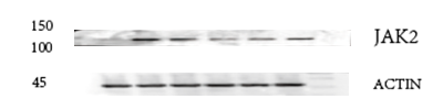
| Sample | rat colon tissue |
| Sample Processing Description | RIPA lysis buffer with protease inhibitor PMSF (100:1) was used to lyse the sample for 10 minutes, followed by centrifugation at 12,000 rpm for 15 minutes. The supernatant was mixed with 5× loading buffer, denatured at 100°C for 10 minutes, and then loaded onto SDS-PAGE. |
| Other Reagents | Blocking buffer |
| Primary Antibody | JAK2 Monoclonal Antibody |
| Primary Incubation | 1:1000, overnight at 4 ℃ |
| Secondary Antibody | HRP Conjugated AffiniPure Goat Anti-Rabbit IgG (H+L) |
| Secondary Incubation | 1:5000, 1 hour in room temperature |
| Detection | Substrate: ECL, Imaging system:ChemiDoc MP |
| Results Summary | The figure shows the Western blot results of the target protein JAK2 and the internal control Actin in rat colon tissue across the following groups: normal, disease model, low/middle/high dose traditional Chinese medicine treatment, and Western medicine treatment. The target bands are clear and well defined, and the experimental results are satisfactory. |
Shiyu Zhang, LUTCM
Verified customer
Submitted 2026-01-06
We promise Anti-JAK2 Monoclonal Antibody performs excellently in relevant applications for IP, IF, IHC, ICC, WB. If it doesn't perform as promised, we will refund the product. Learn more


We promise all of our products perform as described in datasheets.
This product is for Research Use Only. Not for use in diagnostic procedures.