Click image to see more details
-

-
-
-
-
+2
Product Info Summary
| SKU: | PA1642 |
|---|---|
| Size: | 100 μg/vial |
| Reactive Species: | Human |
| Host: | Rabbit |
| Application: | Flow Cytometry, IF, IHC, ICC, WB |
Customers Who Bought This Also Bought

Product info
Product Name
Anti-Ku70/XRCC6 Antibody Picoband®
SKU/Catalog Number
PA1642
BA1810 is an alternative SKU for this antibody, used in previous lots.
Size
100 μg/vial
Form
Lyophilized
Description
Boster Bio Anti-Ku70/XRCC6 Antibody catalog # PA1642. Tested in Flow Cytometry, IF, IHC, ICC, WB applications. This antibody reacts with Human. The brand Picoband indicates this is a premium antibody that guarantees superior quality, high affinity, and strong signals with minimal background in Western blot applications. Only our best-performing antibodies are designated as Picoband, ensuring unmatched performance.
Storage & Handling
Store at -20˚C for one year from date of receipt. After reconstitution, at 4˚C for one month. It can also be aliquotted and stored frozen at -20˚C for six months. Avoid repeated freeze-thaw cycles.
Cite This Product
Anti-Ku70/XRCC6 Antibody Picoband® (Boster Biological Technology, Pleasanton CA, USA, Catalog # PA1642)
Host
Rabbit
Contents
Each vial contains antibody formulated with stabilizing components, 0.9mg NaCl, 0.2mg Na2HPO4, 0.05mg Thimerosal, 0.05mg NaN3.
*This antibody is supplied in a stabilized formulation.
Compatibility with conjugation reactions depends on the chemistry of the conjugation method used.
For conjugation methods that are not compatible with the stabilizing components present in this formulation, a carrier-free antibody format is required.
Clonality
Polyclonal
Isotype
Rabbit IgG
Immunogen
A synthetic peptide corresponding to a sequence at the C-terminus of human Ku70.
Cross-reactivity
No cross-reactivity with other proteins
Reactive Species
PA1642 is reactive to XRCC6 in Human
Observed Molecular Weight
70 kDa
Calculated molecular weight
69.8 kDa
Background of XRCC6
XRCC6 (X-Ray Repair, Complementing Defective, In Chinese Hamster, 6), also called Ku70, G22P1 or TLAA, is a protein that in humans, is encoded by the XRCC6 gene. In addition, the XRCC6 gene encodes subunit p70 of the p70/p80 autoantigen which consists of 2 proteins of molecular mass of approximately 70,000 and 80,000 daltons that dimerize to form a 10 S DNA-binding complex. The XRCC6 gene is mapped to 22q13.2. XRCC6 and Mre11 are differentially expressed during meiosis. XRCC6 interacts with Baxa, a mediator of mitochondrial-dependent apoptosis. Disruption of both FANCC and XRCC6 suppressed sensitivity to crosslinking agents, diminished chromosome breaks, and reversed defective homologous recombination. Ku70 binds directly to free DNA ends, committing them to NHEJ repair. In early meiotic prophase, however, when meiotic recombination is most probably initiated, Mre11 was abundant, whereas XRCC6 was not detectable.
Antibody Validation
Boster validates all antibodies on WB, IHC, ICC, Immunofluorescence, and ELISA with known positive control and negative samples to ensure specificity and high affinity, including thorough antibody incubations.
Application & Images
Applications
PA1642 is guaranteed for Flow Cytometry, IF, IHC, ICC, WB Boster Guarantee
Recommend Dilution
| Application | Dilution | Species |
|---|---|---|
| Western blot | 0.1-0.5μg/ml | Human |
| Immunohistochemistry (Paraffin-embedded Section) | 0.5-1μg/ml | Human |
| Immunocytochemistry | 0.5-1μg/ml | Human, - |
| Immunocytochemistry/Immunofluorescence | 5μg/ml | Human |
| Flow Cytometry (Fixed) | 1-3μg/1x106 cells | Human |
Tested application
Suggested blocking solution with 5% non-fat milk or BSA; (*)Recommended protein loading: 20-40 µg per lane
Use TE buffer pH 9.0 for antigen retrieval; (*) citrate buffer pH 6.0 is an alternative.
Validation Images & Assay Conditions
Click image to see more details
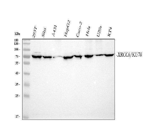
Western blot analysis of XRCC6 using anti-XRCC6 antibody (PA1642).
Electrophoresis was performed on a 5-20% SDS-PAGE gel at 70V (Stacking gel) / 90V (Resolving gel) for 2-3 hours. The sample well of each lane was loaded with 30 ug of sample under reducing conditions.
Lane 1: human 293T whole cell lysates,
Lane 2: human SiHa whole cell lysates,
Lane 3: human A431 whole cell lysates,
Lane 4: human HepG2 whole cell lysates,
Lane 5: human CACO-2 whole cell lysates,
Lane 6: human Hela whole cell lysates,
Lane 7: human U2OS whole cell lysates,
Lane 8: human RT4 whole cell lysates.
After electrophoresis, proteins were transferred to a nitrocellulose membrane at 150 mA for 50-90 minutes. Blocked the membrane with 5% non-fat milk/TBS for 1.5 hour at RT. The membrane was incubated with rabbit anti-XRCC6 antigen affinity purified polyclonal antibody (Catalog # PA1642) at 0.5 μg/mL overnight at 4°C, then washed with TBS-0.1%Tween 3 times with 5 minutes each and probed with a goat anti-rabbit IgG-HRP secondary antibody at a dilution of 1:5000 for 1.5 hour at RT. The signal is developed using an Enhanced Chemiluminescent detection (ECL) kit (Catalog # EK1002) with Tanon 5200 system. A specific band was detected for XRCC6 at approximately 70 kDa. The expected band size for XRCC6 is at 70 kDa.

Click image to see more details
IHC analysis of XRCC6 using anti-XRCC6 antibody (PA1642).
XRCC6 was detected in a paraffin-embedded section of Human Placenta tissue. Heat mediated antigen retrieval was performed in EDTA buffer (pH 8.0, epitope retrieval solution). The tissue section was blocked with 10% goat serum. The tissue section was then incubated with 1 μg/ml rabbit anti-XRCC6 Antibody (PA1642) overnight at 4°C. Peroxidase Conjugated Goat Anti-rabbit IgG was used as secondary antibody and incubated for 30 minutes at 37°C. The tissue section was developed using HRP Conjugated Rabbit IgG Super Vision Assay Kit (Catalog # SV0002) with DAB as the chromogen.

Click image to see more details
ICC analysis of XRCC6 using anti-XRCC6 antibody (PA1642).
XRCC6 was detected in an immunocytochemical section of A549 cells. Enzyme antigen retrieval was performed using IHC enzyme antigen retrieval reagent (AR0022) for 15 mins. The cells were blocked with 10% goat serum. And then incubated with 1 μg/ml rabbit anti-XRCC6 Antibody (PA1642) overnight at 4°C. Biotinylated goat anti-rabbit IgG was used as secondary antibody and incubated for 30 minutes at 37°C. The section was developed using Strepavidin-Biotin-Complex (SABC)(Catalog # SA1022) with DAB as the chromogen.

Click image to see more details
ICC analysis of XRCC6 using anti-XRCC6 antibody (PA1642).
XRCC6 was detected in an immunocytochemical section of Hela cells. Enzyme antigen retrieval was performed using IHC enzyme antigen retrieval reagent (AR0022) for 15 mins. The cells were blocked with 10% goat serum. And then incubated with 1 μg/ml rabbit anti-XRCC6 Antibody (PA1642) overnight at 4°C. Biotinylated goat anti-rabbit IgG was used as secondary antibody and incubated for 30 minutes at 37°C. The section was developed using Strepavidin-Biotin-Complex (SABC)(Catalog # SA1022) with DAB as the chromogen.

Click image to see more details
IF analysis of XRCC6 using anti-XRCC6 antibody (PA1642).
XRCC6 was detected in immunocytochemical section of A549 cells. Enzyme antigen retrieval was performed using IHC enzyme antigen retrieval reagent (AR0022) for 15 mins. The cells were blocked with 10% goat serum. And then incubated with 5μg/mL rabbit anti-XRCC6 Antibody (PA1642) overnight at 4°C. DyLight®488 Conjugated Goat Anti-Rabbit IgG (BA1127) was used as secondary antibody at 1:100 dilution and incubated for 30 minutes at 37°C. The section was counterstained with DAPI. Visualize using a fluorescence microscope and filter sets appropriate for the label used.

Click image to see more details
Flow Cytometry analysis of MCF-7 cells using anti-XRCC6 antibody (PA1642).
Overlay histogram showing MCF-7 cells stained with PA1642 (Blue line). To facilitate intracellular staining, cells were fixed with 4% paraformaldehyde and permeabilized with permeabilization buffer. The cells were blocked with 10% normal goat serum. And then incubated with rabbit anti-XRCC6 Antibody (PA1642, 1μg/1x106 cells) for 30 min at 20°C. DyLight®488 conjugated goat anti-rabbit IgG (BA1127, 5-10μg/1x106 cells) was used as secondary antibody for 30 minutes at 20°C. Isotype control antibody (Green line) was rabbit IgG (1μg/1x106) used under the same conditions. Unlabelled sample without incubation with primary antibody and secondary antibody (Red line) was used as a blank control.
Specific Publications For Anti-Ku70/XRCC6 Antibody Picoband® (PA1642)
Loading publications
Recommended Resources
Here are featured tools and databases that you might find useful.
- Boster's Pathways Library
- Protein Databases
- Bioscience Research Protocol Resources
- Data Processing & Analysis Software
- Photo Editing Software
- Scientific Literature Resources
- Research Paper Management Tools
- Molecular Biology Software
- Primer Design Tools
- Bioinformatics Tools
- Phylogenetic Tree Analysis
Customer Reviews
Have you used Anti-Ku70/XRCC6 Antibody Picoband®?
Share your experimental results or join a short interview to earn up to $1,000 in product credits or other rewards.
0 Reviews For Anti-Ku70/XRCC6 Antibody Picoband®
Customer Q&As
Have a question?
Find answers in Q&As, reviews.
Can't find your answer?
Submit your question
