Click image to see more details
Product Info Summary
| SKU: | PB9658 |
|---|---|
| Size: | 100 μg/vial |
| Reactive Species: | Mouse |
| Host: | Rabbit |
| Application: | ELISA, WB |
Customers Who Bought This Also Bought


Product info
Product Name
Anti-Leptin Antibody Picoband™
SKU/Catalog Number
PB9658
Size
100 μg/vial
Form
Lyophilized
Description
Boster Bio Anti-Leptin Antibody Picoband™ catalog # PB9658. Tested in ELISA, WB applications. This antibody reacts with Mouse.
Storage & Handling
Store at -20˚C for one year from date of receipt. After reconstitution, at 4˚C for one month. It can also be aliquotted and stored frozen at -20˚C for six months. Avoid repeated freeze-thaw cycles.
Cite This Product
Anti-Leptin Antibody Picoband™ (Boster Biological Technology, Pleasanton CA, USA, Catalog # PB9658)
Host
Rabbit
Contents
Each vial contains 5mg BSA, 0.9mg NaCl, 0.2mg Na2HPO4, 0.05mg NaN3.
Clonality
Polyclonal
Isotype
Rabbit IgG
Immunogen
A synthetic peptide corresponding to a sequence in the middle region of mouse Leptin, different from the related human sequence by five amino acids, and from the related rat sequence by two amino acids.
*Blocking peptide can be purchased. Costs vary based on immunogen length. Contact us for pricing.
Cross-reactivity
No cross-reactivity with other proteins
Reactive Species
PB9658 is reactive to LEP in Mouse
Applications
PB9658 is guaranteed for ELISA, WB Boster Guarantee
Observed Molecular Weight
16 kDa
Calculated molecular weight
18.641kDa
Background of Leptin/OB
Leptin is a protein product of the mouse obese gene. Mice with mutations in the obese gene that block the synthesis of Leptin have been found to be obese and diabetic and to have reduced activity, metabolism and body temperature. cDNA clones encoding Leptin have been isolated from human, simian, mouse, and rat cells. The expression of Leptin mRNA has been shown to be restricted to adipose tissue. Although regulation of fat stores is deemed to be the primary function of leptin, it also plays a role in other physiological processes, as evidenced by its multiple sites of synthesis other than fat cells, and the multiple cell types beside hypothalamic cells that have leptin receptors. Many of these additional functions are yet to be defined.
Antibody Validation
Boster validates all antibodies on WB, IHC, ICC, Immunofluorescence, and ELISA with known positive control and negative samples to ensure specificity and high affinity, including thorough antibody incubations.
Innovating Scientists Reward
If you are the first to review this product, or if you have results for a special sample, species or application this product is not validated in, share your results with us and receive product credits you can use towards any Boster products! Applicable to all scientists worldwide.
Submit A Review
Assay dilution & Images
Reconsitution
Add 0.2ml of distilled water will yield a concentration of 500ug/ml.
Assay Dilutions Recommendation
The recommendations below provide a starting point for assay optimization. The actual working concentration varies and should be decided by the user.
ELISA , 0.1-0.5μg/ml, Mouse, -
Western blot, 0.1-0.5μg/ml, Mouse
Validation Images & Assay Conditions
Click image to see more details
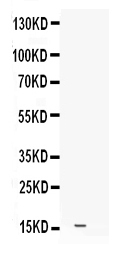
Figure 1. Western blot analysis of Leptin using anti-Leptin antibody (PB9658).
Electrophoresis was performed on a 5-20% SDS-PAGE gel at 70V (Stacking gel) / 90V (Resolving gel) for 2-3 hours.
Lane 1: NIH3T3 Whole Cell Lysate at 40ug.
After electrophoresis, proteins were transferred to a nitrocellulose membrane at 150 mA for 50-90 minutes. Blocked the membrane with 5% non-fat milk/TBS for 1.5 hour at RT. The membrane was incubated with rabbit anti-Leptin antigen affinity purified polyclonal antibody (Catalog # PB9658) at 0.5 μg/mL overnight at 4°C, then washed with TBS-0.1%Tween 3 times with 5 minutes each and probed with a goat anti-rabbit IgG-HRP secondary antibody at a dilution of 1:5000 for 1.5 hour at RT. The signal is developed using an Enhanced Chemiluminescent detection (ECL) kit (Catalog # EK1002) with Tanon 5200 system. A specific band was detected for Leptin at approximately 16 kDa. The expected band size for Leptin is at 16 kDa.
Protein Target Info & Infographic
Gene/Protein Information For LEP (Source: Uniprot.org, NCBI)
Gene Name
LEP
Full Name
Leptin
Weight
18.641kDa
Superfamily
leptin family
Alternative Names
FLJ94114; LEP; leptin (murine obesity homolog); leptin (obesity homolog, mouse); Leptin; OB; Obese protein; obese, mouse, homolog of; Obesity factor; OBOBS LEP LEPD, OB, OBS leptin leptin|leptin (murine obesity homolog)|leptin (obesity homolog, mouse)|obese protein|obese, mouse, homolog of|obesity factor
*If product is indicated to react with multiple species, protein info is based on the gene entry specified above in "Species".For more info on LEP, check out the LEP Infographic

We have 30,000+ of these available, one for each gene! Check them out.
In this infographic, you will see the following information for LEP: database IDs, superfamily, protein function, synonyms, molecular weight, chromosomal locations, tissues of expression, subcellular locations, post-translational modifications, and related diseases, research areas & pathways. If you want to see more information included, or would like to contribute to it and be acknowledged, please contact [email protected].
Specific Publications For Anti-Leptin Antibody Picoband™ (PB9658)
Hello CJ!
PB9658 has been cited in 4 publications:
*The publications in this section are manually curated by our staff scientists. They may differ from Bioz's machine gathered results. Both are accurate. If you find a publication citing this product but is missing from this list, please let us know we will issue you a thank-you coupon.
lncRNA Ftx promotes aerobic glycolysis and tumor progression through the PPAR? pathway in hepatocellular carcinoma
Cheng Ys, Dai Dz, Dai Y. Reprod Toxicol. 2010 Jul;29(4):421-6. Doi: 10.1016/J.Reprotox.2010.03.001. Epub 2010 Mar 20. Testis Dysfunction By Isoproterenol Is Mediated By Upregulating Endothelin Receptor A, Leptin And Protein Kinase Cvarepsilon And ...
Yuan Y, Zhang J, Cai L, Ding C, Wang X, Chen H, Wang X, Yan J, Lu J. Oncol Rep. 2013 Jun;29(6):2291-6. Doi: 10.3892/Or.2013.2390. Epub 2013 Apr 8. Leptin Induces Cell Proliferation And Reduces Cell Apoptosis By Activating C-Myc In Cervical Cancer.
Qin Jh, Ma Jz, Yang Xw, Hu Yj, Zhou J, Fu Lc, Tian Rh, Liu S, Xu G, Shen Xl. Nat Prod Bioprospect. 2015 Jun;5(3):159-66. Doi: 10.1007/S13659-015-0063-5. Epub 2015 Jun 16. A Triterpenoid Inhibited Hormone-Induced Adipocyte Differentiation And Allev...
Recommended Resources
Here are featured tools and databases that you might find useful.
- Boster's Pathways Library
- Protein Databases
- Bioscience Research Protocol Resources
- Data Processing & Analysis Software
- Photo Editing Software
- Scientific Literature Resources
- Research Paper Management Tools
- Molecular Biology Software
- Primer Design Tools
- Bioinformatics Tools
- Phylogenetic Tree Analysis
Customer Reviews
Have you used Anti-Leptin Antibody Picoband™?
Submit a review and receive an Amazon gift card.
- $30 for a review with an image
Be the first to review Anti-Leptin Antibody Picoband™
*The first user to submit a review for a product is eligible for Boster's Innovating Scientists Reward, which gives product credits. This is in addition to the gift card reward.
Customer Q&As
Have a question?
Find answers in Q&As, reviews.
Can't find your answer?
Submit your question
5 Customer Q&As for Anti-Leptin Antibody Picoband™
Question
We are currently using anti-Leptin antibody PB9658 for mouse tissue, and we are well pleased with the WB results. The species of reactivity given in the datasheet says mouse. Is it likely that the antibody can work on feline tissues as well?
Verified Customer
Verified customer
Asked: 2019-12-06
Answer
The anti-Leptin antibody (PB9658) has not been tested for cross reactivity specifically with feline tissues, but there is a good chance of cross reactivity. We have an innovator award program that if you test this antibody and show it works in feline you can get your next antibody for free. Please contact me if I can help you with anything.
Boster Scientific Support
Answered: 2019-12-06
Question
We were well pleased with the WB result of your anti-Leptin antibody. However we have been able to see positive staining in adipose tissue of abdominal region secreted. using this antibody. Is that expected? Could you tell me where is LEP supposed to be expressed?
Verified Customer
Verified customer
Asked: 2019-10-30
Answer
From literature, adipose tissue of abdominal region does express LEP. Generally LEP expresses in secreted. Regarding which tissues have LEP expression, here are a few articles citing expression in various tissues:
Placenta, Pubmed ID: 15489334
Boster Scientific Support
Answered: 2019-10-30
Question
We bought anti-Leptin antibody for WB on placenta in a previous project. I am using mouse, and I plan to use the antibody for ELISA next. I am looking for examining placenta as well as adipose tissue of abdominal region in our next experiment. Could you please give me some suggestion on which antibody would work the best for ELISA?
Verified Customer
Verified customer
Asked: 2019-09-23
Answer
I took a look at the website and datasheets of our anti-Leptin antibody and it appears that PB9658 has been tested on mouse in both WB and ELISA. Thus PB9658 should work for your application. Our Boster satisfaction guarantee will cover this product for ELISA in mouse even if the specific tissue type has not been validated. We do have a comprehensive range of products for ELISA detection and you can check out our website bosterbio.com to find out more information about them.
Boster Scientific Support
Answered: 2019-09-23
Question
We have observed staining in mouse adipose tissue of abdominal region. What should we do? Is anti-Leptin antibody supposed to stain adipose tissue of abdominal region positively?
Verified Customer
Verified customer
Asked: 2019-09-10
Answer
According to literature adipose tissue of abdominal region does express LEP. According to Uniprot.org, LEP is expressed in adipose tissue of abdominal region, placenta, among other tissues. Regarding which tissues have LEP expression, here are a few articles citing expression in various tissues:
Placenta, Pubmed ID: 15489334
Boster Scientific Support
Answered: 2019-09-10
Question
I would like using your anti-Leptin antibody for negative regulation of cartilage development studies. Has this antibody been tested with western blotting on nih3t3 whole cell lysate? We would like to see some validation images before ordering.
Verified Customer
Verified customer
Asked: 2018-06-20
Answer
We appreciate your inquiry. This PB9658 anti-Leptin antibody is validated on nih3t3 whole cell lysate. It is guaranteed to work for ELISA, WB in mouse. Our Boster guarantee will cover your intended experiment even if the sample type has not been be directly tested.
Boster Scientific Support
Answered: 2018-06-20
