Click image to see more details
Product Info Summary
| SKU: | PA1923 |
|---|---|
| Size: | 100 μg/vial |
| Reactive Species: | Human, Mouse, Rat |
| Host: | Rabbit |
| Application: | Flow Cytometry, IHC, WB |
Customers Who Bought This Also Bought

Product info
Product Name
Anti-MEKK1/MAP3K1 Antibody Picoband®
SKU/Catalog Number
PA1923
BA1252-1 is an alternative SKU for this antibody, used in previous lots.
Size
100 μg/vial
Form
Lyophilized
Description
Boster Bio Anti-MEKK1/MAP3K1 Antibody catalog # PA1923. Tested in Flow Cytometry, IHC, WB applications. This antibody reacts with Human, Mouse, Rat. The brand Picoband indicates this is a premium antibody that guarantees superior quality, high affinity, and strong signals with minimal background in Western blot applications. Only our best-performing antibodies are designated as Picoband, ensuring unmatched performance.
Storage & Handling
Store at -20˚C for one year from date of receipt. After reconstitution, at 4˚C for one month. It can also be aliquotted and stored frozen at -20˚C for six months. Avoid repeated freeze-thaw cycles.
Cite This Product
Anti-MEKK1/MAP3K1 Antibody Picoband® (Boster Biological Technology, Pleasanton CA, USA, Catalog # PA1923)
Host
Rabbit
Contents
Each vial contains 4 mg Trehalose, 0.9 mg NaCl and 0.2 mg Na2HPO4.
Clonality
Polyclonal
Isotype
Rabbit IgG
Immunogen
A synthetic peptide corresponding to a sequence at the C-terminus of human MEKK1, identical to the related rat and mouse sequences.
Cross-reactivity
No cross-reactivity with other proteins
Reactive Species
PA1923 is reactive to MAP3K1 in Human, Mouse, Rat
Observed Molecular Weight
164 kDa
Calculated molecular weight
164.5 kDa
Background of MAP3K1
MAP3K1 (Mitogen-activated protein kinase kinase kinase 1), also known as MEKK1, MAPKKK1, MEK KINASE or MAP/ERK KINASE KINASE 1, is an enzyme that in humans is encoded by the MAP3K1 gene. Vinik et al. (1995) identified DNA sequence and size polymorphisms in intronic and 3-prime untranslated regions of the mouse Map3k1 gene and the human MAP3K1 homolog. Using these allele-specific polymorphisms, they mapped the Map3k1 gene in an intersubspecific backcross to mouse chromosome 13. They mapped the human MAP3K1 gene to chromosome 5 by somatic cell hybrid analysis. By assaying transfected COS-1 cells, Xia et al. (1998) showed that human MEKK1 activated JNK1 (MAPK8) robustly and p38-alpha (MAPK14) less efficiently, but it had only a marginal effect on ERK2 (MAPK1). MEKK1 directly and specifically interacted with JNKK1 (MAP2K4) and activated JNKK1 in cells and in vitro. Phosphorylation of JNKK1 by MEKK1 disrupted their interaction. MEKK1 and JNK1 competed for binding to JNKK1. Xia et al. (1998) concluded that JNKK1 is the preferred MEKK1 substrate.
Antibody Validation
Boster validates all antibodies on WB, IHC, ICC, Immunofluorescence, and ELISA with known positive control and negative samples to ensure specificity and high affinity, including thorough antibody incubations.
Application & Images
Applications
PA1923 is guaranteed for Flow Cytometry, IHC, WB Boster Guarantee
Recommend Dilution
| Application | Dilution | Species |
|---|---|---|
| Western blot | 0.1-0.5μg/ml | Human, Mouse, Rat |
| Immunohistochemistry (Paraffin-embedded Section) | 2-5μg/ml | Human |
| Flow Cytometry(Fixed) | 1-3 μg/1x106 cells | Human |
Tested application
Suggested blocking solution with 5% non-fat milk or BSA; (*)Recommended protein loading: 20-40 µg per lane
Use TE buffer pH 9.0 for antigen retrieval; (*) citrate buffer pH 6.0 is an alternative.
Validation Images & Assay Conditions
Click image to see more details
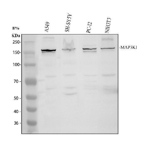
Western blot analysis of MAP3K1 using anti-MAP3K1 antibody (PA1923).
Electrophoresis was performed on a 8% SDS-PAGE gel at 80V (Stacking gel) / 120V (Resolving gel) for 2 hours. The sample well of each lane was loaded with 30 ug of sample under reducing conditions.
Lane 1: human A549 whole cell lysates,
Lane 2: human SH-SY5Y whole cell lysates,
Lane 3: rat PC-12 whole cell lysates,
Lane 4: mouse NIH/3T3 whole cell lysates.
After electrophoresis, proteins were transferred to a nitrocellulose membrane at 150 mA for 50-90 minutes. Blocked the membrane with 5% non-fat milk/TBS for 1.5 hour at RT. The membrane was incubated with rabbit anti-MAP3K1 antigen affinity purified polyclonal antibody (PA1923) at 0.5 μg/mL overnight at 4°C, then washed with TBS-0.1%Tween 3 times with 5 minutes each and probed with a goat anti-rabbit IgG-HRP secondary antibody (Catalog # BA1054) at a dilution of 1:5000 for 1.5 hour at RT. The signal is developed using an ECL Plus Western Blotting Substrate (Catalog # AR1196-200) with Tanon 5200 system. A specific band was detected for MAP3K1 at approximately 164 kDa. The expected band size for MAP3K1 is at 164 kDa.

Click image to see more details
IHC analysis of MAP3K1 using anti-MAP3K1 antibody (PA1923).
MAP3K1 was detected in a paraffin-embedded section of human breast cancer tissue. Heat mediated antigen retrieval was performed in EDTA buffer (pH 8.0, epitope retrieval solution). The tissue section was blocked with 10% goat serum. The tissue section was then incubated with 2 μg/ml rabbit anti-MAP3K1 Antibody (PA1923) overnight at 4°C. Peroxidase Conjugated Goat Anti-rabbit IgG was used as secondary antibody and incubated for 30 minutes at 37°C. The tissue section was developed using HRP Conjugated Rabbit IgG Super Vision Assay Kit (Catalog # SV0002) with DAB as the chromogen.

Click image to see more details
Flow Cytometry analysis of SH-SY5Y cells using anti-MAP3K1 antibody (PA1923).
Overlay histogram showing SH-SY5Y cells stained with PA1923 (Blue line). The cells were fixed with 4% paraformaldehyde and blocked with 10% normal goat serum. And then incubated with rabbit anti-MAP3K1 Antibody (PA1923, 1 μg/1x106 cells) for 30 min at 20°C. DyLight®488 conjugated goat anti-rabbit IgG (BA1127, 5-10 μg/1x106 cells) was used as secondary antibody for 30 minutes at 20°C. Isotype control antibody (Green line) was rabbit IgG (1 μg/1x106) used under the same conditions. Unlabelled sample without incubation with primary antibody and secondary antibody (Red line) was used as a blank control.
Specific Publications For Anti-MEKK1/MAP3K1 Antibody Picoband® (PA1923)
Loading publications
Recommended Resources
Here are featured tools and databases that you might find useful.
- Boster's Pathways Library
- Protein Databases
- Bioscience Research Protocol Resources
- Data Processing & Analysis Software
- Photo Editing Software
- Scientific Literature Resources
- Research Paper Management Tools
- Molecular Biology Software
- Primer Design Tools
- Bioinformatics Tools
- Phylogenetic Tree Analysis
Customer Reviews
Have you used Anti-MEKK1/MAP3K1 Antibody Picoband®?
Share your experimental results or join a short interview to earn up to $1,000 in product credits or other rewards.
0 Reviews For Anti-MEKK1/MAP3K1 Antibody Picoband®
Customer Q&As
Have a question?
Find answers in Q&As, reviews.
Can't find your answer?
Submit your question
1 Customer Q&As for Anti-MEKK1/MAP3K1 Antibody Picoband®
Question
We are currently using anti-MEKK1/MAP3K1 antibody PA1923 for human tissue, and we are content with the IHC results. The species of reactivity given in the datasheet says human, mouse, rat. Is it true that the antibody can work on primate tissues as well?
Verified Customer
Verified customer
Asked: 2019-09-10
Answer
The anti-MEKK1/MAP3K1 antibody (PA1923) has not been validated for cross reactivity specifically with primate tissues, but there is a good chance of cross reactivity. We have an innovator award program that if you test this antibody and show it works in primate you can get your next antibody for free. Please contact me if I can help you with anything.
Boster Scientific Support
Answered: 2019-09-10
