
Click image to see more details
-

-
-
-
-
+11
Product Info Summary
| SKU: | A00109-1 |
|---|---|
| Size: | 0.1 mg |
| Reactive Species: | Human, Mouse, Rat |
| Host: | Rabbit |
| Application: | ELISA, Flow Cytometry, IF, IHC-P, WB |
Customers Who Bought This Also Bought

Product info
Product Name
Anti-PD-L1 CD274 Antibody
SKU/Catalog Number
A00109-1
Size
0.1 mg
Form
Liquid
Description
Boster Bio Anti-PD-L1 CD274 Antibody (Catalog # A00109-1). Tested in ELISA, WB, IHC-P, IF, Flow Cytometry applications. This antibody reacts with Human, Mouse, Rat.
Storage & Handling
PD-L1 antibody can be stored at 4°C for three months and -20°C, stable for up to one year. Avoid repeated freeze-thaw cycles. Antibodies should not be exposed to prolonged high temperatures.
Cite This Product
Anti-PD-L1 CD274 Antibody (Boster Biological Technology, Pleasanton CA, USA, Catalog # A00109-1)
Host
Rabbit
Contents
PD-L1 Antibody is supplied in PBS containing 0.02% sodium azide.
Clonality
Polyclonal
Isotype
IgG
Immunogen
Anti-PD-L1 antibody was raised against a peptide corresponding to 17 amino acids near the center of human PD-L1 isoform 1. The immunogen is located within amino acids 60 - 110 of PD-L1.
*Blocking peptide can be purchased. Costs vary based on immunogen length. Contact us for pricing.
Cross-reactivity
PD-L1 antibody has no cross-reactivity to PD-L2.
Reactive Species
A00109-1 is reactive to CD274 in Human, Mouse, Rat
Applications
A00109-1 is guaranteed for ELISA, Flow Cytometry, IF, IHC-P, WB Boster Guarantee
Observed Molecular Weight
68 kDa
Calculated molecular weight
33.275kDa
Background of PD-L1
PD-L1 plays a critical role in induction and maintenance of immune tolerance to self. As a ligand for the inhibitory receptor PDCD1/CD279, PD-L1 modulates the activation threshold of T-cells and limits T-cell effector response (1). The PDCD1/CD279-mediated inhibitory pathway is exploited by tumors to attenuate anti-tumor immunity and facilitate tumor survival (2,3). Through a yet unknown activating receptor, it may costimulate T-cell subsets that predominantly produce interleukin-10 (IL10) (4).
Antibody Validation
Boster validates all antibodies on WB, IHC, ICC, Immunofluorescence, and ELISA with known positive control and negative samples to ensure specificity and high affinity, including thorough antibody incubations.
Innovating Scientists Reward
If you are the first to review this product, or if you have results for a special sample, species or application this product is not validated in, share your results with us and receive product credits you can use towards any Boster products! Applicable to all scientists worldwide.
Submit A Review
Assay dilution & Images
Assay Dilutions Recommendation
The recommendations below provide a starting point for assay optimization. The actual working concentration varies and should be decided by the user.
WB: 1-2 μg/mL; IHC-P: 2.5-5 μg/mL; IF: 20 μg/mL; Flow Cyt: 0.5 μg/mL.
Antibody validated: Western Blot in human and mouse samples; Immunohistochemistry in human and rat samples; Immunofluorescence in human and rat samples and Flow Cytometry in mouse samples. All other applications and species not yet tested. Optimal dilutions for each application should be determined by the researcher.
Validation Images & Assay Conditions
Click image to see more details
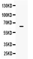
Western Blot Validation of PD-L1 in HeLa Cells
Loading: 15 μg of lysates per lane.Antibodies: A00109-1 (A: 1 μg/mL, B: 2 μg/mL), 1 h incubation at RT in 5% NFDM/TBST.Secondary: Goat anti-rabbit IgG HRP conjugate at 1:10000 dilution.

Click image to see more details
Independent Antibody Validation (IAV) via Protein Expression Profile in Human and Mouse cell lines
Loading: 15 μg of lysates per lane.Antibodies: A00109-1 (2 μg/mL), (2 μg/mL), and beta-actin (1 μg/mL), 1 h incubation at RT in 5% NFDM/TBST.Secondary: Goat anti-rabbit and or anti-mouse IgG HRP conjugate at 1:10000 and 1:5000 dilution, respectively.

Click image to see more details
Validation with PD-L1 siRNA Knockdown in HeLa Cells
HeLa cells were transfected with control siRNAs (lane 1) or PD-L1 siRNAs (lane 2) Loading: 10 μg of HeLa whole cell lysates per lane. Antibodies: (2 μg/mL) and GAPDH (0.02 μg/mL), 1 h incubation at RT in 5% NFDM/TBST. Secondary: Goat anti-mouse IgG HRP conjugate at 1:5000 dilution.

Click image to see more details
Validation with PD-L1 overexpression in 293 cells
Loading: Lysates/proteins at 15 μg per lane.Lane 1: non-transfected 293 cellsLane 2: PD-L1 overexpressed 293 cellsAntibodies: A00109-1 (1 μg/mL). 1 h incubation at RT in 5% NFDM/TBST. Secondary: Goat anti-rabbit IgG HRP conjugate at 1:10000 dilution.

Click image to see more details
Immunohistochemistry Validation of PD-L1 in Human Tonsil Cells
Immunohistochemical analysis of paraffin-embedded human tonsil tissue using anti-PD-L1 antibody (A00109-1) at 5 μg/ml. Tissue was fixed with formaldehyde and blocked with 10% serum for 1 h at RT; antigen retrieval was by heat mediation with a citrate buffer (pH6). Samples were incubated with primary antibody overnight at 4˚C. A goat anti-rabbit IgG H&L (HRP) at 1/250 was used as secondary. Counter stained with Hematoxylin.

Click image to see more details
Immunofluorescence Validation of PD-L1 in Human Heart
Immunofluorescent analysis of 4% paraformaldehyde-fixed human heart tissue labeling PD-L1 with A00109-1 at 20 μg/mL, followed by goat anti-rabbit IgG secondary antibody at 1/500 dilution (red). Image showing both membrane and cytoplasmic staining on human heart tissue.

Click image to see more details
Immunofluorescence Validation of PD-L1 in Rat Heart
Immunofluorescence analysis of 4% paraformaldehyde-fixed rat heart tissue labeling PD-L1 with A00109-1 at 20 μg/ml, followed by goat anti-rabbit IgG secondary antibody at 1/250 dilution (red).

Click image to see more details
Immunohistochemistry Validation of PD-L1 in Human thyroid cancer (Angell et al., 2014)
Immunohistochemical analysis of PD-L1 expression in human thyroid cancer with anti-PD-L1 antibodeis (A00109-1). Placenta was used a positive control.

Click image to see more details
Immunohistochemistry Validation of PD-L1 in Human Lung Cancer (Ilie et al., 2015)
Surgical specimens (left panel) and matched biopsy specimens (right panel). PD-L1-positive (A,B) and PD-L1-negative (C,D) tumors.

Click image to see more details
Flow Cytometry Validation of PD-L1
Overlay histogram showing A-20 cells stained with A00109-1 (red line, 1μg/1x106 cells). 1 h incubation at 4˚C in 2% FBS/PBS. Followed by secondary antibody 488 goat anti-rabbit IgG (H+L) at 1/500 dilution for 1 h 4˚C. Isotype control antibody (Green line) was mouse IgG1 (1μg/1x106 cells) used under the same conditions. Acquisition of >10,000 events was performed.

Click image to see more details
Immunohistochemistry Validation of PD-L1 in Rat Heart
Immunohistochemical analysis of paraffin-embedded rat heart tissue using anti-PD-L1 antibody (A00109-1) at 5 μg/ml. Tissue was fixed with formaldehyde and blocked with 10% serum for 1 h at RT; antigen retrieval was by heat mediation with a citrate buffer (pH6). Samples were incubated with primary antibody overnight at 4˚C. A goat anti-rabbit IgG H&L (HRP) at 1/250 was used as secondary. Counter stained with Hematoxylin.

Click image to see more details
Immunohistochemistry Validation of PD-L1 in Human Heart
Immunohistochemical analysis of paraffin-embedded human heart tissue using anti-PD-L1 antibody (A00109-1) at 2.5 ˚g/ml. Tissue was fixed with formaldehyde and blocked with 10% serum for 1 h at RT; antigen retrieval was by heat mediation with a citrate buffer (pH6). Samples were incubated with primary antibody overnight at 4˚C. A goat anti-rabbit IgG H&L (HRP) at 1/250 was used as secondary. Counter stained with Hematoxylin.

Click image to see more details
Immunofluorescence Validation of PD-L1 in tumors in Human Cells (Dhar et al., 2018)
(A) Several antibody brands were first tested with RBCs, WBCs, and HeLa cells. A00109-1 was chosen as it provided the highest staining intensity
. (B, C) Using the optimal conditions of anti-PDL1 at a concentration of 50μg/mL, following by goat anti-rabbit Alexa Fluor 647, PDL-1 staining was tested on several lung cancer cell lines: A549 (adenocarcinoma), H1703 (adenocarcinoma), H3255 (squamous) and WBCs as a control. (D) Once validated, patient samples were stained for PD-L1, CK, CD45, DAPI.

Click image to see more details
Immunohistochemistry Validation of PD-L1 in Human Tumors (Gadiot et al., 2011)
Immunohistochemical analysis of patient tumors labeling PD-L1 with anti-PD-L1 antibodies (A00109-1). Several anti-PD-L1 antibodies were tested for staining, “Only 1 antibody gave no background staining and was competitively blocked by the addition of PD-L1Fc protein ”.

Click image to see more details
Immunohistochemistry Validation of PD-L1 in Human Lung adenocarcinoma (Heymann et al., 2017)
HE staining (left panel) and PD-L1 expression (right panel) in the tumor and pleural fluid for a patient with lung adenocarcinoma. PD-L1 expression detected by anti-PD-L1 antibodies (A00109-1) demonstrated membranous staining in approximately 80% of tumor cells (C) and in approximately 75% of tumor cells (D), respectively.
Protein Target Info & Infographic
Gene/Protein Information For CD274 (Source: Uniprot.org, NCBI)
Gene Name
CD274
Full Name
Programmed cell death 1 ligand 1
Weight
33.275kDa
Superfamily
immunoglobulin superfamily
Alternative Names
B7-H; B7H1; B7-H1; B7H1PDCD1L1; CD274 antigenMGC142294; CD274 molecule; CD274; PDCD1L1; PDCD1LG1; PDCD1LG1MGC142296; PDL1; PD-L1; PD-L1B7 homolog 1; PDL1PDCD1 ligand 1; programmed cell death 1 ligand 1; Programmed death ligand 1 CD274 B7-H, B7H1, PD-L1, PDCD1L1, PDCD1LG1, PDL1, hPD-L1 CD274 molecule programmed cell death 1 ligand 1|B7 homolog 1|CD274 antigen|PDCD1 ligand 1
*If product is indicated to react with multiple species, protein info is based on the gene entry specified above in "Species".For more info on CD274, check out the CD274 Infographic

We have 30,000+ of these available, one for each gene! Check them out.
In this infographic, you will see the following information for CD274: database IDs, superfamily, protein function, synonyms, molecular weight, chromosomal locations, tissues of expression, subcellular locations, post-translational modifications, and related diseases, research areas & pathways. If you want to see more information included, or would like to contribute to it and be acknowledged, please contact [email protected].
Specific Publications For Anti-PD-L1 CD274 Antibody (A00109-1)
Hello CJ!
No publications found for A00109-1
*Do you have publications using this product? Share with us and receive a reward. Ask us for more details.
Recommended Resources
Here are featured tools and databases that you might find useful.
- Boster's Pathways Library
- Protein Databases
- Bioscience Research Protocol Resources
- Data Processing & Analysis Software
- Photo Editing Software
- Scientific Literature Resources
- Research Paper Management Tools
- Molecular Biology Software
- Primer Design Tools
- Bioinformatics Tools
- Phylogenetic Tree Analysis
Customer Reviews
Have you used Anti-PD-L1 CD274 Antibody?
Submit a review and receive an Amazon gift card.
- $30 for a review with an image
Be the first to review Anti-PD-L1 CD274 Antibody
*The first user to submit a review for a product is eligible for Boster's Innovating Scientists Reward, which gives product credits. This is in addition to the gift card reward.
Customer Q&As
Have a question?
Find answers in Q&As, reviews.
Can't find your answer?
Submit your question
