
Click image to see more details
Product Info Summary
| SKU: | P02067 |
|---|---|
| Size: | 100μl |
| Reactive Species: | Human, Mouse, Rat |
| Host: | Rabbit |
| Application: | ELISA, WB |
Customers Who Bought This Also Bought

Product info
Product Name
Anti-Phospho-RPA p32 (S33) Antibody
SKU/Catalog Number
P02067
Size
100μl
Form
Liquid
Description
Boster Bio Anti-Phospho-RPA p32 (S33) Antibody catalog # P02067. Tested in ELISA, WB applications. This antibody reacts with Human, Mouse, Rat.
Storage & Handling
Store at -20°C for one year. For short term storage and frequent use, store at 4°C for up to one month. Avoid repeated freeze-thaw cycles.
Cite This Product
Anti-Phospho-RPA p32 (S33) Antibody (Boster Biological Technology, Pleasanton CA, USA, Catalog # P02067)
Host
Rabbit
Contents
Liquid in PBS containing 50% glycerol, 0.5% BSA and 0.02% sodium azide.
Clonality
Polyclonal
Clone Number
608
Isotype
IgG
Immunogen
Synthesized peptide derived from human RPA p32 around the phosphorylation site of S33.
*Blocking peptide can be purchased. Costs vary based on immunogen length. Contact us for pricing.
Cross-reactivity
No cross reactivity with other proteins.
Reactive Species
P02067 is reactive to RPA2 in Human, Mouse, Rat
Applications
P02067 is guaranteed for ELISA, WB Boster Guarantee
Observed Molecular Weight
52 kDa
Calculated molecular weight
29.247kDa
Background of RPA2
REDD1, Regulated in Development and DNA damage responses 1, is induced by hypoxia, cell stress, and apoptosis. Reduced REDD1 levels can sensitize cells towards apoptosis, where elevated levels of REDD1 induced by hypoxia can desensitize cells to apoptotic stimuli (Schwarzer et al, 2005). REDD1 has a crucial role in inhibiting mammalian rapamycin complex 1 (mTORC1) signaling during hypoxic stress (Katiyar et al, 2009). It has been shown that the rapid degradation of REDD1 is mediated by the CUL4A–DDB1–ROC1–b-TRCP E3 ligase complex and is regulated by REDD1 phosphorylation at Thr-25, Thr-23 and Ser-19 through the activity of GSK3b (Katiyar et al, 2009).
Antibody Validation
Boster validates all antibodies on WB, IHC, ICC, Immunofluorescence, and ELISA with known positive control and negative samples to ensure specificity and high affinity, including thorough antibody incubations.
Innovating Scientists Reward
If you are the first to review this product, or if you have results for a special sample, species or application this product is not validated in, share your results with us and receive product credits you can use towards any Boster products! Applicable to all scientists worldwide.
Submit A Review
Assay dilution & Images
Assay Dilutions Recommendation
The recommendations below provide a starting point for assay optimization. The actual working concentration varies and should be decided by the user.
WB, 1:500-1:2000
ELISA 1:10000
Validation Images & Assay Conditions
Click image to see more details
Western Blot (WB) analysis of MCF-7 cells using PhosphoRPA p32 (S33) Polyclonal antibody

Click image to see more details
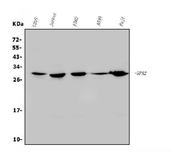
Western Blot (WB) analysis of specific cells using PhosphoRPA p32 (S33) Polyclonal antibody.

Click image to see more details
Western blot analysis of lysates from NIH/3T3 cells treated with Adriamycin 0.5 µg/ml 24h, using RFA2 (Phospho-Ser33) Antibody. The lane on the right is blocked with the phospho peptide.
Protein Target Info & Infographic
Gene/Protein Information For RPA2 (Source: Uniprot.org, NCBI)
Gene Name
RPA2
Full Name
Replication protein A 32 kDa subunit
Weight
29.247kDa
Superfamily
replication factor A protein 2 family
Alternative Names
REPA2; Replication factor A protein 2; replication protein A 32 kDa subunit; Replication protein A 34 kDa subunit; replication protein A2 (32kD); replication protein A2, 32kDa; RF-A protein 2; RP-A p32; RP-A p34; RPA2; RPA32; RPA34 RPA2 REPA2, RP-A p32, RP-A p34, RPA32 replication protein A2 replication protein A 32 kDa subunit|RF-A protein 2|replication factor A protein 2|replication protein A 34 kDa subunit
*If product is indicated to react with multiple species, protein info is based on the gene entry specified above in "Species".For more info on RPA2, check out the RPA2 Infographic

We have 30,000+ of these available, one for each gene! Check them out.
In this infographic, you will see the following information for RPA2: database IDs, superfamily, protein function, synonyms, molecular weight, chromosomal locations, tissues of expression, subcellular locations, post-translational modifications, and related diseases, research areas & pathways. If you want to see more information included, or would like to contribute to it and be acknowledged, please contact [email protected].
Specific Publications For Anti-Phospho-RPA p32 (S33) Antibody (P02067)
Hello CJ!
No publications found for P02067
*Do you have publications using this product? Share with us and receive a reward. Ask us for more details.
Recommended Resources
Here are featured tools and databases that you might find useful.
- Boster's Pathways Library
- Protein Databases
- Bioscience Research Protocol Resources
- Data Processing & Analysis Software
- Photo Editing Software
- Scientific Literature Resources
- Research Paper Management Tools
- Molecular Biology Software
- Primer Design Tools
- Bioinformatics Tools
- Phylogenetic Tree Analysis
Customer Reviews
Have you used Anti-Phospho-RPA p32 (S33) Antibody?
Submit a review and receive an Amazon gift card.
- $30 for a review with an image
Be the first to review Anti-Phospho-RPA p32 (S33) Antibody
*The first user to submit a review for a product is eligible for Boster's Innovating Scientists Reward, which gives product credits. This is in addition to the gift card reward.
Customer Q&As
Have a question?
Find answers in Q&As, reviews.
Can't find your answer?
Submit your question
