Click image to see more details
-

-
-
-
-
+3
Product Info Summary
| SKU: | M00105-1 |
|---|---|
| Size: | 100 μl |
| Reactive Species: | Human, Mouse, Rat |
| Host: | Rabbit |
| Application: | IF, IHC, ICC, WB |
Customers Who Bought This Also Bought


Product info
Product Name
Anti-SOX2 Rabbit Monoclonal Antibody
SKU/Catalog Number
M00105-1
Size
100 μl
Form
Liquid
Description
Boster Bio Anti-SOX2 Rabbit Monoclonal Antibody catalog # M00105-1. Tested in WB, IHC, ICC/IF applications. This antibody reacts with Human, Mouse, Rat.
Storage & Handling
Store at -20°C for one year. For short term storage and frequent use, store at 4°C for up to one month. Avoid repeated freeze-thaw cycles.
Cite This Product
Anti-SOX2 Rabbit Monoclonal Antibody (Boster Biological Technology, Pleasanton CA, USA, Catalog # M00105-1)
Host
Rabbit
Contents
Rabbit IgG in phosphate buffered saline, pH 7.4, 150mM NaCl, 0.02% sodium azide and 50% glycerol, 0.4-0.5mg/ml BSA.
Clonality
Monoclonal
Clone Number
BIO-19
Isotype
Rabbit IgG
Immunogen
A synthesized peptide derived from human SOX2
*Blocking peptide can be purchased. Costs vary based on immunogen length. Contact us for pricing.
Reactive Species
M00105-1 is reactive to SOX2 in Human, Mouse, Rat
Applications
M00105-1 is guaranteed for IF, IHC, ICC, WB Boster Guarantee
Observed Molecular Weight
34 kDa
Calculated molecular weight
34.31kDa
Background of SOX2
Phosphoinositide-3-kinase (PI3K) that phosphorylates PtdIns (Phosphatidylinositol), PtdIns4P (Phosphatidylinositol 4-phosphate) and PtdIns (4,5) P2 (Phosphatidylinositol 4,5-bisphosphate) to generate phosphatidylinositol 3,4,5-trisphosphate (PIP3) . PIP3 plays a key role by recruiting PH domain-containing proteins to the membrane, including AKT1 and PDPK1, activating signaling cascades involved in cell growth, survival, proliferation, motility and morphology. Participates in cellular signaling in response to various growth factors.
Antibody Validation
Boster validates all antibodies on WB, IHC, ICC, Immunofluorescence, and ELISA with known positive control and negative samples to ensure specificity and high affinity, including thorough antibody incubations.
Innovating Scientists Reward
If you are the first to review this product, or if you have results for a special sample, species or application this product is not validated in, share your results with us and receive product credits you can use towards any Boster products! Applicable to all scientists worldwide.
Submit A Review
Assay dilution & Images
Reconsitution
Restore with deionized water (or equivalent) for reconstitution volume of 1.0 mL
Assay Dilutions Recommendation
The recommendations below provide a starting point for assay optimization. The actual working concentration varies and should be decided by the user.
WB 1:1000-1:2000
IHC 1:50-1:200
ICC/IF 1:50-1:200
Validation Images & Assay Conditions
Click image to see more details
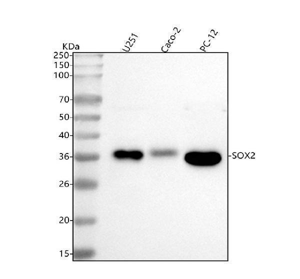
Figure 1. Western blot analysis of SOX2 using anti-SOX2 antibody (M00105-1).
Electrophoresis was performed on a 5-20% SDS-PAGE gel at 70V (Stacking gel) / 90V (Resolving gel) for 2-3 hours. The sample well of each lane was loaded with 30 ug of sample under reducing conditions.
Lane 1: human U251 whole cell lysates,
Lane 2: human CACO-2 whole cell lysates,
Lane 3: rat PC-12 whole cell lysates.
After electrophoresis, proteins were transferred to a nitrocellulose membrane at 150 mA for 50-90 minutes. Blocked the membrane with 5% non-fat milk/TBS for 1.5 hour at RT. The membrane was incubated with rabbit anti-SOX2 antigen affinity purified monoclonal antibody (Catalog # M00105-1) at 1:1000 overnight at 4°C, then washed with TBS-0.1%Tween 3 times with 5 minutes each and probed with a goat anti-rabbit IgG-HRP secondary antibody at a dilution of 1:500 for 1.5 hour at RT. The signal is developed using an Enhanced Chemiluminescent detection (ECL) kit (Catalog # EK1002) with Tanon 5200 system. A specific band was detected for SOX2 at approximately 34 kDa. The expected band size for SOX2 is at 34 kDa.

Click image to see more details
All lanes use the Antibody at 1:1K dilution for 1 hour at room temperature.

Click image to see more details
Immunohistochemical analysis of paraffin-embedded human cervix carcinoma, using SOX2 Antibody.

Click image to see more details
Immunofluorescent analysis of NCCIT cells, using SOX2 Antibody .

Click image to see more details
Immunofluorescent analysis using the Antibody at 1:50 dilution.

Click image to see more details
Immunofluorescent analysis using the Antibody at 1:50 dilution.

Click image to see more details
Immunofluorescent analysis using the Antibody at 1:500 dilution.
Protein Target Info & Infographic
Gene/Protein Information For SOX2 (Source: Uniprot.org, NCBI)
Gene Name
SOX2
Full Name
Transcription factor SOX-2
Weight
34.31kDa
Alternative Names
ANOP3; MCOPS3; MGC2413; SOX2; SRY (sex determining region Y)-box 2; SRY-related HMG-box gene 2; transcription factor SOX2; transcription factor SOX-2 SOX2 ANOP3, MCOPS3 SRY-box transcription factor 2 transcription factor SOX-2|SRY (sex determining region Y)-box 2|SRY-box 2|SRY-related HMG-box gene 2|sex determining region Y-box 2|transcription factor SOX2
*If product is indicated to react with multiple species, protein info is based on the gene entry specified above in "Species".For more info on SOX2, check out the SOX2 Infographic

We have 30,000+ of these available, one for each gene! Check them out.
In this infographic, you will see the following information for SOX2: database IDs, superfamily, protein function, synonyms, molecular weight, chromosomal locations, tissues of expression, subcellular locations, post-translational modifications, and related diseases, research areas & pathways. If you want to see more information included, or would like to contribute to it and be acknowledged, please contact [email protected].
Specific Publications For Anti-SOX2 Rabbit Monoclonal Antibody (M00105-1)
Hello CJ!
No publications found for M00105-1
*Do you have publications using this product? Share with us and receive a reward. Ask us for more details.
Recommended Resources
Here are featured tools and databases that you might find useful.
- Boster's Pathways Library
- Protein Databases
- Bioscience Research Protocol Resources
- Data Processing & Analysis Software
- Photo Editing Software
- Scientific Literature Resources
- Research Paper Management Tools
- Molecular Biology Software
- Primer Design Tools
- Bioinformatics Tools
- Phylogenetic Tree Analysis
Customer Reviews
Have you used Anti-SOX2 Rabbit Monoclonal Antibody?
Submit a review and receive an Amazon gift card.
- $30 for a review with an image
Be the first to review Anti-SOX2 Rabbit Monoclonal Antibody
*The first user to submit a review for a product is eligible for Boster's Innovating Scientists Reward, which gives product credits. This is in addition to the gift card reward.
Customer Q&As
Have a question?
Find answers in Q&As, reviews.
Can't find your answer?
Submit your question
4 Customer Q&As for Anti-SOX2 Rabbit Monoclonal Antibody
Question
We have seen staining in human fetal brain. Any tips? Is anti-SOX2 Rabbit Monoclonal antibody supposed to stain fetal brain positively?
Verified Customer
Verified customer
Asked: 2019-09-13
Answer
Based on literature fetal brain does express SOX2. Based on Uniprot.org, SOX2 is expressed in cerebral cortex, fetal brain, retina, lung, among other tissues. Regarding which tissues have SOX2 expression, here are a few articles citing expression in various tissues:
Fetal brain, Pubmed ID: 7849401
Lung, Pubmed ID: 15489334
Boster Scientific Support
Answered: 2019-09-13
Question
My boss were satisfied with the WB result of your anti-SOX2 Rabbit Monoclonal antibody. However we have seen positive staining in fetal brain nucleus. using this antibody. Is that expected? Could you tell me where is SOX2 supposed to be expressed?
Verified Customer
Verified customer
Asked: 2019-07-29
Answer
From what I have seen in literature, fetal brain does express SOX2. Generally SOX2 expresses in nucleus. Regarding which tissues have SOX2 expression, here are a few articles citing expression in various tissues:
Fetal brain, Pubmed ID: 7849401
Lung, Pubmed ID: 15489334
Boster Scientific Support
Answered: 2019-07-29
Question
We bought anti-SOX2 Rabbit Monoclonal antibody for IHC on retina in a previous experiment. I am using mouse, and We intend to use the antibody for IF next. you antibody examining retina as well as lung in our next experiment. Do you have any suggestion on which antibody would work the best for IF?
Verified Customer
Verified customer
Asked: 2019-07-22
Answer
I viewed the website and datasheets of our anti-SOX2 Rabbit Monoclonal antibody and it seems that M00105-1 has been tested on mouse in both IHC and IF. Thus M00105-1 should work for your application. Our Boster satisfaction guarantee will cover this product for IF in mouse even if the specific tissue type has not been validated. We do have a comprehensive range of products for IF detection and you can check out our website bosterbio.com to find out more information about them.
Boster Scientific Support
Answered: 2019-07-22
Question
We are currently using anti-SOX2 Rabbit Monoclonal antibody M00105-1 for human tissue, and we are happy with the IF results. The species of reactivity given in the datasheet says human, mouse. Is it true that the antibody can work on feline tissues as well?
Verified Customer
Verified customer
Asked: 2019-02-15
Answer
The anti-SOX2 Rabbit Monoclonal antibody (M00105-1) has not been validated for cross reactivity specifically with feline tissues, though there is a good chance of cross reactivity. We have an innovator award program that if you test this antibody and show it works in feline you can get your next antibody for free. Please contact me if I can help you with anything.
Boster Scientific Support
Answered: 2019-02-15
