This website uses cookies to ensure you get the best experience on our website.
- Table of Contents
ELISA (enzyme-linked immunosorbent assay) is a convenient and simple method to quantitatively or qualitatively detect peptides, proteins, antibodies, and hormones in samples, rendering it as one of the most widely used immunoassays. Many laboratories perform these assays using standardized elisa kits, which help streamline assay setup and improve reproducibility. To derive accurate concentrations from measured signals, most ELISA protocols rely on constructing an standard curve from known standards, which serves as the reference for sample interpolation. Despite the many advantages of conducting ELISA—or more complex formats like those used in Multiplex Assay Services and multiplex E...
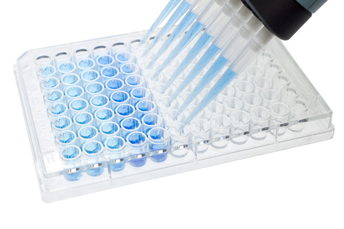
The ELISA (enzyme-linked immunosorbent assay) is recognized by scientists for its many advantages. The assay is convenient, quick, and simple to execute. ELISA’s versatility to detect peptides, proteins, antibodies, and hormones, and its ability to generate quantitative and qualitative data make it one of the most popular and powerful immunoassays available, with further efficiency supported by quick elisa kits in modern laboratory workflows.
In response to popular demand, numerous commercial ELISA kits are offered in the market, but not all ELISA kits are created equal.
How do we sift through the masses and choose a good ELISA kit?
Keep in mind the following points next time you go hunting for an ELISA kit.

First described in 1979, the technique of western blotting has since become one of the most commonly used analytical methods in life science research. Just last week, we received a few questions from confused researchers about weird band sizes in their western blot results:
Accurate western...

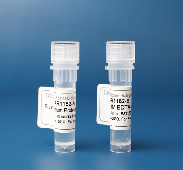
Protease inhibitor cocktails are chemical compounds that act to protect and maintain cellular protein composition after lysis of a cell, preventing natural degradation. They play an important role in protein quantification analysis, rendering the protease ineffective while obtaining appropriate protein purification yields. Protease inhibitors are classified by either the type of protease they act on or their mechanism of actions.
Proteases are enzymes that degrade proteins, playing an important role in cellular protein catabolism. Through their interaction with the proteins, they influence their activity and production of bioactive molecules, cellular repair, and the degradation of extracellular material. Proteases are also vital for food...
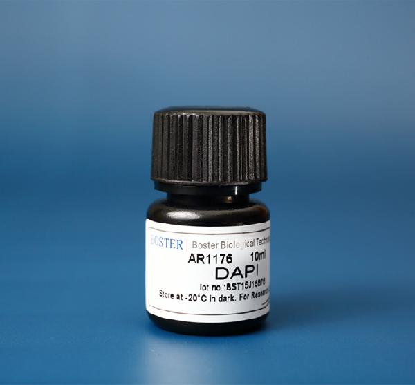
DAPI, or 4',6-Diamidino-2-Phenylindole, Dihydrochloride, is a commonly used fluorescent dye that binds to double-stranded DNA (dsDNA).
DAPI binds to and ‘stains’ double-stranded DNA, preferably binding to A-T-rich regions in DNA. DAPI stain is excited by ultraviolet (UV) light, with its largest excitation wavelength at ~360nm, and it produces a vibrant blue color with its largest emission wavelength at ~460nm when bound to DNA. Due to its fluorescent properties and rich blue color, it is readily used for visualization in fluorescence microscopy and other assays. Because it can pass through the cell membrane and stain DNA, DAPI is a useful dye for nuclear quantification and has been utilized in numerous assays, such as live or fixed cell staining, cell viability assays, flow cytometry, cell cycle analysis, mycoplasma contamination detection and in fluorescence microscopy. In fluorescence-based tissue analysis, especially where nuclear visualization guides diagnostic interpretation, accurate identification of morphological patterns is often supported by expert Pathology Review, complementing the use of nuclear dyes like DAPI. Because of its wide range of applications, Boster offers an affordable DAPI stain solution (Catalog# AR1176) that has been validated and cited in several publications.
Cell cycle analysis & flow cytometry
Since DAPI binds to DNA, it can be used to determine the relative amount of DNA in cells for cell cycle analysis. Cells currently in the G2 phase of mitosis will have twice the amount of DNA as cells in the G1 phase of mitosis, which will be reflected in the amount of fluorescence from DAPI in each cell. Apoptotic cells will have less DNA than a single cell, since DNA is being deg...
Proper fixation workflows support reliable antibody performance when experiments depend on monoclonal antibodies or Polyclonal antibodies.
Sample fixation is a required and crucial step for every successful IHC/ICC experiment. Appropriate fixation of samples provides the following benefits during the tissue preparation process. Without proper fixation, cellular structures can collapse, antigens may degrade, and staining results become unreliable. Reliable fixation helps achieve specific staining while minimizing background staining that can interfere with interpretation. Within standardized immunohistochemistry services, fixation parameters are carefully controlled to support consistent staining performance across diverse sample types. Controlled protocols also help preserve protein-protein interaction, enabling more accurate biological analysis.
Fixatives work by stabilizing proteins and other cellular components, either by creating cross-links or by precipitating molecules to maintain structural integrity. This chemical stabilization preserves the spatial arrangement of cells and tissues, allowing accurate localization of antigens and reliable analysis.
Maintaining structural integrity is especially important for studies evaluating protein-protein interaction within complex tissue environments.
Choosing which fixing solution to use depends on your sample type and antigen. Since there is no standard fixing solution for all samples, we recommend testing to determine which specific type of solution will be most appropriate and effective for antigen immobilization in your sample. Optimization often includes adjusting Incubation time to balance antigen preservation with antibody accessibility. This step is particularly important when preparing slides for a multiplex IHC service, where inconsistent fixation can compromise the detection of multiple targets within the same section. Improper fixation may increase background staining and reduce specific staining clarity. For samples undergoing diagnostic evaluation or complex biomarker analysis, an expert Pathology Review Service ensures tissue quality and fixation adequacy before interpretation, improving experimental accuracy. Expert review can help confirm compatibility with monoclonal antibodies and Polyclonal antibodies.
As an example, compare the morphologies demonstrated below using different fixatives, both photographed at the same magnification.
Differences in morphology often reflect variations in incubation time and fixation chemistry.

On the left: A paraffin section of the small intestine mucosa that has been fixed in neutral buffered formalin, a cross-linking fixative. Nuclear and cytoplasmic preservation is satisfactory but some cellular shrinkage is present.
Cross-linking fixatives typically support strong specific staining with reduced background staining when protocols are optimized.
On the right: A paraffin section of the small intestine mucosa that has been fixed in 95% ethanol, a denaturing fixative. While nuclear preservation is fair, there is substantial shrinkage of cytoplasmic and extracellular elements.
Denaturing fixatives may alter protein-protein interaction if fixation conditions are not carefully controlled.
Several fixing solutions are available for use and should be chosen based on the sample type or antigen studied in the experiment. Below are the 3 different categories of fixatives:
Selection should consider antibody type, Incubation time, and the need to limit background staining.
Aldehyde fixatives are di-functional cross-linking agents, which are widely used due to their strong penetrability, low contractibility, and low background. They help keep the cross-linking between tissues and maintain the antigen.
These properties make aldehydes compatible with both monoclonal antibodies and Polyclonal antibodies.

Proper Incubation time with aldehyde fixatives is necessary to prevent excessive background staining.
Formaldehyde and formalin are often referred to interchangeably. They are similar, but their chemical compositions are in fact different. Formalin (aka NBF) is a saturated water solution consisting of 37 to 40% (w/v) formaldehyde, which is diluted with a phosphate buffer to...
At Boster, one common question we get from researchers is, “How do I prepare the ELISA standard?” We’re glad you asked because proper construction of the standard curve is the very first step for every ELISA experiment.The standard curve can help confirm that the quality of th...
Immunohistochemistry (IHC) is a popular protein detection method that utilizes antibody-antigen interactions to visualize the distribution and localization of specific cellular components within cells and in their proper tissue context. This methodology also forms the basis for more complex techniques like Multiplex...


