This website uses cookies to ensure you get the best experience on our website.


- Table of Contents
Antigen retrieval is essential in immunohistochemistry (IHC) due to protein cross-linking caused by formalin fixation, which masks antigenic sites and hinders antibody binding. Formaldehyde fixation usually generates methylene bridges which cross-link...
Nitrocellulose membranes are one of the top matrices used in protein blotting in Western Blotting, offering significant advantages over simple filter paper in terms of protein retention and signal clarity. These membrane filters used as a Western blotting membrane have high protein-binding capacity, strong affinity for proteins of varying protein size, compatibility with a variety of protein detection methods, and the ability to immobilize proteins, glycoproteins, or nucleic acids. This variety of detection methods include chemiluminescent detection, fluorescence detection, and chromogenic detection systems commonly used in Western blot analysis. It is proven to produce excellent signal-to-noise results when used for amino acid analysis, protein sequencing and western, northern, and Southern blotting.
Western blotting (also called Protein Immunoblotting) is an analytical technique used to detect specific proteins in the given sample. A thorough understanding of the western blot principle, including protein separation by SDS-PAGE gel electrophoresis and subsequent protein transfer efficiency, is crucial to successfully applying this method and...

In order to get the best results from your ELISA assay, the dilution factors of the sample and the detection antibodies must be optimized—this principle also applies broadly across many types of assay services where precision and optimization a...
Are you familiar with the multiple methods you could use to perform an ELISA, including the wide range of commercially available elisa kits designed for different targets and sensitivity requirements? Among the standard
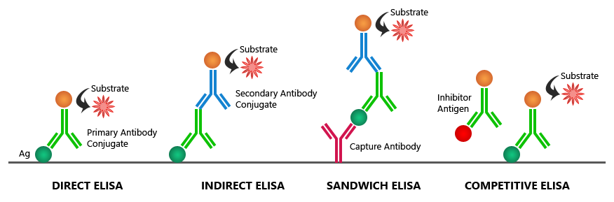
If you are having trouble with saturated signals in your ELISA data results, check out this table for Boster’s possible solutions to your problem:
| Possible Causes | Possible Solutions |
|---|---|
| High sample concentration | Use higher sample dilutions (Determine the optimal dilutions by titration assay). In parallel, construct an ELISA standard curve using known concentrations to more accurately map signal intensities to antigen levels. |
| Excessive substrate | Decrease concentration or amount of substrate: Follow manufacturer guidelines (The substrate provided with the ELISA kit might require further dilution) |
| Substrate color changed before use | Make substrate immediately before use |
| Non-specific antibody binding | Try different formulations in coating solutions; Ensure wells are pre-processed to prevent non-specific binding; Use affinity-purified antibody and preferably one that is pre- adsorbed; Use serum (5-10%) from same species as secondary antibody (bovine serum is also recommended). |
| Incubation time too long | Follow the manufacturer guidelines (If the problem persists, try incubating samples at 4°C overnight) |
| Excess antibody | Repeat the assay with lower antibody concentrations to find the optimal one for your experiment |
| Contaminated buffers with metals or HRP | Make and use fresh buffers |
ELISA (enzyme-linked immunosorbent assay) is a plate-based assay used to detect the concentration of a specific protein in a liquid sample and is frequently customized as part of custom assay development services tailored to specific research goals, including workflows that use validated elisa antibody pairs for target-specific detection. In applications that require simultaneous measurement of multiple targets, researchers may also utilize multiplex ELISA kits to analyze several proteins within a single sample. It is also commonly performed using standardized elisa kits to ensure consistency across experiments. Three different types of data output can be obtained:
ELISA (enzyme-linked immunosorbent assay) is a convenient and simple method to quantitatively or qualitatively detect peptides, proteins, antibodies, and hormones in samples, rendering it as one of the most widely used immunoassays. Many laboratories perform these assays using standardized elisa kits, which help streamline assay setup and improve reproducibility. To derive accurate concentrations from measured signals, most ELISA protocols rely on constructing an standard curve from known standards, which serves as the reference for sample interpolation, alongside the use of quick elisa kits to support faster and more efficient assay workflows. Despite the many advantages of conducting ELISA—or more complex formats like those used in <...
Accurate protein band size prediction starts with the primary sequence, but real gels reflect biology. PTMs, processing, isoforms, and experimental conditions shift apparent molecular weight. Use the estimate, then verify with the diagnostic steps below.
Western blotting separates proteins by size via gel electrophoresis (SDS-PAGE...
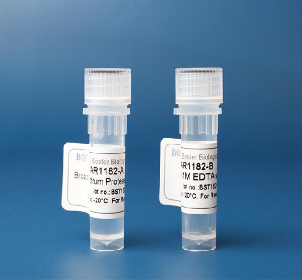
Protease inhibitor cocktails are chemical compounds that act to protect and maintain cellular protein composition after lysis of a cell, preventing natural degradation. They play an important role in protein quantification analysis, rendering the protease ineffective while obtaining appropriate protein purification yields. Protease inhibitors are classified by either the type of protease they act on or their mechanism of actions.
Proteases are enzymes that degrade proteins, playing an important role in cellular protein catabolism. Through their interaction with the proteins, they influence their activity and production of bioactive molecules, cellular repair, and the degradation of extracellular material. Proteases are also vital for food...
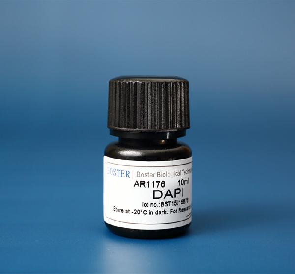
DAPI, or 4',6-Diamidino-2-Phenylindole, Dihydrochloride, is a commonly used fluorescent dye that binds to double-stranded DNA (dsDNA).
DAPI binds to and ‘stains’ double-stranded DNA, preferably binding to A-T-rich regions in DNA. DAPI stain is excited by ultraviolet (UV) light, with its largest excitation wavelength at ~360nm, and it produces a vibrant blue color with its largest emission wavelength at ~460nm when bound to DNA. Due to its fluorescent properties and rich blue color, it is readily used for visualization in fluorescence microscopy and other assays. Because it can pass through the cell membrane and stain DNA, DAPI is a useful dye for nuclear quantification and has been utilized in numerous assays, such as live or fixed cell staining, cell viability assays, flow cytometry, cell cycle analysis, mycoplasma contamination detection and in fluorescence microscopy. In fluorescence-based tissue analysis, especially where nuclear visualization guides diagnostic interpretation, accurate identification of morphological patterns is often supported by expert Pathology Review, complementing the use of nuclear dyes like DAPI. Because of its wide range of applications, Boster offers an affordable DAPI stain solution (Catalog# AR1176) that has been validated and cited in several publications.
Cell cycle analysis & flow cytometry
Since DAPI binds to DNA, it can be used to determine the relative amount of DNA in cells for cell cycle analysis. Cells currently in the G2 phase of mitosis will have twice the amount of DNA as cells in the G1 phase of mitosis, which will be reflected in the amount of fluorescence from DAPI in each cell. Apoptotic cells will have less DNA than a single cell, since DNA is being deg...