This website uses cookies to ensure you get the best experience on our website.


- Table of Contents


Immunohistochemistry (IHC) is a vital technique in biomedical research and clinical diagnostics, enabling the visualization and localization of specific proteins within tissue samples. In this blog, we outline the different types of IHC staining, including direct and indirect...

Blotting techniques are essential tools in molecular biology and biochemistry, allowing researchers to detect and analyze nucleic acids and proteins. These techniques enable scientists to uncover intricate details about genetic material, gene expression, and protein interactions. From the foundational Southern blotting, which revolutionized DNA analysis, to the versatile Western blotting, a staple in protein research, blotting methods have become indispensable tools in the laboratory.
This guide introduces ten blotting types, summarizing their principles, applications, and distinguishing features. Whether you are new to these methods or seeking a quick reference, the following sections outline how each technique contributes to DNA, RNA, and protein studies.
Southern blotting, the original blotting technique, is named after its inventor Edwin Southern who developed the method in 1975 for transferring DNA from a gel to a membrane, enabling the identification of specific DNA sequences. The naming convention for subsequent blotting techniques was influenced by this original name.
To this day, Southern blotting remains a common technique for DNA analysis and identification. The process involves digesting DNA with restriction enzymes, separating the fragments by gel electrophoresis, and transferring them onto a membrane. Labeled DNA or RNA probes then hybridize to the target sequences. Popular in genetics and molecular biology, Southern blotting is used for the detection of specific DNA sequences, study of DNA methylation patterns, RFLP (Restriction Fragment Length Polymorphism) analysis for genetic fingerprinting, identification of gene mutations and polymorphisms, and gene mapping and cloning.
Northern blotting was named by James Alwine, David Kemp, and George Stark in a 1977 paper as a playful pun on Southern blotting, indicating its use for RNA rather than DNA.
This technique is similar to Southern blotting, but focuses on RNA analysis. RNA samples are separated by gel electrophoresis, transferred to a membrane, and hybridized with labeled DNA or RNA probes to detect and quantify specific RNA transcripts. Northern blotting is commonly used to analyze gene expression patterns, determine mRNA size and abundance, study RNA processing and degradation, and detect alternative splicing events. To quantify RNA expression levels beyond blotting techniques, a solid qpcr service provides a highly sensitive and accurate alternative for transcript analysis.
Western blotting, also called immunoblotting, is widely used for protein detection and analysis, making it the most popular blotting technique. For streamlined and reproducible results, many researchers turn to a dedicated Western Blotting Service to handle the full workflow with optimized protocols. Proper western blot sample preparation is essential to ensure reliable and reproducible protein detection throughout the process. Understanding the western blot principle is key to effectively applying this method and interpreting its results accurately. The method was first described by Towbin et al. in 1979 and the term "western" was coined by W. Neal Burnette in 1981 as a tongue-in-cheek reference to the direction naming theme established by Southern and Northern blotting, this time focusing on proteins instead of nucleic acids.
This technique detects specific proteins by separating them via gel electrophoresis (typically SDS-PAGE), transferring them to a membrane (usually PVDF or nitrocellulose: AR0135-02, AR0135-04), and using primary and secondary antibodies for detection. The secondary antibody is usually conjugated to an enzyme or a fluorescent tag that produces a detectable signal when exposed to a substrate or under specific conditions. For your western blot experiment, you can explore Boster’s catalog to browse primary antibodies and secondary antibodies validated for western blot as well as find western blot reagents you will need.
Before electrophoresis begins, sample extraction quality already shapes the final blot. This guide on which lysis buffer to use for Western blot helps match buffer choice to target protein type, solubility, and downstream blot performance.
After protein extraction and electrophoresis, transfer is the next major checkpoint. This practical guide on how to check transfer quality in Western blot is useful for confirming whether weak or uneven signal comes from transfer failure rather than from the antibody step alone.
If you are deciding between PVDF and nitrocellulose for sensitivity, background, binding capacity, or handling preference, this guide on which membrane to choose for Western blot offers a more practical side-by-side comparison for real workflow decisions.
For researchers moving from detection to measurement, this guide on western blot quantification provides a practical workflow for densitometry, normalization, and fold-change calculation, with particular emphasis on staying in the linear range and avoiding saturation-related errors.
If the same membrane needs to be reprobed for another target or for loading-control confirmation, this Western blot stripping buffer protocol provides a practical starting point for removing bound antibodies while preserving usable blot signal.
In protein research and diagnostics, Western blot is used for the detection and quantification of specific proteins in a sample, analysis of protein expression levels across different conditions or treatments, detection of post-translational modifications (e.g., phosphorylation, glycosylation), study of protein-protein interactions, and confirmation of protein identity and purity in recombinant protein production. For researchers interested in analyzing protein expression directly within intact cells, consider our in-cell western blot service, which offers a high-throughput, quantitative alternative to traditional blotting techniques.
To learn more about western blotting, download our Western Blot eBook, which discusses the principle, protocol, troubleshooting tips, and FAQs for western blot.
Following the directional theme set by Southern and Western blotting, Eastern blotting is used to analyze post-translational modifications (PTMs) of proteins, such as glycosylation or phosphorylation. Many scientists deem Eastern blotting as a variation of Western blotting.
The Eastern blot technique involves transferring proteins separated by gel electrophoresis onto a membrane, followed by detection using specialized probes or antibodies targeting the PTM of interest. Though less frequently performed than other blotting techniques, Eastern blotting is useful for analyzing PTMs (e.g., glycosylation), studying protein modifications (e.g., phosphorylation, lipidation), and detecting and characterizing glycoproteins and other modified proteins.
Far-Western blotting, derived from Western blotting, focuses on protein-protein interactions. It involves transferring proteins separated by gel electrophoresis onto a membrane and identifies interactions using labeled proteins or peptides to probe for binding with the immobilized target proteins.
Growing in popularity, Far-Western blotting is valuable for identifying and studying protein-protein interactions, mapping interaction domains, and screening potential binding partners.
Southwestern blotting combines features of Southern and Western blotting techniques, focusing on DNA-binding protein detection. This technique involves transferring denatured proteins from a gel onto a membrane, followed by incubation with labeled DNA probes to identify proteins that bind to specific DNA sequences.
Though less common than Southern and Western blotting, Southwestern blotting is useful for detecting DNA-binding proteins, studying protein-DNA interactions, and identifying transcription factors and other regulatory proteins.
Reverse Northern blotting indicates the reversal of the Northern blotting process. Instead of transferring RNA and probing with DNA, Reverse Northern blotting transfers DNA and probes with labeled RNA.
This technique involves immobilizing DNA on a membrane and hybridizing it with labeled RNA or cDNA probes to detect DNA sequences. Although less common than standard Northern blotting, it is used for gene expression analysis using cDNA or genomic DNA arrays, screening differentially expressed genes in various conditions, and studying changes in transcript levels in response to treatments.
Colony blotting is named for its application in screening microbial colonies, such as bacterial or yeast colonies. It involves transferring entire colonies from a culture plate onto a membrane, where specific nucleic acids or proteins are detected through DNA hybridization analysis.
Primarily used in microbiology and cloning, colony blotting helps screen colonies for target DNA or RNA sequences, identify recombinant clones containing specific genetic inserts, and rapidly detect plasmid-containing colonies.
Dot blotting is frequently used as a quick and simple screening method for the presence or absence of nucleic acids and proteins. Named for the dot-like application of samples on the membrane, it is a simplified version of Western blotting, where samples are directly spotted onto a membrane for detection without prior gel electrophoresis.
Popular for rapid screening, dot blotting is used to screen specific nucleic acids or proteins, relatively quantify target molecules, and analyze large numbers of samples simultaneously. It should be noted that dot blots do not provide information about molecular weight, so false positive signals or the presence of modified proteins are difficult to identify.
Slot blotting is similar to dot blotting, but less commonly used. Named for the slot-like application of samples, slot blotting involves applying samples in rectangular slots on a membrane, allowing more uniform application and the quantification of target molecules in a sample without the need for gel electrophoresis.
This technique is helpful for analyzing nucleic acids or proteins without electrophoresis, comparing the relative abundance of target molecules in different samples, and screening multiple samples in high-throughput formats. As with dot blots, slot blots are also unable to provide information about the size of the target protein.
We have provided a table below that highlights the features of each blotting type.
| Blotting Type | Target Molecule | Detection Method | Technique Description | Applications |
|---|---|---|---|---|
| Southern Blotting | DNA | Labeled DNA/RNA probes | DNA fragments are separated by electrophoresis, transferred to a membrane, and probed. | Detection of DNA sequences, genetic fingerprinting, gene mapping |
| Northern Blotting | RNA | Labeled DNA/RNA probes | RNA is separated by electrophoresis, transferred to a membrane, and hybridized with probes. | Analysis of gene expression, RNA processing, alternative splicing |
| Western Blotting | Proteins | Primary/secondary antibodies | Proteins are separated by SDS-PAGE, transferred to a membrane, and detected with antibodies. | Protein detection, expression analysis, post-translational modifications |
| Eastern Blotting | Modified proteins | Specific probes/antibodies | Proteins are transferred to a membrane and probed for post-translational modifications. | Analysis of glycosylation and other post-translational modifications |
| Far-Western Blotting | Proteins (interactions) | Labeled proteins/peptides | Proteins are transferred to a membrane, probed with labeled proteins to detect interactions. | Study of protein-protein interactions, mapping interaction domains |
| Southwestern Blotting | DNA-binding proteins | Labeled DNA probes | Proteins are transferred to a membrane and probed with labeled DNA to detect binding. | Detection of DNA-binding proteins, study of protein-DNA interactions |
| Reverse Northern Blotting | DNA | Labeled RNA/cDNA probes | DNA is immobilized on a membrane and hybridized with labeled RNA/cDNA probes. | Gene expression profiling, screening for differentially expressed genes |
| Colony Blotting | DNA/RNA in colonies | Labeled probes | Microbial colonies are transferred to a membrane and probed for specific sequences. | Screening for specific sequences, identifying recombinant clones |
| Dot Blotting | Nucleic acids/proteins | Labeled probes/antibodies | Samples are spotted directly onto a membrane for rapid detection. | Rapid screening, quantification of target molecules |
| Slot Blotting | Nucleic acids/proteins | Labeled probes/antibodies | Samples are applied in slots on a membrane for quantitative analysis. | Quantitative analysis, comparing relative abundance of target molecules |
Blotting techniques are important tools in molecular biology and biochemistry. Each blotting method caters to different experimental needs, enabling detailed analysis of DNA, RNA, proteins, and their interactions. By utilizing each technique’s unique advantages and applications, researchers can deepen our understanding of molecular interactions and processes. Boster Bio supports these workflows with advanced solutions like our Recombinant Ant...
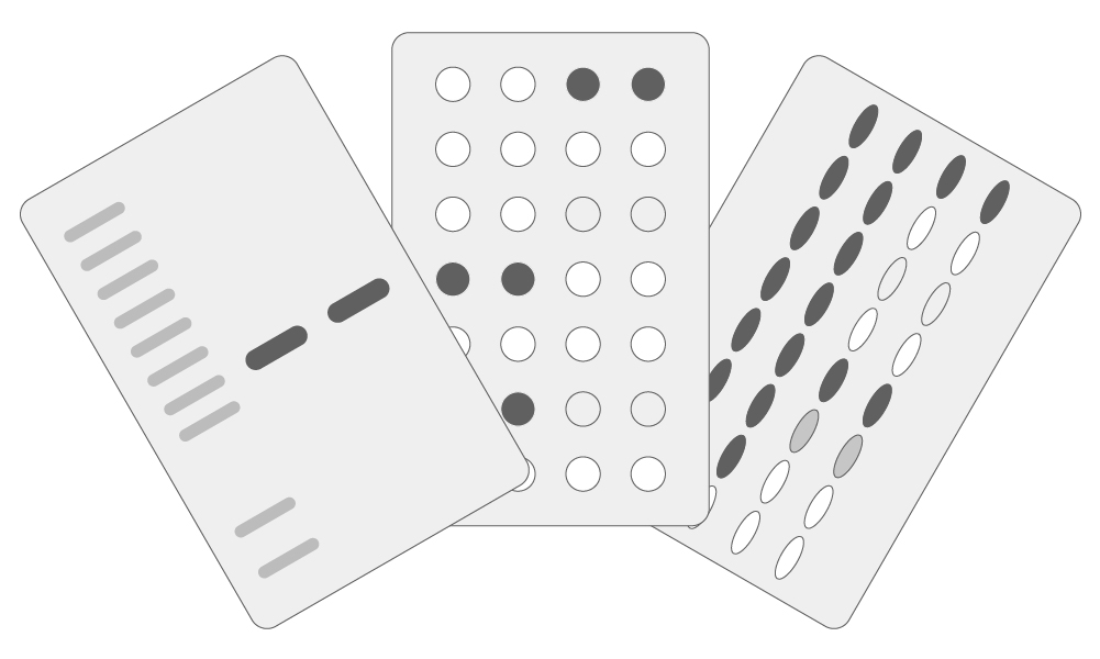
Enzyme-Linked Immunosorbent Assay (ELISA) is a versatile and widely used biochemical technique for detecting and quantifying specific molecules such as proteins, peptides, antibodies, and small molecules. These assays are commonly performed using standardized elisa kits, which help ensure consistency and reproducibility across experiments. Researchers may...

Understanding the relative abundance of target proteins and effectively normalizing data are crucial aspects of Western blot analysis. These critical aspects are deeply rooted in the western blotting principle and procedure, which outline how proteins are separated...
Sodium citrate buffer, also known as citrate buffer, is a common reagent used for antigen retrieval in immunohistochemistry. Immunohistochemistry (IHC) is a technique that enables high-quality visualization of proteins in a variety of tissues. IHC is a multi-step process that comprises fixation, embedding, and sectioning of a target tissue followed by incubation with an antibody targeting a protein of interest.
Once the tissue is fixed, embedded in paraffin, and sectioned on slides, researchers can utilize antibodies to target and identify their protein of interest. However, fixation and embedding of tissues, often with 10% formalin and paraffin respectively, initiates cross-linking of proteins that can mask antigen epitopes. Antigen masking may reduce the sensitivity of antibodies and decrease the detection of certain proteins of interest. A process known as antigen retrieval breaks the bonds between formalin and proteins, removing the effects of cross-linking, thereby enabling better detection of proteins. Sodium citrate buffer is highly used in the process of antigen retrieval, specifically for heat-induced antigen retrieval. Boster Bio offers a simple-to-use Sodium Citrate Buffer (Catalog# AR0024) for heat-induced antigen retrieval to remove the effects of antigen masking in your IHC experiments.
Antigen retrieval is a process of breaking the bonds from cross-linking, enabling proteins to interact with antibodies during immunohistochemistry. The purpose of antigen retrieval in immunohistochemistry is to unmask antigens in fixed tissues, which can significantly improve the quality of staining and detection of proteins. While optimization of this method may add additional steps to an already multi-step, complex procedure, antigen retrieval has improved IHC...

Western blotting (also called Protein Immunoblotting) is an analytical technique used to detect specific proteins in the given sample. A deep understanding of the western blot principle provides the essential foundation for applying this method successfully in protein detection. It uses SDS-polyacrylamide gel electrophoresis (SDS-PAGE) to separate various proteins contained in the sample. The separated proteins are then transferred or blotted onto a matrix, where they are stained with antibodies specific to the target protein. Expression details of the target proteins in the given cells or tissue homogenate can then be obtained through analyzing the location and intensity of the specific reaction. Western blotting analysis can detect target protein as low as 1 ng due to high resolution of the gel electrophoresis and strong specificity and high sensitivity of the immunoassay. This method is used in the fields of molecular biology, biochemistry, immunogenetics and other molecular biology disciplines for various experiments. Researchers who prefer standardized, high-sensitivity workflows often rely on a professional western blot service to ensure consistent and reproducible results.
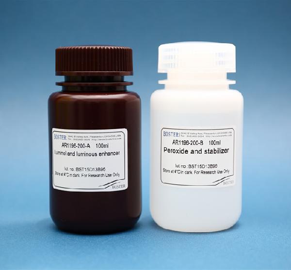
Enhanced Chemiluminescence (ECL) Western Blot Substrate is a very sensitive, non-radioactive, enhanced luminol-based chemiluminescent substrate that allows for easy detection of horseradish peroxidase (HRP) on immunoblots. HRP is a common molecule conjugated to antibodies. ECL Western Blot Substrate has the capability...
Western blot filter paper stabilizes the transfer stack and regulates the buffer flow, helping proteins transfer efficiently from the gel to the membrane. Correct paper choice, hydration, and stacking pressure reduce artifacts and improve band clarity.
Western blotting (also called Protein Immunoblotting) is used to detect specific proteins separated by SDS-polyacrylamide gel electrophoresis (SDS-PAGE) and transferred to a membrane where antibodies bind the target. The workflow includes transfer, blocking, antibody incubation, and detection—each step affecting signal quality and background. For researchers looking to streamline or standardize their workflow, a credible Western Blotting Service can help ensure consistent, high-quality results.
Western blotting works best when the basics are solid and the sample is prepared well. After SDS-PAGE separates the proteins, they’re transferred to a membrane and probed with antibodies that recognize the target. The band position and intensity indicate expression levels in cells or tissue lysates, with sensitivity down to about 1 ng under optimized conditions. Because of its resolution and antibody specificity, the technique is widely used in molecular biology, biochemistry, and immunogenetics for both qualitative and quantitative protein analysis.
Filter paper (or blotting paper) is important to ensure quick and efficient transfer of molecules from the electrophoresis gel to the matrix membrane. It is used for transfer sandwiches and cassettes during Western blotting and assists with transferring proteins unto polyvinylidene fluoride (PVDF membranes), nitrocellulose membranes, and other types of transfer membranes for accurate protein detection. Western blot filter paper is a semi-permeable paper barrier used to separate fine solid particles from liquids or gases.
It can be purchased as pre-cut blotting paper sheets for wet, semi-dry, passive, or electrophoretic transfer of proteins. The thickness of the paper determines the flow rate of the protein or nucleic acid.
During the process of western blotting, Mahmood et al., 2012 des...

According to a report on GEN, 41% of researchers admit that their Western blots are unsuccessful at least 25% of the time.
Yikes! Western blotting (WB) is a widely practiced analytical technique to detect target proteins within samples using antigen-specific antibodies, and when properly optimized, it can be significantly enhanced by a professional Western Blotting Service that ensures reproducibility and high-quality results. A solid grasp of the western blot principle is essential to maximize the success rate and minimize common experimental failures. , while meticulous western blot sample preparation lays the foundation for accurate and reliable protein detection. When it fails to perform as expected, it can really be a downer. Common issues like high background or weak signals often stem from early-stage variables related to western blot optimization.
We’re here to help you succeed. Next time you encounter another problem with Western blot, we’ve compiled a checklist to help you troubleshoot your experiment.
High background frequently occurs due to...





