This website uses cookies to ensure you get the best experience on our website.
- Table of Contents
Affinity gauges the binding strength of a single bond, while avidity measures the combined stability of multivalent interactions. Understanding the difference enhances assay design, vaccine evaluation, and therapeutic efficacy.

Affinity vs avidity describes how tightly antibody attach to antigens, and together, these values predict real-world performance. High-affinity IgG molecules often show a low dissociation rate constants (Kd) below 10⁻⁹ M, yet even weaker binding sites can yield strong avidity when arranged on a pentameric IgM. Roughly 80% of early infection tests rely on avidity principles, whereas most monoclonal antibodies undergo affinity maturation before approval, a process supported by our antibody production services. This reflects a fundamental aspect of molecular interactions essential in diagnostics, drug development, and biomedical research.
A single antigen-antibody reaction can decide whether a pathogen infects a cell membrane or is cleared from it. Strong binding reduces the likelihood of escape, enhances diagnostic sensitivity, and can prolong drug residence time. Therefore, scientists track both affinity—the force at a single binding site—and avidity—the collective force from immune complexes formed by multiple bonds.
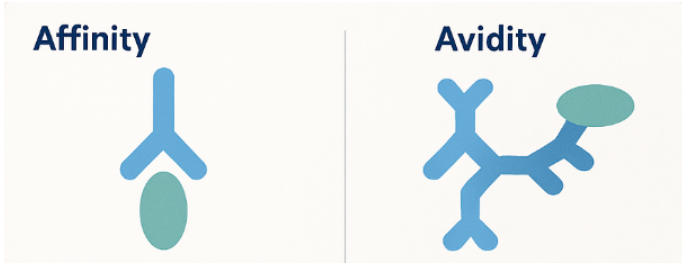
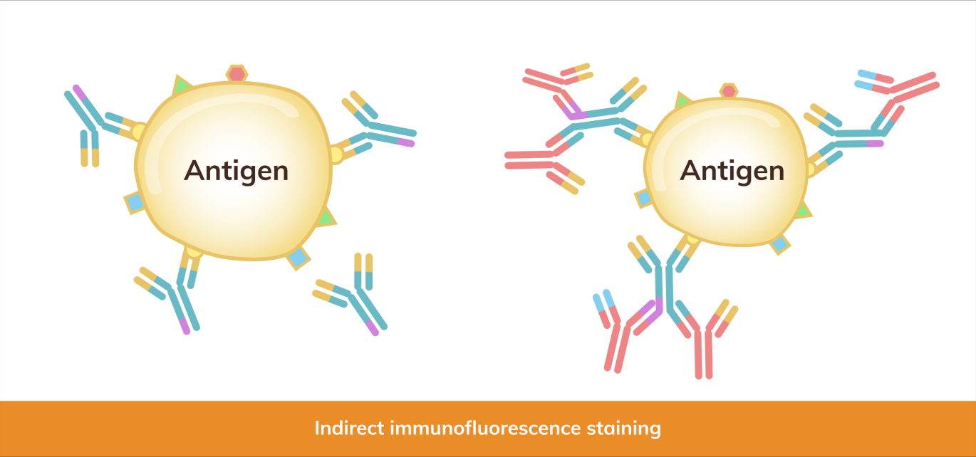
Fig.1 Illustrating antigen-antibody interaction, comparing affinity and avidity. Shows a monovalent antibody binding to a single epitope (affinity) alongside a multivalent antibody binding to multiple epitopes on a pathogen surface (avidity).
Affinity is the attraction between one antigen-binding site and one epitope. It reflects how quickly the complex forms (association rate constant)and how slowly it breaks apart (dissociation rate constant). Strong affinity implies a low Kd, often 10⁻⁹ M or lower for mature IgG. Small mutations in complementarity-determining regions can shift affinity by a thousand-fold due to changes in hydrogen bonds and hydrophobic interactions at the binding interface.
Researchers choose different tools depending on throughput and sensitivity:
High-affinity antibodies improve assay specificity because weak binders wash away during stringent steps in Western blotting or ELISA.
Avidity combines multiple affinities. An IgM pentamer, with ten identical Fab fragments, can latch onto a multivalent antigen structure even if each site binds weakly. Cooperative binding stabilizes complexes, especially when antigens display repeating epitopes. This process integrates Bond Strength with structural arrangement to resist dissociation.
Laboratories often use an avidity ELISA in which samples bound to the antigen are washed with a chaotropic agent, such as 6 M urea. The avidity index equals the signal retained after treatment divided by the signal before treatment, expressed as a %.
Clinicians apply this in diagnosing rubella, toxoplasmosis, and other infections, differentiating acute from past exposure in the humoral immune response.
Affinity and avidity describe related but distinct aspects of molecular binding, and understanding their differences is essential for interpreting experimental results.
| Factor | Affinity | Avidity |
|---|---|---|
| Definition | Strength of a single binding site | Combined strength of multiple sites |
| Unit | Dissociation rate constant (Kd) | Avidity index or functional strength |
| Influenced by | Amino-acid contacts, shape fit | Valency, epitope density, flexibility |
| Main role | Determines specificity | Enhances binding stability |
| Primary assay | SPR, BLI, equilibrium ELISA | Avidity ELISA, functional tests |
These practical examples show how affinity and avidity influence diagnostic accuracy, therapeutic design, and vaccine assessment in real-world applications.
Rapid tests for early infection often detect IgM because its high avidity yields visible agglutination. For confirmation, labs switch to IgG affinity tests that discriminate true positives from antibody cross reactivity and false signals.
Drug developers raise affinity through mutagenesis and selection cycles. Values below 10⁻¹¹ M increase target occupancy at lower doses. In contrast, bispecific antibodies sometimes leverage avidity, arranging two weaker arms to reduce off-target effects while maintaining strong overall attachment, an approach enabled by custom monoclonal antibodies.
This process considers antibody concentration, functional affinity, and how each antibody molecule binds and remains stable within antibody-antigen complexes.
After vaccination, affinity matures through somatic hypermutation, and avidity rises as antibodies switch from IgM to IgG. Monitoring the avidity index helps researchers predict the durability of protection and decide whether booster shots are necessary.
Selecting the appropriate assay depends on the research objective. When detailed binding kinetics are required, techniques such as Surface plasmon resonance (SPR) or bio-layer interferometry (BLI) provide real-time measurements of the association rate constant and dissociation rate constant. For high-throughput screening of multiple antibody clones, equilibrium ELISA is often the first step, with promising candidates later validated by SPR for precise affinity data. In infectious disease studies, avidity ELISAs that incorporate chaotropic washes help distinguish recent infections from long-term immunity by analyzing the stability of antigen–antibody complexes. For mucosal immunity, IgA assays are essential, as secretory IgA exhibits unique resistance to enzymatic digestion, offering insights into protective responses at epithelial barriers.
Affinity and avidity together describe how antibodies interact with antigens, guiding numerous decisions in diagnostics, research, and medicine. Focusing solely on one value can be misleading; a balanced evaluation ensures accurate conclusions and successful product development.
Are you looking to refine your assays, develop high-performance antibodies, or showcase new findings?
Submit an inquiry to connect with our expert team. Discover how tailored assay design, collaborative research, and exhibition opportunities can enhance your projects and foster new partnerships.
Western blot “failures” are often blamed on antibodies, transfer, or blocking. But in many cases, the real bottleneck happens earlier: lysis choice plus lysate handling. A buffer that’s too mild can leave your target behind. A buffer that’s too harsh can produce viscous, debris-rich lysate that smears lanes and raises background. The goal is not “the strongest lysis possible,” but the mildest system that reliably extracts your target with the best signal-to-background—and then handling it in a way that keeps lanes clean.
This post is the very beginning for our Western blot experiment starting from sample prep. If you want broader context (or want to move downstream after lysate quality is solid), these internal hubs are designed to be your next clicks:

Recombinant DNA technology is used to produce antibodies by assembling, expressing, and purifying antibody genes in vitro. This approach supports consistent, scalable antibody production without the need for animal immunogen preparation. It is widely applied in research, diagnostics, and therapeutic development, offering advantages in assay reliability and customization across the immune system.
Recombinant DNA technology involves combining DNA sequences from different sources to create new genetic constructs. In the context of antibody production, this means taking the genetic code that encodes an antibody’s variable and constant regions, inserting it into an expression vector, and producing the antibody in a host cell system such as mammalian cells, yeast, or bacteria.
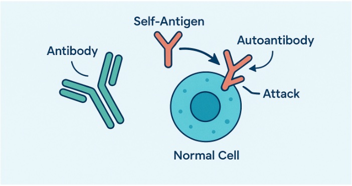
Roughly 10% of people worldwide live with an autoimmune condition, and more than 80 of these disorders trace at least part of their pathology to antibodies that have turned on the body. These rogue molecules—often referred to as autoantibodies—trigger inflammation, degrade healthy tis
Proteins are the workhorse molecules that drive virtually every biological system. With the increasing recognition of the role of proteins in various research and manufacturing activities, simply isolating them from their natural host cells cannot meet the escalating demand of the market. Chemical synthesis is also not a viable option for this endeavor due to the size and complexity of proteins. Instead...
The domestic chicken (Gallus gallus domesticus) has been a model organism for scientific research due to its accessibility, ease of breeding, and large-sized eggs, which are particularly useful for embryological studies.
In this blog, we describe a brief history and some key breakthroughs of chicken as a model organism. We discuss the research advantages and limitations of chickens, and describe some research areas where scientists have explored using chickens. For researchers interested in working with chickens, we have provided a list of resources and guiding questions. If you’re considering choosing chickens for your research studies or simply want to learn more about chickens, this blog is for you!
Feel free to jump to a specific section about chicken:
Gallus gallus domesticus, commonly known as the domestic chicken, is a bird species widely used in developmental biology and genetic research. As a subspecies of the red junglefowl, which is native to southern Asia, the domestic chicken is versatile and economically significant. Adult chickens typically measure between 40 to 60 centimeters in length from beak to tail and weigh around 2 to 4 kilograms. They are characterized by their feathered bodies, beaks, and a range of plumage colors and patterns.
Gallus gallus domesticus are chosen as research models due to its well-understood developmental processes and genetic makeup. Chickens have a relatively long incubation period of about 21 days from egg fertilization to hatching, during which the embryos can be observed and manipulated. This extended embryonic development allows for detailed studies of developmental biology, including organ formation and genetic regulation. Furthermore, the chicken genome was sequenced in 2004, presenting a comprehensive resource for genetic studies. This genome facilitates research into gene function, evolution, and disease mechanisms.
The chicken is examined in immunology and vaccine development due to its production of large amounts of antibodies. Additionally, its large eggs make it a favored model for studying early embryonic development and the effects of genetic and environmental factors on growth and differentiation. Overall, Gallus gallus domesticus offers valuable insights into vertebrate biology and developmental processes, bridging the gap between simpler model organisms and more complex mammalian systems.
Gallus gallus domesticus, the domestic chicken, has been instrumental in several key scientific breakthroughs, especially in embryology, virology, genetics, and developmental biology. We discuss some of the most notable milestones below.
In 1910, Peyton Rous discovered the Rous sarcoma virus (RSV) in chickens, marking one of the first demonstrations that viruses could cause cancer. This discovery crucially linked viruses and cancer, fundamentally changing our comprehension of carcinogenesis. The identification of RSV in chickens eventually led to the discovery of oncogenes, which are genes that can cause normal cells to become cancerous.1 Rous's work was so influential that he was awarded the Nobel Prize in Physiology or Medicine in 1966.2
The chicken embryo became a key model in embryology starting in the early 20th century. In the 1920s and 1930s, Viktor Hamburger’s work with chicken embryos was foundational in determining the stages of vertebrate development. His research, in collaboration with Howard L. Hamilton, led to the establishment of the Hamburger-Hamilton stages in 1951, a detailed series of 46 stages that describe the chronological development of chick embryos from the laying of the egg to hatching. This work became a standardized system for describing embryonic development in chickens and presented a vital framework for studying developmental processes across vertebrates.3,4
Chickens have played a critical role in immunology, notably in the development of vaccines. For instance, the use of chicken eggs in the production of vaccines for diseases such as influenza has been a significant breakthrough.5 The ability to produce large quantities of virus in chicken eggs has been crucial for the rapid development and distribution of vaccines, especially during pandemics.
In the 20th century, chickens gained prominence in genetics research. The establishment of inbred chicken strains empowered scientists to investigate genetic variation and inheritance patterns.6 Studies on chickens uncovered quantitative genetics related to traits such as disease resistance and growth.7,8
The sequencing of the chicken genome in 2004 was a major milestone in genetic research. The chicken was the first bird and the first agricultural animal to have its genome fully sequenced.9 This sequencing clarified the evolution of vertebrates, revealing how birds, including chickens, evolved from dinosaur ancestors. The chicken genome has also been used to examine gene function and genetic diseases, providing a comparative framework for understanding human genetics.
Chickens have been employed in genetic engineering and transgenics. The development of transgenic chickens, which carry foreign genes inserted into their genome, has allowed researchers to unravel gene function and regulation in a vertebrate model.10 This technology has implications for both basic research and the development of biopharmaceuticals.
The domestic chicken has been a valuable model organism, contributing to major scientific breakthroughs in various fields. From developmental biology to cancer research and vaccine production, the use of Gallus gallus domesticus has deepened our comprehension of fundamental biological processes and their applications in medicine and agriculture.
Gallus gal
...
Antibodies, also known as immunoglobulins, are specialized proteins produced by the immune system to identify and neutralize foreign invaders such as bacteria, viruses, and toxins. Their ability to recognize and bind to specific antigens make them play a critical role in the immune response and serve as key tools in biomedical research, including advanced applications like...

Antibodies do more than stick to germs. They block infection, label threats for cleanup, and spark immune system responses that keep the body safe.
The antibody action mechanism refers to the step-by-step process an antibody follows once it binds to a target antigen. The protein’s antibody structure enables each molecule to recognize a target with pinpoint precision and then trigger a robust immune response. Over the past several years, monoclonal antibodies have become a dominant format in therapeutic development, highlighting the importance of antibody engineering and antibody production in clinical applications. To understand why, we can examine four main mechanisms: neutralization, opsonization, complement-dependent cytotoxicity, and antibody-dependent cellular cytotoxicity (ADCC)
Binding of antibodies is only the first move. The antibody’s variable region grabs the antigen, while the Fc region faces outward—a process often studied using recombinant protein expression systems. That outward portion connects to immune receptors, starts signaling pathways, and guides the next steps. Signals can recruit other proteins, attract B cells or T cell populations, or alter the shape of the antibody itself. These downstream events decide whether the invader is blocked, swallowed, or destroyed. Such outcomes vary depending on the antibody class, the type of antigen, and the surrounding tissue environment.
Antibodies protect the body through the following mechanisms that block, tag, or destroy harmful targets:
Neutralization acts like closing a door before a burglar walks in. An antibody covers the exact part of a virus, toxin, or bacterium that would usually latch onto a host receptor. Viruses use viral proteins to attach to and fuse with tumor cells or human tissues. Toxins require antigen-binding site access to en...


