This website uses cookies to ensure you get the best experience on our website.


- Table of Contents
Plan chromogenic ADAM10 IHC on paraffin sections using an IHC-validated antibody (datasheet M00566). Score the mainly cytoplasmic tissue staining (HPA tissue IHC), while considering membrane and junctional localization (UniProt).

Expected localisation, validated protocols, controls and antibodies — the at-a-glance facts below, then the full design guide.
| Expected localisation | Mainly cytoplasmic (HPA tissue IHC); membrane and junctional (UniProt) | |
| Staining pattern | Cytoplasmic staining across several tissue cell types (HPA tissue IHC) | |
| Antigen retrieval | EDTA pH 8.0 HIER, heat-mediated (datasheet M00566) | |
| Positive control | Lung+4 more · see all | |
| Negative control | Adrenal gland+4 more · see all |
| Fixation | Keep fixation consistent across sections (standard IHC practice; not target-specific) | |
| Caveat | Mature ADAM10 can cluster at adherens junctions (UniProt) | |
| Regulation | TSPAN14 promotes cell-surface expression (UniProt) | |
| Isoform / epitope | Two isoforms; check whether the epitope is extracellular or cytoplasmic (UniProt) |
The catalog antibody has an IHC-P protocol (datasheet M00566); the published examples below cover hepatocellular carcinoma and pancreatic cancer tissue (PMC3829731; PMC5342537).
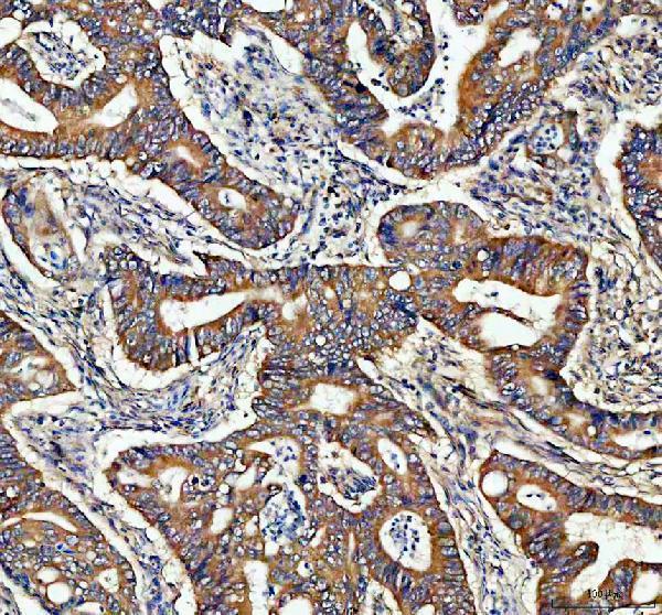
| Sample | Paraffin-embedded human rectum adenocarcinoma tissue; fixative not specified (datasheet M00566) |
| Fixation | Image fixative and duration unreported (datasheet M00566); verify before use. |
| Sectioning | 4–5 µm sections on charged slides (standard) |
| Deparaffinisation | Xylene, graded ethanol series to water (standard) |
| Antigen retrieval | Heat retrieval: EDTA pH 8.0 (datasheet M00566); 20 min, 95–100 °C (standard) |
| Peroxidase block | 3% H2O2, 10 min, room temperature (standard) |
| Blocking | 10% goat serum (datasheet M00566) |
| Primary antibody | Rabbit monoclonal (clone AAIC-1) anti-ADAM10, 1:50 recommended; image 1:200 (datasheet M00566) |
| Primary incubation | Overnight at 4 °C (datasheet M00566) |
| Detection | HRP-conjugated secondary, DAB chromogen (datasheet M00566) |
| Counterstain | Hematoxylin, blue, dehydrate and mount (standard) |
| Expected result | ADAM10-positive staining in macrophages of lung (HPA tissue IHC: Medium). HPA tissue profile: Mainly cytoplasmic expression in several tissues. No signal in the no-primary control. |
ADAM10 spans the cell membrane and also localizes to Golgi-associated vesicles and cytoplasm (UniProt O14672 topology and localization). In tissue IHC, HPA describes mainly cytoplasmic staining and reports medium staining in lung macrophages, prostate and stomach glandular cells, and bladder urothelial cells (HPA tissue IHC). HPA rates its tissue staining Approved but notes low consistency with RNA expression (HPA tissue IHC).
| Cytoplasmic staining in lung macrophages or prostate, stomach, or seminal-vesicle glandular cells. | This fits HPA's mainly cytoplasmic tissue profile and its medium staining calls for these cells (HPA tissue IHC). Assess the named cell population rather than treating every cell in the section as an expected positive. |
| Membrane-enriched staining, possibly alongside cytoplasmic puncta, in an appropriate cell population. | ADAM10 is a membrane protein also found in Golgi-associated and clathrin-coated vesicles (UniProt O14672 localization). HPA tissue IHC describes mainly cytoplasmic staining, so membrane enrichment alone is not the sole required tissue pattern (HPA tissue IHC). |
| Predominantly nuclear staining with little cytoplasmic or membrane signal. | Nuclear localization is absent from the supplied UniProt and HPA localization records (UniProt O14672; HPA subcellular). Treat this as a possible artefact; review controls and antibody specificity before scoring it as ADAM10. |
| Strong staining across cell types that HPA calls not detected, such as adrenal glandular cells. | The distribution conflicts with that HPA observation and warrants checks for cross-reactivity or endogenous detection activity (HPA tissue IHC; standard IHC practice). A single HPA negative call does not establish universal biological absence. |
| Diffuse color throughout tissue or no signal in an HPA medium-staining cell population. | Neither result cleanly reproduces the cell-resolved HPA pattern (HPA tissue IHC). Use controls to separate nonspecific background or a detection problem from genuine sample variation (standard IHC practice). |
| Cellular compartment | The single transmembrane segment places ADAM10 at membranes, while UniProt also lists Golgi-associated vesicles (UniProt O14672 topology and localization). HPA's mainly cytoplasmic tissue pattern should guide the IHC readout (HPA tissue IHC). |
| Epitope position and processing | ADAM10 has an extracellular region at residues 20–672, a cytoplasmic tail at 694–748, and a cleaved propeptide at 20–213 (UniProt O14672). Interpret any compartment difference in light of the antibody's documented epitope, if known. |
| Shedding activity | ADAM10 sheds ectodomains of multiple membrane proteins (UniProt O14672 function). This activity does not by itself predict where a particular antibody will stain in a paraffin section; the antibody epitope and tissue result remain decisive. |
| Isoforms | UniProt lists 2 ADAM10 isoforms (UniProt O14672). The supplied evidence gives no isoform-specific tissue pattern, so avoid assigning a staining difference to an isoform without epitope or other supporting data. |
| Strength of tissue evidence | HPA rates the tissue IHC pattern Approved but reports low consistency between antibody staining and RNA expression (HPA tissue IHC). Its not detected calls are useful comparisons, not definitive absence controls. |
| Antibody validation | CAB001709 is IHC Approved; HPA050670 has no IHC status in the supplied list (HPA antibodies). Do not transfer an ICC/IF validation label to paraffin-section IHC. |
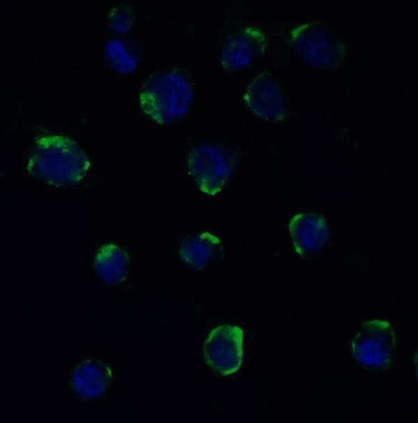
| IF/ICC Q: What localization is expected? | A: Plasma membrane and vesicles are the supported main locations; images are listed for A-431, U-251MG, and U2OS (HPA subcellular). This ICC/IF observation is separate from HPA's tissue IHC pattern. |
| Situation | Likely cause | Next action |
|---|---|---|
| No stain in lung macrophages or prostate glandular cells. | These cells have medium HPA staining, but the result may vary by sample or assay (HPA tissue IHC). | Check a section with identifiable expected cells, then review retrieval, antibody dilution, and detection controls as general IHC variables (standard IHC practice). |
| Color covers the section without clear cellular boundaries. | Diffuse color does not match HPA's cell-specific observations (HPA tissue IHC); background or endogenous detection activity may contribute (standard IHC practice). | Compare a no-primary control and inspect blocking, washes, and chromogen development (standard IHC practice). |
| Signal is mainly nuclear. | The supplied localization records emphasize membranes, vesicles, and cytoplasm (UniProt O14672; HPA subcellular). | Do not score nuclear-only color as a confirmed positive; compare controls and reassess antibody specificity (standard IHC practice). |
| Adrenal glandular cells stain strongly. | HPA calls that population not detected in tissue IHC (HPA tissue IHC). | Inspect morphology and no-primary controls for misplaced cell identification, cross-reactivity, or endogenous detection activity (standard IHC practice). |
| Membrane staining appears without a strong cytoplasmic pattern. | Membrane localization is supported, although HPA describes tissue staining as mainly cytoplasmic (UniProt O14672; HPA tissue IHC). | Record the compartment and cell type, then compare them with the antibody epitope and an HPA-listed positive population before interpreting the difference. |
| A bone-marrow section shows no hematopoietic-cell staining. | HPA calls these cells not detected, while UniProt lists bone-marrow expression without resolving this cell-level IHC result (HPA tissue IHC; UniProt O14672 tissue specificity). | Do not use this section alone to judge assay failure; check an HPA medium-staining cell population and its controls (HPA tissue IHC; standard IHC practice). |
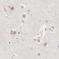


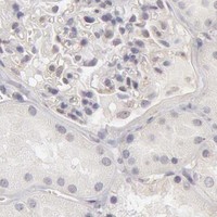


Comprehensive Human Protein Atlas IHC scoring per tissue (reliability: Approved — Low consistency between antibody staining and RNA expression data.). Rows are taken directly from the HPA tissue chart — click any row's HPA link to view the source.
| Tissue | Cell type | Level | Evidence | Source |
|---|---|---|---|---|
| Lung | Macrophages | Medium | Protein (IHC) | HPA → |
| Prostate | Glandular cells | Medium | Protein (IHC) | HPA → |
| Seminal vesicle | Glandular cells | Medium | Protein (IHC) | HPA → |
| Stomach | Glandular cells | Medium | Protein (IHC) | HPA → |
| Urinary bladder | Urothelial cells | Medium | Protein (IHC) | HPA → |
| Tissue | Cell type | Level | Evidence | Source |
|---|---|---|---|---|
| Adrenal gland | Glandular cells | Not detected | Protein (IHC) | HPA → |
| Bone marrow | Hematopoietic cells | Not detected | Protein (IHC) | HPA → |
| Caudate | Glial cells | Not detected | Protein (IHC) | HPA → |
| Cerebellum | Cells in granular layer | Not detected | Protein (IHC) | HPA → |
| Cerebral cortex | Glial cells | Not detected | Protein (IHC) | HPA → |
Troubleshoot ADAM10 staining in paraffin sections by checking retrieval, detection controls, and whether the signal matches its documented cellular distribution.
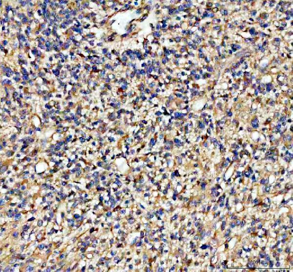
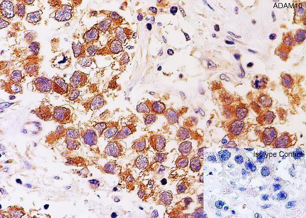
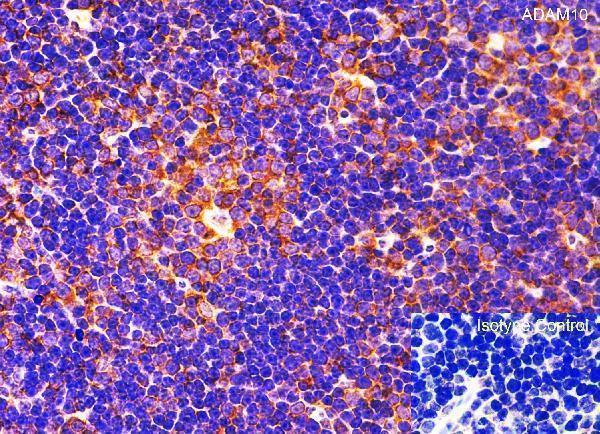
Validated anti-ADAM10 antibodies have real IHC images from human and mouse paraffin sections and IF images from MOLT4 cells and mouse/rat tissues (M00566 and A00566-2 image captions).
M00566 has IHC images from human paraffin tumor sections and lists human, mouse and rat reactivity (M00566 IHC captions; catalog). A00566-2 has IHC images from human testis and mouse thymus, plus IF images from MOLT4 cells and mouse/rat tissues; ICC is listed as an application (A00566-2 IHC/IF captions; catalog).
Which to pick: For paraffin tissue IHC, choose M00566 when a rabbit monoclonal with human tumor-section examples fits; its caption describes EDTA retrieval but does not report the fixative (M00566 catalog; IHC captions). Choose A00566-2 for IF/ICC because both applications are listed and IF images are supplied; its IHC captions document paraffin-section human testis and mouse thymus with citrate retrieval (A00566-2 catalog; IHC/IF captions). For cross-species IHC, A00566-2 has human and mouse image examples, while its rat examples are IF; M00566 lists human, mouse and rat reactivity but supplies human IHC examples (A00566-2 IHC/IF captions; M00566 catalog and IHC captions). The selected M00566 tissue-IHC caption documents paraffin sections, but does not specify the fixative (selected-SKU IHC image M00566).