ANP32A / Acidic leucine-rich nuclear phosphoprotein 32 family member A · IHC design guide

Design Immunohistochemistry for ANP32A

Plan ANP32A staining in paraffin sections around its reported ubiquitous nuclear pattern (HPA tissue IHC). This guide covers fixation, retrieval, antibody titration, chromogenic detection and scoring, with attention to possible staining from more than one gene product (HPA tissue IHC).

Evidence assembled Oct 2026 · For research use; verify linked source records and product datasheet before use
Immunohistochemistry protocol sheet for ANP32A (IHC for ANP32A): expected localisation Nuclear staining in tissue (HPA tissue IHC), antibody M03625, validated IHC image, and IHC protocol steps
Printable ANP32A IHC protocol sheet — expected localisation Nuclear staining in tissue (HPA tissue IHC), antibody M03625, controls and protocol steps. Open the full ANP32A IHC guide →

ANP32A Immunohistochemistry Experimental Design Guide

Expected localisation, validated protocols, controls and antibodies — the at-a-glance facts below, then the full design guide.

Must know before staining
Expected localisation Nuclear staining in tissue (HPA tissue IHC)
Staining pattern Ubiquitous nuclear staining across tissue cell types (HPA tissue IHC)
Antigen retrieval Tris-EDTA pH 9.0 HIER, 95–98 °C, 20 min (rule: nuclear antigen)
Positive control ⓘ Adrenal gland+4 more · see all
Negative control ⓘ None in HPA (detected in all 45 tissues); use no-primary + isotype controls
Important caveats
Reasons your staining may differ from the expected pattern.
Fixation Keep fixation conditions consistent across sections. (standard IHC practice; not target-specific)
Caveat Antibody may recognize products of more than one gene (HPA tissue IHC)
Regulation No staining regulator specified (UniProt)
Isoform / epitope No isoforms listed; one 1–249 chain (UniProt)
Section 1

Recommended ANP32A IHC & IF Protocols

Compare the catalog antibody’s IHC-P protocol with published ANP32A staining protocols for oral squamous cell carcinoma (PMC4905446) and developing brain (PMC4383345).

Recommended immunohistochemistry (IHC-P) protocol parameters
SampleParaffin-embedded Human Kidney tissue; fixative not specified (datasheet M03625)
FixationImage fixative and duration unreported (datasheet M03625); verify before use.
Sectioning4–5 µm sections on charged slides (standard)
DeparaffinisationXylene, graded ethanol series to water (standard)
Antigen retrievalHeat-induced epitope retrieval in Tris-EDTA buffer, pH 9.0, 20 min at 95–98 °C (standard rule: nuclear antigen)
Peroxidase block3% H2O2, 10 min, room temperature (standard)
Blocking10% normal serum of the secondary host, 30 min, room temperature (standard)
Primary antibodyMouse monoclonal (clone 8D2) anti-ANP32A, 1:200-1:1000 (datasheet M03625)
Primary incubationOvernight at 4 °C (standard)
DetectionHRP-polymer secondary, DAB chromogen 5–10 min (standard)
CounterstainHematoxylin, blue, dehydrate and mount (standard)
Expected resultANP32A-positive staining in glandular cells of adrenal gland (HPA tissue IHC: High). HPA tissue profile: Ubiquitous nuclear expression. No signal in the no-primary control.
💡Decision noteStart with Tris-EDTA pH 9.0 HIER at 95–98 °C for 20 min (page retrieval rule: nuclear antigen); citrate buffer is a published alternative (PMC4905446; PMC4383345).
Section 2

What Is the Expected ANP32A Staining Pattern?

ANP32A should stain mainly nuclei across many cell types in paraffin sections, including high staining in colon glandular cells, bone marrow hematopoietic cells and cerebral cortex neurons (HPA: ubiquitous nuclear expression; High in these cells). Cytoplasmic staining can occur (UniProt P39687: nuclear–cytoplasmic shuttling). ANP32A has no transmembrane segment (UniProt P39687: topology). HPA rates tissue IHC Supported, with medium agreement between staining and RNA and a warning that antibodies target proteins from more than one gene (HPA: tissue IHC reliability).

What am I looking at on my slide?
Clear nuclear staining in colon glandular cells or bone marrow hematopoietic cells, with low surrounding background.This fits the reported compartment and two high-staining cell populations (HPA: ubiquitous nuclear expression; High in colon glandular and bone marrow hematopoietic cells). Judge signal within the relevant cells rather than by whole-section color (general IHC practice). HPA's multi-gene targeting warning limits attribution of the stain to ANP32A alone (HPA: tissue IHC caution).
Strong membrane-rim staining dominates while nuclei remain unstained.A membrane-only pattern conflicts with the reported nuclear pattern and the absence of a transmembrane segment (HPA: ubiquitous nuclear expression; UniProt P39687: topology). Check morphology and chromogen placement, then review antibody specificity and controls before interpreting it as ANP32A (general IHC practice). Cytoplasmic signal alone is not proof of an artefact because ANP32A can shuttle (UniProt P39687: subcellular location).
The strongest signal lies outside the expected positive cell population in a reference tissue.For example, colon glandular cells are reported High; signal confined to unrelated structures does not reproduce that reference pattern (HPA: High in colon glandular cells). Consider antibody cross-reactivity or endogenous detection activity, and inspect matched controls (general IHC practice). HPA's multi-gene targeting warning makes specificity assessment especially relevant (HPA: tissue IHC caution).
Weak, diffuse brown haze spans nuclei, cytoplasm and tissue spaces.Poor compartment contrast prevents a confident positive call against the reported nuclear pattern (HPA: ubiquitous nuclear expression). Background can arise from nonspecific antibody binding or endogenous detection activity (general IHC practice). Compare the primary-antibody omission control and tissue morphology before assigning any cellular signal (general IHC practice).
No cellular signal appears in a section containing a reported high-staining population.Absent staining in colon glandular cells or cerebral cortex neurons fails the reported High reference pattern (HPA: High in those cells). First assess whether the cells are present and intact; then check the staining run, detection reagents and antibody conditions (general IHC practice). A single failed section does not establish biological absence (general IHC practice).
💡Expected ANP32A appearanceCall a positive result when identifiable reference cells show clear, predominantly nuclear staining—High in colon glandular cells or bone marrow hematopoietic cells (HPA: tissue IHC)—while treating membrane-only color or diffuse, cell-independent haze as a reason to investigate artefact (UniProt P39687: no transmembrane segment; general IHC practice).
How each factor affects the staining
Cellular compartmentUse nuclear signal as the principal IHC landmark (HPA: ubiquitous nuclear expression). Some cytoplasmic signal is biologically plausible because ANP32A shuttles between nucleus and cytoplasm (UniProt P39687: subcellular location). Its reported cytoplasmic translocation during neuritogenesis is by similarity, so that note alone should not determine an IHC call (UniProt P39687: subcellular note).
Reference cell selectionHPA reports High staining in adrenal glandular cells, bronchial respiratory epithelial cells, cerebellar molecular-layer cells and several other listed populations (HPA: tissue IHC). It reports Low staining in caudate neurons, oral mucosa squamous epithelium and stomach glandular cells (HPA: tissue IHC). Low is a weak reference, not a verified negative (HPA: tissue IHC levels).
Strength of staining evidenceThree listed antibodies have Supported IHC status, while the tissue profile has medium staining–RNA consistency and a multi-gene targeting caution (HPA: antibody validation; tissue IHC reliability). Reproduction of the reported pattern supports interpretation, but the supplied evidence does not establish ANP32A-exclusive staining for any one reagent (HPA: tissue IHC caution).
Protein topology and processingThe recorded protein has no transmembrane segment, signal peptide or propeptide, and its listed chain spans residues 1–249 (UniProt P39687: topology and processing). These annotations support a non-membranous expectation; they do not specify antigen-retrieval conditions or prove how paraffin processing affects the epitope (UniProt P39687: topology; general IHC practice).
Why is my staining missing, weak or wrong?
SituationLikely causeNext action
Reference cells are blank across the section.The run may have failed, or staining conditions may give insufficient signal (general IHC practice); the result conflicts with High reference-cell staining (HPA: tissue IHC).Confirm the reference cells on the counterstained section, then check detection reagents and optimize retrieval and primary-antibody conditions within the laboratory's IHC workflow (general IHC practice). No ANP32A-specific retrieval requirement is supplied (HPA: tissue IHC; UniProt P39687).
Nuclei and tissue spaces share uniform brown haze.Diffuse color may reflect nonspecific binding or endogenous enzyme activity (general IHC practice), rather than the reported nuclear pattern (HPA: tissue IHC profile).Compare a primary-antibody omission control; assess blocking, washes and endogenous enzyme control for the detection system (general IHC practice). Reassess staining only where cell boundaries and nuclei remain interpretable (general IHC practice).
Only membrane outlines stain.That distribution conflicts with nuclear IHC and the absence of a transmembrane segment (HPA: tissue IHC profile; UniProt P39687: topology).Review section morphology and control staining, then compare with an independently validated antibody or orthogonal evidence if available (general IHC practice). Do not score membrane-only color as the expected ANP32A pattern (HPA: ubiquitous nuclear expression).
A presumed negative tissue has weak staining.The supplied HPA record lists Low populations but no negative population; weak color may be compatible with a Low call (HPA: tissue IHC levels).Check the named cell type and compartment against the HPA reference, and use a staining control to assess background (HPA: tissue IHC; general IHC practice). Avoid treating caudate neurons, oral mucosa epithelium or stomach glands as verified negatives (HPA: Low in these cells).
Two IHC antibodies give different cell patterns.HPA lists Supported IHC status for HPA063133, HPA067561 and CAB005231, but warns about targeting proteins from more than one gene (HPA: antibody validation; tissue IHC caution).Compare nuclear localization and the same HPA reference cell populations across sections; investigate discordant staining with independent specificity evidence before assigning it to ANP32A (HPA: tissue IHC profile and caution; general IHC practice).
What should a separate IF/ICC image show?HPA reports nucleoplasm as the main location, with supported cytosol and approved centrosome locations; its subcellular summary carries a multi-gene antibody caution (HPA: ICC-IF subcellular).Expect chiefly nucleoplasmic signal and interpret additional cytosolic or centrosomal signal with that caveat (HPA: ICC-IF subcellular). Use the separate IF/ICC guide for its workflow; these location data supply no IHC protocol conditions (HPA: ICC-IF subcellular).

Sample controls for ANP32A IHC & IF

🧪Run breast first: glandular cells should stain (HPA: High in breast glandular cells). HPA detects ANP32A in all 45 scored tissues and lists no negative tissue (HPA: no negative rows), so no-primary and isotype controls carry the negative comparison; no cell population on the positive slide is established as an internal antigen-negative control, and unstained cells should not be assumed negative.
Positive control tissue: Adrenal gland (Glandular cells, HPA High)
Negative control tissue: None in HPA: ANP32A is detected in all 45 scored tissues. Use a no-primary (secondary-only) and an isotype control instead.
ICC-IF cell lines (HPA subcellular resource): HPA ICC-IF images show ANP32A in MCF-7, Rh30, U2OS, HEK293, SK-MEL-30, with annotated localisation: Nucleoplasm (supported) (HPA subcellular).
Technical controls: Include a no-primary, secondary-only section; an isotype control matched to the primary antibody’s host species and clonality; and, where available, an ANP32A knockout biological control. For chromogenic IHC, block endogenous peroxidase and assess pigment or other tissue background against the no-primary section (standard IHC practice).
⚠️Feasibility: A target-specific fixation window and fixation effect are unreported in the supplied evidence, and the selected M03625 paraffin-section caption does not state a fixative (M03625 tissue-IHC caption). Antigen retrieval requirements are likewise unreported, so evaluate retrieval conditions empirically for paraffin sections (standard IHC practice). The supplied evidence does not establish that frozen sections or IF are easier; IF/ICC images exist for listed cell lines (HPA: subcellular), while tissue-specific artefacts should be checked with the chromogenic background control (standard IHC practice).

HPA tissue IHC evidence for ANP32A

Comprehensive Human Protein Atlas IHC scoring per tissue (reliability: Supported — Medium consistency between antibody staining and RNA expression data. Caution, targets protein from more than one gene.). Rows are taken directly from the HPA tissue chart — click any row's HPA link to view the source.

Positive expression · recommended positive controls

TissueCell typeLevelEvidenceSource
Adrenal gland Glandular cells High Protein (IHC) HPA →
Bone marrow Hematopoietic cells High Protein (IHC) HPA →
Breast Glandular cells High Protein (IHC) HPA →
Bronchus Respiratory epithelial cells High Protein (IHC) HPA →
Cerebellum Cells in molecular layer High Protein (IHC) HPA →

Undetected expression · recommended negative controls

TissueCell typeLevelEvidenceSource
None in HPA: ANP32A is detected in all 45 scored tissues. Use a no-primary (secondary-only) and an isotype control instead.
Section 3

Advanced ANP32A IHC Tips

Troubleshoot ANP32A staining in paraffin sections by checking retrieval, nuclear localisation, antibody specificity, and consistent scoring (UniProt P39687; HPA tissue IHC).

How should I retrieve ANP32A in paraffin sections with weak nuclear staining?
Start with Tris-EDTA at pH 9.0, heated to 95–98 °C for 20 min (page retrieval setting: nuclear antigen). Allow slides to cool in retrieval buffer before washing, and run a known positive section alongside the test section (standard IHC practice). Judge improvement by nuclear signal because the reported tissue profile is ubiquitously nuclear, while the selected M03625 image shows DAB staining in paraffin-embedded human kidney (HPA tissue IHC; M03625 caption). If staining remains weak, test an alternative retrieval buffer as a fallback on matched sections, changing one variable at a time (standard IHC practice). Compare signal and tissue integrity before adopting that fallback (standard IHC practice).
Could fixation explain inconsistent ANP32A staining between paraffin blocks?
Target-specific ANP32A fixation sensitivity is unknown: the selected M03625 caption identifies paraffin-embedded human kidney and DAB staining but does not report a fixative (M03625 caption). Record each block’s fixative and fixation duration when available, then compare sections processed with the same retrieval and detection conditions (standard IHC practice). For new material, a documented 10% neutral buffered formalin process provides a reproducible starting point, without establishing an ANP32A-specific optimum (standard IHC practice). Include a control section in each run so a processing difference is easier to distinguish from staining failure (standard IHC practice). Do not infer fixation tolerance from the HPA expression pattern or ANP32A topology (HPA tissue IHC; UniProt P39687 topology).
What should I check when ANP32A staining appears mostly cytoplasmic?
First inspect nuclei against the counterstain: the HPA tissue profile reports ubiquitous nuclear expression, and its subcellular profile supports nucleoplasmic localisation (HPA tissue IHC; HPA subcellular). Cytoplasmic staining can still be biologically plausible because ANP32A shuttles between nucleus and cytoplasm; cytoplasmic translocation during neuritogenesis is annotated by similarity (UniProt P39687 subcellular). Compare the cytoplasmic pattern with matched controls, section morphology, and the fraction of cells retaining nuclear staining (standard IHC practice). Repeat staining with controlled retrieval and detection if diffuse colour obscures compartment boundaries (standard IHC practice). Treat an exclusively cytoplasmic pattern cautiously, particularly because the HPA antibody evidence can include proteins from multiple genes (HPA subcellular caution).
How can I assess whether an ANP32A epitope is inaccessible or cross-reactive?
The supplied UniProt record lists no annotated isoforms, so an unexpected staining pattern should not be assigned to a specific ANP32A isoform without further evidence (UniProt P39687 isoforms). ANP32A has no transmembrane segment and contains an LRRCT region at residues 123–161; phosphorylation is annotated at several residues (UniProt P39687 topology, domains, modified residues). If epitope information becomes available, map it against that sequence and compare retrieval conditions on adjacent sections (UniProt P39687; standard IHC practice). Include an independent specificity control, such as a validated second antibody or genetic loss control, when interpretation matters (standard IHC practice). HPA warns that its antibody staining may reflect proteins from multiple genes, so agreement with HPA alone cannot establish specificity (HPA tissue IHC).
How should I extend an ANP32A tissue finding to multiplex IF?
Treat multiplex IF as a separate assay and pair ANP32A with a marker of the expected cell type in the section being examined (standard IF practice). Select fluorophores after checking unstained tissue autofluorescence, and place the weaker target in a channel with low background and good detector separation (standard IF practice). ANP32A lacks a transmembrane segment and is reported mainly in the nucleoplasm, so permeabilisation must give antibody access to intracellular and nuclear epitopes (UniProt P39687 topology; HPA subcellular). A brief 0.1% Triton X-100 treatment for 5 min is a general starting condition to optimise against morphology and background, not an ANP32A-validated setting (standard IF practice). Include single-colour controls to identify bleed-through before interpreting colocalisation (standard IF practice).
What causes diffuse DAB colour or apparent ANP32A positivity across the section?
Inspect a no-primary control and tissue morphology before calling diffuse DAB colour ANP32A signal (standard IHC practice). In chromogenic sections, an endogenous peroxidase block, for example 3% hydrogen peroxide for 10 min, and an appropriate protein block are general workflow controls (standard IHC practice). Titrate the primary antibody and shorten DAB development if colour obscures nuclei, keeping the control section under identical conditions (standard IHC practice). The selected M03625 caption demonstrates DAB staining in paraffin-embedded human kidney but supplies no antibody dilution or blocking conditions (M03625 caption). Nuclear enrichment is a useful pattern check, though the HPA multi-gene caution means morphology alone cannot prove specificity (HPA tissue IHC).
How should I quantify ANP32A when nuclear intensity varies across a section? ⚠ ANSWER MARKED FOR VERIFICATION
Define the analysed cell population and count only morphologically intact cells within a prespecified region of interest (standard IHC practice). Report the percentage of positive nuclei and an H-score from nuclear intensity categories, using the same threshold and counterstain criteria across sections (standard IHC practice; HPA tissue IHC: ubiquitous nuclear expression). If cell abundance differs between regions, normalise positive counts to the total eligible nuclei or report positive-cell density per mm² (standard IHC practice). Score cytoplasmic staining separately because ANP32A can shuttle between nucleus and cytoplasm (UniProt P39687 subcellular). Exclude folds, edges, and necrotic areas consistently, and compare batches through a shared control section (standard IHC practice).
How do I distinguish convincing ANP32A staining from artefact?
A convincing result shows interpretable nuclear signal in intact cells, consistent with the HPA tissue profile and supported nucleoplasmic localisation (HPA tissue IHC; HPA subcellular). Assess any cytoplasmic signal separately because nucleus-to-cytoplasm shuttling is reported, while the HPA tissue assay warns of possible multi-gene detection (UniProt P39687 subcellular; HPA tissue IHC). Reject colour confined to section edges, folds, necrotic tissue, or the no-primary control as evidence of target expression (standard IHC practice). Check endogenous enzyme activity when DAB appears without a plausible cellular pattern (standard IHC practice). For consequential conclusions, confirm the pattern with an independent specificity control and comparable processing across sections (standard IHC practice).
Boster reagents

Best ANP32A / Acidic leucine-rich nuclear phosphoprotein 32 family member A IHC Antibodies

Anti-ANP32A antibodies have human paraffin-section IHC data (M03625 image caption) and Raji-cell IF/ICC data (A03625-1 and A03625-2 image captions); catalog reactivity also lists mouse and rat (A03625-1/A03625-2 catalog).

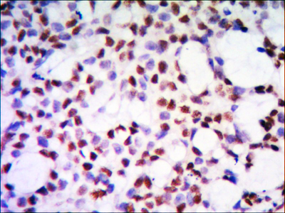
Real IHC data Immunohistochemistry validation of ANP32A using Anti-ANP32A Monoclonal Antibody (M03625). Immunohistochemistry (IHC) analysis of paraffin-embedded Human Kidney tissue with DAB staining using ANP32A Monoclonal Antibody. For more protocol information of IHC
Anti-ANP32A Monoclonal Antibody
Cat # M03625
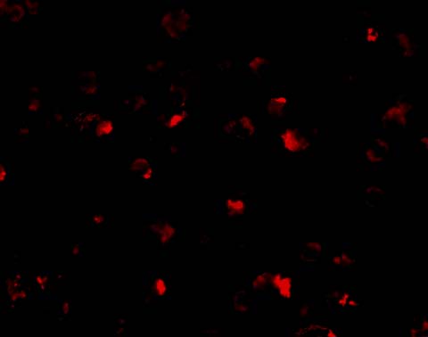
Real IF data Immunofluorescence Validation of PHAP I in Raji Cells Immunofluorescent analysis of 4% paraformaldehyde-fixed Raji Cells labeling PHAP I with A03625-1 at 10 μg/mL, followed by goat anti-rabbit IgG secondary antibody at 1/500 dilution (red).
Anti-PHAP I ANP32A Antibody
Cat # A03625-1
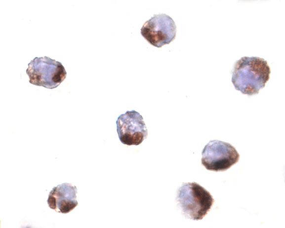
Real IF data Immunocytochemistry of PHAP in Raji cells with PHAP antibody at 1 μg/mL.
Anti-PHAP ANP32A Antibody
Cat # A03625-2

M03625 shows DAB IHC in paraffin-embedded human kidney, while A03625-1 shows IF and ICC in Raji cells (M03625 image caption; A03625-1 image captions). A03625-2 lists IHC-P and human, mouse and rat reactivity, but its displayed image shows Raji-cell ICC (A03625-2 catalog; A03625-2 image caption).

Which to pick: For human tissue IHC, choose M03625, a mouse monoclonal (clone 8D2) shown on paraffin-embedded human kidney; its caption does not report the fixative (M03625 catalog; M03625 image caption). For IF/ICC, choose A03625-1, a rabbit antibody with Raji-cell IF and ICC images (A03625-1 catalog; A03625-1 image captions). For cross-species tissue IHC, A03625-2 lists rabbit-host IHC-P and human, mouse and rat reactivity, starting at 1 μg/mL; its displayed image shows Raji-cell ICC rather than tissue IHC (A03625-2 catalog; A03625-2 image caption).

Each figure is that product's own IHC / IF validation image from its datasheet.

References

  1. UniProt Consortium. UniProt entry P39687 (AN32A_HUMAN, Acidic leucine-rich nuclear phosphoprotein 32 family member A).
  2. Human Protein Atlas. ANP32A tissue IHC expression (reliability: Supported).
  3. Human Protein Atlas. ANP32A subcellular location (ICC-IF): Mainly localized to the nucleoplasm. In addition localized to the cytosol and centrosome. Caution: Based on antibodies targeting proteins from multiple genes..
  4. Human Protein Atlas. ANP32A antibody validation summary (3 antibodies).
  5. Acidic leucine-rich nuclear phosphoprotein-32A (ANP32A) association with lymph node metastasis predicts poor survival in oral squamous cell carcinoma patients. Oncotarget 2016 — PMC4905446.
  6. The expression and distributions of ANP32A in the developing brain. BioMed research international 2015 — PMC4383345.
  7. Prion disease induced alterations in gene expression in spleen and brain prior to clinical symptoms. Advances and applications in bioinformatics and chemistry : AABC 2008 — PMC3169940.
  8. Anp32a Promotes Neuronal Regeneration after Spinal Cord Injury of Zebrafish Embryos. International journal of molecular sciences 2022 — PMC9786895.
  9. PubMed PMID:8192856 — UniProt-cited evidence.
  10. PubMed PMID:8679524 — UniProt-cited evidence.
  11. PubMed PMID:8970164 — UniProt-cited evidence.