This website uses cookies to ensure you get the best experience on our website.


- Table of Contents
Plan chromogenic BCL2L1 IHC around the cytoplasmic staining reported in select tissues (HPA tissue IHC). This guide covers fixation consistency, tissue controls and antibody epitope coverage across 3 isoforms (standard IHC practice; UniProt).

Expected localisation, validated protocols, controls and antibodies — the at-a-glance facts below, then the full design guide.
| Expected localisation | Cytoplasmic tissue staining (HPA tissue IHC) | |
| Staining pattern | Cytoplasmic signal in marrow, lymph node and tubule cells (HPA tissue IHC) | |
| Antigen retrieval | EDTA pH 8.0 HIER, heat-mediated (datasheet PA1232) | |
| Positive control | Adrenal gland+4 more · see all | |
| Negative control | Adipose tissue+4 more · see all |
| Fixation | Keep fixation consistent across paraffin sections (standard IHC practice; not target-specific) | |
| Caveat | Antibody staining and RNA show low consistency (HPA tissue IHC) | |
| Regulation | Bcl-X(S) high in developing lymphocytes (UniProt) | |
| Isoform / epitope | 3 splice isoforms; confirm antibody epitope coverage (UniProt) |
The catalog antibody’s IHC-P protocol (datasheet: PA1232) is accompanied by one published colorectal tumor IHC protocol (PMC4456718).
| Sample | Tissue sections; selected-image fixative not specified (standard IHC workflow) |
| Fixation | Image fixative and duration unreported (datasheet PA1232); verify before use. |
| Sectioning | 4–5 µm sections on charged slides (standard) |
| Deparaffinisation | Xylene, graded ethanol series to water (standard) |
| Antigen retrieval | Heat retrieval: EDTA pH 8.0 (datasheet PA1232); 20 min, 95–100 °C (standard) |
| Peroxidase block | 3% H2O2, 10 min, room temperature (standard) |
| Blocking | 10% goat serum (datasheet PA1232) |
| Primary antibody | Rabbit anti-BCL2L1, 0.5-1μg/ml (datasheet PA1232) |
| Primary incubation | Overnight at 4 °C (datasheet PA1232) |
| Detection | HRP-conjugated secondary, DAB chromogen (datasheet PA1232) |
| Counterstain | Hematoxylin, blue, dehydrate and mount (standard) |
| Expected result | BCL2L1-positive staining in glandular cells of adrenal gland (HPA tissue IHC: Medium). HPA tissue profile: Cytoplasmic expression in a few tissues, including bone marrow, lymph node and cells in tubules. No signal in the no-primary control. |
BCL2L1 is a membrane associated protein with a transmembrane segment at residues 210–226 and reported mitochondrial, cytosolic, vesicular, and other locations (UniProt Q07817 topology and subcellular location). In paraffin section IHC, expect mainly cytoplasmic staining in selected cells, including bone marrow hematopoietic cells, lymph node non-germinal center cells, and kidney tubular cells (HPA tissue IHC). HPA rates the tissue staining Approved but reports low consistency with RNA expression (HPA tissue IHC).
| Moderate cytoplasmic staining in kidney tubular cells, bone marrow hematopoietic cells, or lymph node non-germinal center cells (HPA tissue IHC). | This matches the reported positive cell populations and compartment (HPA tissue IHC). Score the named cells rather than calling the entire section positive. HPA reports low consistency between antibody staining and RNA expression, so this pattern supports interpretation without independently proving antibody specificity (HPA tissue IHC). |
| Predominantly nuclear chromogen, with little cytoplasmic staining in the expected cells. | Treat a nuclear dominant IHC pattern as questionable: HPA describes tissue staining as cytoplasmic, while UniProt lists a nuclear membrane location among several locations (HPA tissue IHC; UniProt Q07817 subcellular location). Check the counterstain and detection controls before assigning that pattern to BCL2L1 (general IHC practice). |
| Strong staining in adipocytes or heart cardiomyocytes, with weak staining in the expected positive cells. | HPA reports BCL2L1 as not detected in those two cell populations (HPA tissue IHC). Consider cross-reactivity or endogenous detection activity, especially if a no-primary control also stains (general IHC practice). A single unexpected positive field does not establish a new tissue pattern. |
| Diffuse chromogen across stroma, cell borders, and empty areas rather than within identifiable cells. | This distribution does not resemble HPA's cell-restricted cytoplasmic tissue pattern (HPA tissue IHC). Review the no-primary control for detection background and assess washing and blocking (general IHC practice). Interpret intensity only after background allows individual positive cells to be distinguished. |
| No staining in kidney tubular cells or bone marrow hematopoietic cells. | Both are reported as Medium positive by HPA, so absence of signal calls for a run-level check (HPA tissue IHC). Confirm that a known-positive control worked, then review section handling, antigen retrieval, antibody application, and chromogen detection (general IHC practice). HPA's low RNA concordance limits how much one negative specimen can prove (HPA tissue IHC). |
| Cell population chosen for scoring (HPA tissue IHC). | HPA reports Medium staining in selected cells across several tissues, including respiratory epithelial cells in bronchus and glandular cells in adrenal gland (HPA tissue IHC). It also reports Not detected in specific cells within other tissues; compare like cell populations rather than whole organs (HPA tissue IHC). |
| Tissue IHC evidence and antibody validation (HPA tissue IHC; HPA antibodies). | The HPA tissue assessment is Approved but notes low agreement between staining and RNA data; HPA035734 has Approved IHC status (HPA tissue IHC; HPA antibodies). Use the reported staining as a reference pattern, with a positive and detection control for the run (general IHC practice). |
| Membrane association and cellular compartment (UniProt Q07817 topology and subcellular location). | A transmembrane segment at residues 210–226 accompanies UniProt mitochondrial membrane, synaptic vesicle membrane, cytosolic, and other location annotations (UniProt Q07817). These locations inform interpretation, but HPA's paraffin section reference is cytoplasmic staining (HPA tissue IHC). |
| Isoforms and tissue context (UniProt Q07817 isoforms and tissue specificity). | UniProt lists Bcl-X(L), Bcl-X(S), and Bcl-X(beta); it describes Bcl-X(S) in high-turnover cells such as developing lymphocytes and Bcl-X(L) in long-lived cells such as adult brain (UniProt Q07817). The supplied record gives no antibody epitope or isoform selectivity, so IHC intensity cannot identify an isoform. |
| IF/ICC Q: Should punctate mitochondrial fluorescence be expected? | A: HPA calls mitochondria the approved main ICC-IF location; nucleoli fibrillar center is uncertain, and acrosome is approved (HPA subcellular). That IF observation does not set a required punctate appearance for chromogenic paraffin section IHC, whose HPA tissue description is cytoplasmic (HPA tissue IHC). |
| Situation | Likely cause | Next action |
|---|---|---|
| Expected positive cells are blank while the no-primary control is clean. | The run may have failed at section preparation, retrieval, primary antibody application, or detection (general IHC practice); HPA reports Medium staining in kidney tubular cells and bone marrow hematopoietic cells (HPA tissue IHC). | Check a known-positive section processed in the same run, then review each IHC step and reagent record (general IHC practice). Avoid calling the specimen biologically negative until the control produces interpretable staining. |
| Chromogen appears in the no-primary control. | Detection-system or endogenous activity can produce antibody-independent signal (general IHC practice). Such signal cannot be counted as BCL2L1 even if it overlaps a cell population reported positive by HPA (HPA tissue IHC). | Review endogenous enzyme blocking, secondary detection, and chromogen controls; resolve the control signal before scoring the sample (general IHC practice). |
| Most cells and extracellular areas show similar color. | High nonspecific background can obscure HPA's selective cytoplasmic pattern (HPA tissue IHC; general IHC practice). | Compare with the no-primary control, then review blocking, washes, and antibody concentration according to the antibody's validated IHC instructions (general IHC practice). Score only after cell boundaries and the expected positive populations are distinguishable. |
| Staining is nuclear dominant in every tissue compartment. | The distribution conflicts with HPA's cytoplasmic tissue description; a listed nuclear membrane location does not establish diffuse nuclear IHC staining (HPA tissue IHC; UniProt Q07817 subcellular location). | Check counterstain interpretation and no-primary staining, then compare a validated positive section in the same run (general IHC practice). |
| Adipocytes or cardiomyocytes stain more strongly than the expected positive cells. | HPA reports those populations as Not detected, making cross-reactivity or detection background plausible explanations (HPA tissue IHC; general IHC practice). | Confirm cell identity on the section, inspect the no-primary control, and compare with an HPA-reported positive population before attributing the unexpected signal to BCL2L1 (HPA tissue IHC; general IHC practice). |
| An IF image looks mitochondrial, but the matched IHC section looks broadly cytoplasmic. | HPA assigns mitochondria as the approved main ICC-IF location and describes tissue IHC staining as cytoplasmic (HPA subcellular; HPA tissue IHC). The two readouts have different spatial resolution (general microscopy practice). | Judge each image against its own HPA reference and controls. Use the tissue IHC cell population and cytoplasmic distribution to score the paraffin section (HPA tissue IHC; general IHC practice). |





Comprehensive Human Protein Atlas IHC scoring per tissue (reliability: Approved — Low consistency between antibody staining and RNA expression data.). Rows are taken directly from the HPA tissue chart — click any row's HPA link to view the source.
| Tissue | Cell type | Level | Evidence | Source |
|---|---|---|---|---|
| Adrenal gland | Glandular cells | Medium | Protein (IHC) | HPA → |
| Bone marrow | Hematopoietic cells | Medium | Protein (IHC) | HPA → |
| Bronchus | Respiratory epithelial cells | Medium | Protein (IHC) | HPA → |
| Cerebral cortex | Endothelial cells | Medium | Protein (IHC) | HPA → |
| Fallopian tube | Glandular cells | Medium | Protein (IHC) | HPA → |
| Tissue | Cell type | Level | Evidence | Source |
|---|---|---|---|---|
| Adipose tissue | Adipocytes | Not detected | Protein (IHC) | HPA → |
| Cervix | Glandular cells | Not detected | Protein (IHC) | HPA → |
| Heart muscle | Cardiomyocytes | Not detected | Protein (IHC) | HPA → |
| Hippocampus | Glial cells | Not detected | Protein (IHC) | HPA → |
| Nasopharynx | Respiratory epithelial cells | Not detected | Protein (IHC) | HPA → |
Troubleshoot BCL2L1 staining in paraffin sections by checking retrieval, cell identity, subcellular pattern and controls before comparing signal across samples.
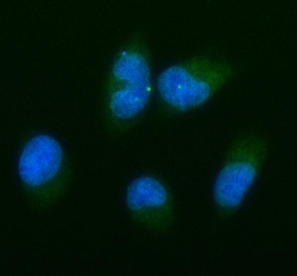
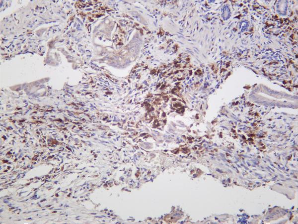
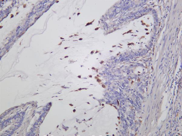
IHC images show human tissues (catalog IHC captions: PA1232, A00181, M00181-1, M00181-4); IF images show TPC-1 and HeLa cells (catalog IF captions: A00181, M00181-1). All four list human, mouse and rat reactivity (catalog reactivity).
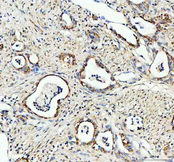
PA1232 shows paraffin-section IHC in human colon cancer, while A00181 shows paraffin-section IHC in human pancreatic cancer and IF in TPC-1 cells (PA1232 and A00181 image captions). M00181-1 shows paraffin-section IHC in human pituitary tumor and liver cancer plus IF in HeLa cells; M00181-4 shows IHC in human colon carcinoma (M00181-1 and M00181-4 image captions).
Which to pick: For tissue IHC targeting Bcl-XL, choose A00181: its paraffin-section caption documents EDTA retrieval at pH 8.0 and primary antibody at 2 μg/ml; the fixative is unreported (A00181 IHC caption). For IF/ICC, A00181 has a TPC-1 cell image at 5 μg/ml (A00181 IF caption). For mouse or rat IHC planning, M00181-4 is a monoclonal option with both species listed as reactive, although its IHC image shows human colon carcinoma (M00181-4 catalog description, reactivity and IHC caption).