This website uses cookies to ensure you get the best experience on our website.
- Table of Contents
2 Citations 9 Q&As
1 Citations 7 Q&As
4 Citations 17 Q&As
1 Citations 3 Q&As
1 Citations 14 Q&As
2 Citations 16 Q&As
1 Citations 1 Q&As
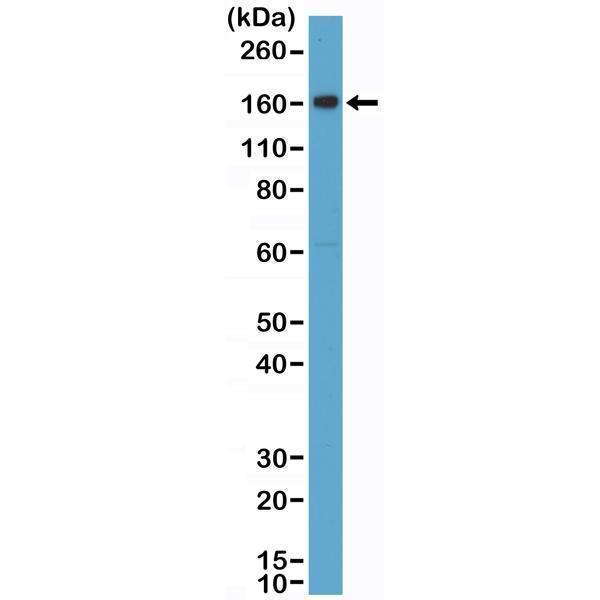
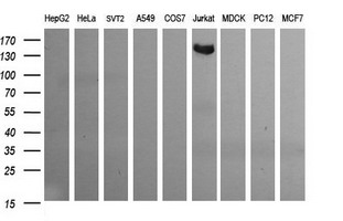
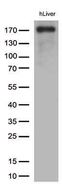
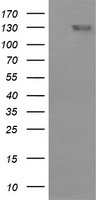
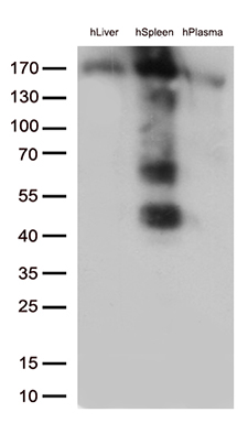
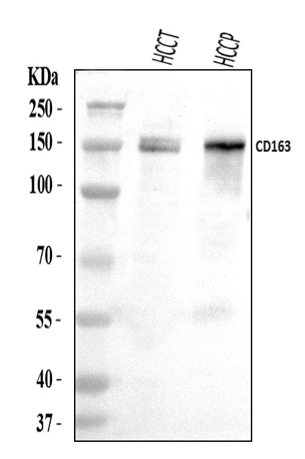
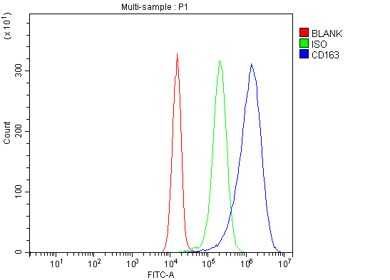
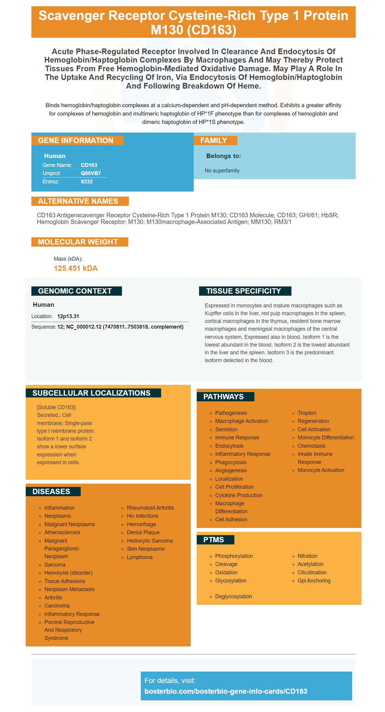
Facts about Scavenger receptor cysteine-rich type 1 protein M130.
Binds hemoglobin/haptoglobin complexes at a calcium-dependent and pH-dependent method. Exhibits a greater affinity for complexes of hemoglobin and multimeric haptoglobin of HP*1F phenotype than for complexes of hemoglobin and dimeric haptoglobin of HP*1S phenotype.
Human | |
---|---|
Gene Name: | CD163 |
Uniprot: | Q86VB7 |
Entrez: | 9332 |
Belongs to: |
---|
No superfamily |
CD163 antigenscavenger receptor cysteine-rich type 1 protein M130; CD163 molecule; CD163; GHI/61; HbSR; Hemoglobin scavenger receptor; M130; M130macrophage-associated antigen; MM130; RM3/1
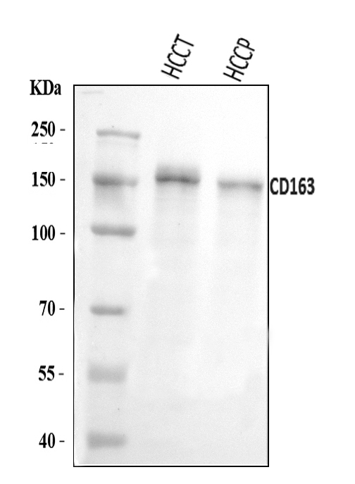
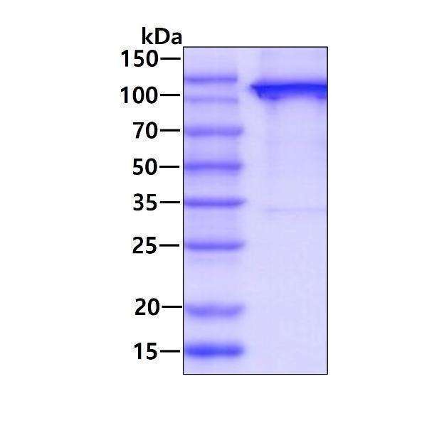
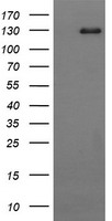
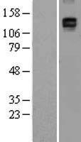
Mass (kDA):
125.451 kDA
Human | |
---|---|
Location: | 12p13.31 |
Sequence: | 12; NC_000012.12 (7470811..7503818, complement) |
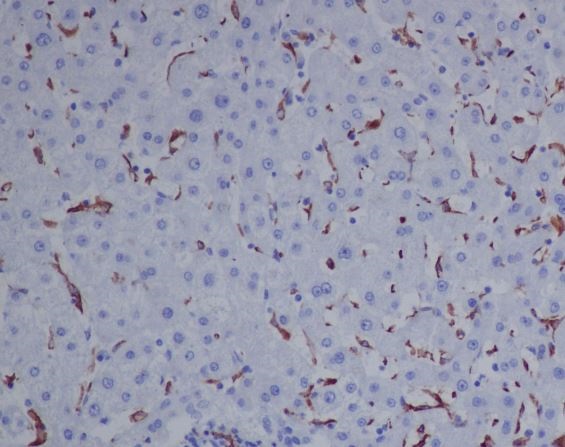
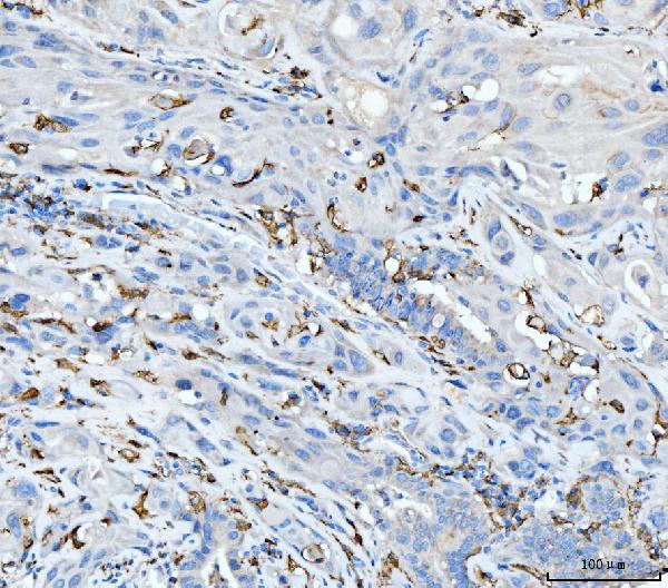
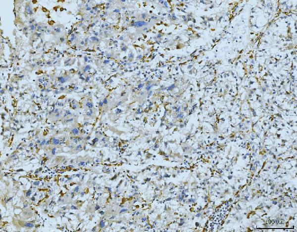
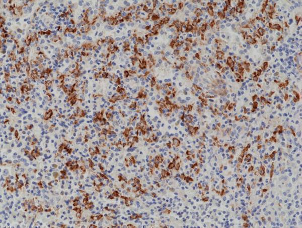
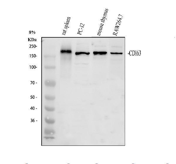
Expressed in monocytes and mature macrophages such as Kupffer cells in the liver, red pulp macrophages in the spleen, cortical macrophages in the thymus, resident bone marrow macrophages and meningeal macrophages of the central nervous system. Expressed also in blood. Isoform 1 is the lowest abundant in the blood. Isoform 2 is the lowest abundant in the liver and the spleen. Isoform 3 is the predominant isoform detected in the blood.
[Soluble CD163]: Secreted.; Cell membrane; Single-pass type I membrane protein. Isoform 1 and isoform 2 show a lower surface expression when expressed in cells.
If you're considering using the CD163 Marker in your research, read this article first to find out more about the CD163 antibody and its clinical applications. The article will also address the methods of validation, Immunofluorescence, and Validation. The CD163 antibody is widely utilized in cancer research, including dermatology, hematology and pediatrics. In addition the antibody has many applications, including cytokines, cell culture, and gene therapy.
Patients suffering from HCV/HIV infection were discovered to have elevated sCD163 levels in both the LDTR (SCOPE) and SCOPE studies. The levels dropped following successful antiviral treatment. The markers were also linked with noninvasive fibrosis scores and necroinflammation. With an increase in Ishak fibrosis score, sCD163 levels were significantly higher in patients. In addition, the amount of periportal CD163+ macrophages grew with Ishak fibrosis. These data were significant for HCV/HCV comorbidity as well as chronic HCV infection.
In addition, HIV/HCV coinfection results in an increase of CD163+ macrophages in the liver. This is due to accelerated the process of fibrogenesis. This study suggests that sCD163 could be a dynamic biomarker for active fibrogenesis in the liver. While further research is required to validate the clinical usage of sCD163 however, these results suggest that the biomarker may be used to detect hepatic inflammation.
FlowJo software was used to analyze the samples. In addition to this, a sample of 1 to 5 x 10⁶ cells was stained using the Biotin-tyramide signal amplification kit with Alexa-555-conjugated streptavidin and 46-diamidino-2-phenyl-indole. Next, the slides were blocked using Discovery Inhibitor. Then, the antibody CD11b was applied at a 1:1000 dilution using Boster Bio's FlowJo program. The slide was covered with the ProLong Gold antifade reagent.
Boster Bio Anti CD163 panel data demonstrate that liver and serum CD163 markers are correlated with both histological and non-invasive measures of liver dysfunction. Furthermore it is known that serum CD163 is also linked to the severity of fibrosis which can be determined by using the Ishak scoring system as well as the Fibrosis 4 index. If the Anti-CD163 marker from Boster Bio is confirmed by biopsy, it will be used to further investigate.
The CD163 marker is located in macrophages as well as monocytes. It may be used in clinical trials for the treatment of inflammatory diseases , such as cancer and other types of inflammation. The presence of pathogens in macrophages and rare genetic disorders are other areas for potential applications. The delivery of drugs has also been made possible through the marker. Although it is not widely used there are numerous reasons that you know more about the clinical uses of the CD163 marker.
One instance of a clinical application of the CD163 marker is its role in preventing oxidative damage in human cells. It is also an receptor of TWEAK which is a member of the TNF superfamily that is involved in angiogenesis. This makes it a possible alternative marker for TNF-a. Additionally the CD163 level rises due to Fc receptor crosslinking and TACE/ADAM17 activation.
In addition to determining if an individual sufferer has an organ that is expressing CD163, researchers also studied how it is related to inflammatory processes and immune responses. They also looked at the role of CD163 in glioma progression and concluded that high levels of CD163 are associated with poor prognosis. However, in the future, CD163 expression could be used to determine potential therapeutic targets for GBM and gliomas.
CD163 is an attractive therapeutic target despite the fact that it is not expressed in the majority of cells. These cells are associated with inflammation and increase the likelihood of developing disease. Anti-TNF-a drugs can be used to treat macrophage-mediated disorders. The proof of concept for the anti-CD163 antibody being a targeted therapy has been proven in a study on rats.
The CD163 marker is part of the multimarker panels for human monocytes. Flow cytometry is a reliable method of studying CD163 expression in human macrophages. Recent findings indicate that CD163's expression is controlled by the microRNA network. Validation methods for the marker CD163 are crucial for studies that study the pathogenesis of hyperinflammatory conditions. There are many methods to test the validity of the CD163 marker.
One of the most common methods used to validate CD163 markers is to use an extensive database of human cell populations. These results are similar to those obtained from the TCGA database. Validation methods for the CD163 marker can be useful in predicting risk in clinical settings and monitoring of disease drug delivery, and drug delivery. The marker can also be used to target macrophages order to deliver drugs. Liposomes are able to deliver drugs directly to cells through using ligands or antibodies that target the cells with liposomes.
Another method involves analyzing mRNA levels of CD163 in human cells. The expression of CD163 mRNA is detected through flow cytometry using the PrimeFlow method. This method is sensitive to changes in mRNA levels . It can therefore detect changes in the expression of surface proteins before significant changes in the protein levels. This technique can be used to determine whether the CD163 signal is sufficient for determining if the cell is monocyte, or macrophage.
There have also been positive findings in the area of CD163 gene expression in patients suffering from glioma. The gene is expressed extensively in gliomas and is observed to be elevated in the tissues from patients suffering from IDH-wild type glioma. Thus, this marker may be a useful biomarker to patients suffering from GBM. This study suggests that CD163 is a reliable marker for GBM.
Cell biology is highly interested in the soluble CD163. This protein's soluble version is positively associated with insulin resistance which is a major problem in type 2 diabetes.
The precise purpose of this biomarker remains unclear. The majority of research is restricted to animal models, which don't allow direct comparison between human and mouse samples. However double immunofluorescence allows researchers to study peripheral CD163-positive microglia in the brain. The results suggest that these cells originate from the peripheral blood but have a specific character that marks them as resident microglia. In addition, these cells may also be able of boosting CD163 in response to stimulation.
Human monocytes express CD163 when stimulated by anti-inflammatory stimuli. Moreover, this receptor is a potent scavenger and is involved in anti-inflammatory responses. Therefore, macrophages from blood can quickly adapt to the CNS. In the same way, pro-inflammatory and anti-inflammatory cytokines may activate this receptor. However, researchers are unclear about its function in the human brain.
The flow cytometry method and immunohistochemistry are two of the most common methods used to examine macrophages. Both of these methods use fluorescently-conjugated antibodies to label specific structures on cells. In both methods, the fluorescence-conjugated cells interact with a laser beam, producing a pulse of photon emission. The photon emission is observed by PMTs and the characteristic visible light can be observed through the fluorescence microscope. Tables 1 and 2 outline the common fluorescent chromes and their emission wavelengths and excitation wavelengths.
There are numerous advantages for using CD163 as an indicator of inflammation. The ability to detect inflammatory cells in various types of tissues is particularly useful in diagnosing diseases connected to the immune system. It can identify blood components that trigger an inflamatory response. To confirm the potential of this use additional clinical studies will be required. Although its research applications are limited by regulatory approval, it does have potential clinical applications.
A novel and effective strategy to fight cancer is to provide anti-inflammatory drugs to macrophages which target the CD163 marker. In this study, we demonstrate that synthetic glucocorticoids selectively target CD163 on macrophages. They also reduce the release of tumor-necrosis-factor-a. We also demonstrate that conjugated dexamethasone is more potent than unconjugated and has a more systemic effect than the equivalent dose. We also demonstrate that combination of antibody-drug combinations targeting CD163 may be a promising option for future anti-inflammatory macrophage targeted therapies.
We have previously shown that the marker CD163 can target macrophages that have the M2 cytokine receptor. In this study, we encapsulated fluorescent dye calcein into modified LCLs to target CD163 and then deliver drugs to the cells. We were able to track and visualize the LCLs in the mouse model using confocal microscopy.
In the mouse model CD163 is expressed on the surface of all monocytes that are positive for CD14. Its expression is greater in monocytes with CD16 expression. These monocytes express CD163 more than IL-6 and IL-10 as well as dexamethasone. Additionally, CD163 plays a significant role in recycling iron and iron uptake. CD163 is expressed by human monocytes, but not by tissue macrophages.
Drug delivery that targets the CD163 marker can also be accomplished via polarization. HA-PEI/pDNA IL4 significantly increased CD206 and CD163 expression in macrophages. The polarized macrophages also showed increased expression of M2 surface markers such as CD163 and the Arg. This suggests that drug delivery could be targeted to the CD163 marker and enhance the efficacy and effectiveness of other therapies.
PMID: 8370408 by Law S.K.A., et al. A new macrophage differentiation antigen which is a member of the scavenger receptor superfamily.
PMID: 10403791 by Ritter M., et al. Genomic organization and chromosomal localization of the human CD163 (M130) gene: a member of the scavenger receptor cysteine-rich superfamily.
*More publications can be found for each product on its corresponding product page