This website uses cookies to ensure you get the best experience on our website.


- Table of Contents
Plan EPHB4 staining in paraffin sections using colon glandular cells or liver hepatocytes as positive tissue examples (HPA tissue IHC). The catalog antibody’s IHC dilution is 1:100–1:300 (datasheet A00690); interpret cytoplasmic staining in light of the uncertain tissue evidence (HPA tissue IHC).

Expected localisation, validated protocols, controls and antibodies — the at-a-glance facts below, then the full design guide.
| Expected localisation | Tissue IHC: cytoplasmic (HPA tissue IHC); membrane protein (UniProt) | |
| Staining pattern | Glandular cells and hepatocytes show cytoplasmic staining (HPA tissue IHC) | |
| Antigen retrieval | Tris-EDTA pH 9.0 HIER, heat-mediated (datasheet A00690) | |
| Positive control | Appendix+4 more · see all | |
| Negative control | Adipose tissue+4 more · see all |
| Fixation | Keep fixation consistent across sections (standard IHC practice; not target-specific) | |
| Caveat | Antibody staining has low consistency with RNA (HPA tissue IHC) | |
| Regulation | Expression regulation unreported (UniProt) | |
| Isoform / epitope | 4 isoforms; map epitopes to extracellular or cytoplasmic regions (UniProt) |
The catalog antibody’s IHC-P protocol uses Tris-EDTA pH 9.0 retrieval (datasheet A00690). Published EPHB4 IHC protocols cover prostate, colon, and oral carcinoma samples (PMC1266025; PMC64642; PMC12044661).
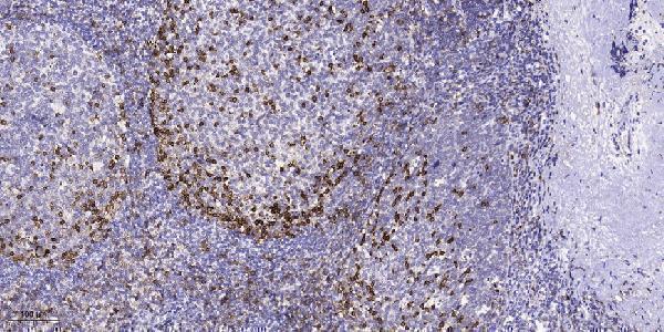
| Sample | Paraffin-embedded human tonsil tissue; fixative not specified (datasheet A00690) |
| Fixation | Image fixative and duration unreported (datasheet A00690); verify before use. |
| Sectioning | 4–5 µm sections on charged slides (standard) |
| Deparaffinisation | Xylene, graded ethanol series to water (standard) |
| Antigen retrieval | Heat retrieval: Tris-EDTA pH 9.0 (datasheet A00690); 20 min, 95–100 °C (standard) |
| Peroxidase block | 3% H2O2, 10 min, room temperature (standard) |
| Blocking | 10% normal serum of the secondary host, 30 min, room temperature (standard) |
| Primary antibody | Rabbit anti-EPHB4, 1:100-1:300 (datasheet A00690) |
| Primary incubation | Overnight at 4 °C (standard) |
| Detection | HRP-polymer secondary, DAB chromogen 5–10 min (standard) |
| Counterstain | Hematoxylin, blue, dehydrate and mount (standard) |
| Expected result | EPHB4-positive staining in glandular cells of appendix (HPA tissue IHC: High). HPA tissue profile: Cytoplasmic expression in several tissues. No signal in the no-primary control. |
EPHB4 is a single-pass cell-membrane receptor with an extracellular region and a cytoplasmic kinase region (UniProt P54760 topology). In paraffin-section IHC, assess staining in the cell types HPA reports as positive, including intestinal glandular cells and hepatocytes (HPA: High). HPA also describes cytoplasmic staining across several tissues, but rates its tissue IHC profile Uncertain because antibody staining and RNA expression show low consistency (HPA: Uncertain).
| Membrane-associated staining, with possible cytoplasmic staining, in intestinal glandular cells or hepatocytes. | This is compatible with EPHB4's membrane topology and HPA's reported cytoplasmic IHC profile (UniProt P54760 topology; HPA: High in glandular cells and hepatocytes). Record membrane and cytoplasmic signal separately; HPA's Uncertain rating limits how confidently either pattern identifies EPHB4 (HPA: Uncertain). |
| Strong, isolated nuclear staining with little membrane or cytoplasmic signal. | Nuclear localisation is not the expected pattern for this receptor (UniProt P54760: cell membrane; HPA: cytoplasmic IHC profile). Treat an isolated nuclear pattern as suspect and compare it with a no-primary control and staining from an independent antibody (general IHC practice). |
| Prominent staining in adipocytes or lymph-node non-germinal-center cells. | HPA reports EPHB4 as Not detected in these cells, so prominent staining warrants a specificity check (HPA: Not detected). Possible contributors include cross-reactivity or endogenous detection activity; controls can help distinguish them (general IHC practice). HPA's Uncertain rating prevents a definitive negative-tissue claim (HPA: Uncertain). |
| Diffuse color across cells and surrounding tissue without clear cellular boundaries. | This is difficult to score as receptor localisation and may reflect nonspecific background or detection chemistry (general IHC practice). Compare with a no-primary control before interpreting weak cytoplasmic color, particularly because HPA reports a cytoplasmic profile with Uncertain reliability (HPA: Uncertain). |
| No signal in a colon glandular-cell section run as a positive reference. | HPA reports High staining in colon glandular cells, making them a useful comparison, but its overall IHC reliability is Uncertain (HPA: High in colon glandular cells; HPA: Uncertain). Check section quality and assay controls before concluding that the sample lacks EPHB4 (general IHC practice). |
| Cellular compartment | EPHB4 spans the membrane at residues 540–560, with extracellular residues 16–539 and cytoplasmic residues 561–987 (UniProt P54760 topology). HPA describes cytoplasmic tissue IHC staining (HPA: cytoplasmic profile). Score membrane and cytoplasmic staining explicitly; neither source establishes that every positive section must show a crisp membrane rim. |
| Choice of comparison tissue | HPA reports High staining in appendix, colon, duodenum and small-intestine glandular cells and in hepatocytes; adipocytes and skeletal-muscle myocytes are Not detected (HPA: tissue IHC). These are observed cell-specific patterns, not guaranteed positive or negative controls, because the tissue profile is rated Uncertain (HPA: Uncertain). |
| Antibody validation | The listed antibody, CAB013537, has an Uncertain IHC validation status (HPA: CAB013537 IHC Uncertain). HPA also reports low consistency between antibody staining and RNA expression (HPA: Uncertain). Interpret unexpected patterns with controls and, where feasible, an independently validated antibody (general IHC practice). |
| Epitope and isoform scope | The receptor has an extracellular region, a cytoplasmic region and 4 listed isoforms (UniProt P54760 topology; UniProt P54760 isoforms). The supplied record does not locate the antibody epitope or establish which isoforms it detects; consult the antibody's documented epitope before attributing compartment differences to an isoform (general IHC practice). |
| ICC/IF Q: Is its localisation established here? | HPA's subcellular summary says Membrane, but supplies no main-location designation or ICC/IF image-bearing cell lines; CAB013537 has no listed ICC validation. Use the separate IF/ICC guide for that application; these records do not establish an IF staining pattern. |
| Situation | Likely cause | Next action |
|---|---|---|
| Expected glandular-cell signal is absent. | The IHC run may have failed, or the sampled cells may differ from HPA's reported High glandular-cell populations (HPA: High in colon and small intestine; general IHC practice). | Confirm the intended cells are present, inspect the counterstain, and compare a concurrently processed reference section and assay controls (general IHC practice). Do not treat HPA's Uncertain profile as proof that every section must stain (HPA: Uncertain). |
| Only nuclei stain strongly. | An isolated nuclear pattern conflicts with UniProt's membrane assignment and HPA's cytoplasmic IHC description (UniProt P54760: cell membrane; HPA: cytoplasmic profile). | Review a no-primary control and compare another antibody or control tissue before scoring it as EPHB4 (general IHC practice). |
| Adipocytes or lymph-node non-germinal-center cells stain strongly. | These cells are reported Not detected by HPA; cross-reactivity or endogenous detection activity is possible (HPA: Not detected; general IHC practice). | Compare no-primary and detection controls and check whether expected positive cells stain in the same run (general IHC practice). Treat the mismatch as a specificity concern, given HPA's Uncertain rating (HPA: Uncertain). |
| Brown color is diffuse across the section. | Background from antibody binding or chromogenic detection can obscure cell boundaries (general IHC practice). | Compare no-primary controls, review blocking and washes, and adjust the detection workflow if background persists (general IHC practice). Score only signal that can be assigned to cells and compartments. |
| Cytoplasmic staining appears without a sharp membrane rim. | HPA describes cytoplasmic tissue IHC staining, while UniProt assigns EPHB4 to the cell membrane; the supplied sources do not resolve that difference (HPA: cytoplasmic profile; UniProt P54760: cell membrane). | Record the compartment as observed and compare controls and an independent antibody before calling it specific (general IHC practice). Report HPA's Uncertain reliability alongside the result (HPA: Uncertain). |
| Staining differs among sections or sampled tissues. | HPA reports different cell-specific levels, including High glandular-cell staining and Not detected adipocytes; its overall IHC profile is Uncertain (HPA: tissue IHC; HPA: Uncertain). | Compare like cell types, section quality and concurrent controls before interpreting a biological difference (general IHC practice). Avoid assigning a change in EPHB4 abundance from staining intensity alone. |


Comprehensive Human Protein Atlas IHC scoring per tissue (reliability: Uncertain — Low consistency between antibody staining and RNA expression data.). Rows are taken directly from the HPA tissue chart — click any row's HPA link to view the source.
| Tissue | Cell type | Level | Evidence | Source |
|---|---|---|---|---|
| Appendix | Glandular cells | High | Protein (IHC) | HPA → |
| Colon | Glandular cells | High | Protein (IHC) | HPA → |
| Duodenum | Glandular cells | High | Protein (IHC) | HPA → |
| Liver | Hepatocytes | High | Protein (IHC) | HPA → |
| Small intestine | Glandular cells | High | Protein (IHC) | HPA → |
| Tissue | Cell type | Level | Evidence | Source |
|---|---|---|---|---|
| Adipose tissue | Adipocytes | Not detected | Protein (IHC) | HPA → |
| Lymph node | Non-germinal center cells | Not detected | Protein (IHC) | HPA → |
| Skeletal muscle | Myocytes | Not detected | Protein (IHC) | HPA → |
| Smooth muscle | Smooth muscle cells | Not detected | Protein (IHC) | HPA → |
| Spleen | Cells in red pulp | Not detected | Protein (IHC) | HPA → |
Use the paraffin-section caption as the starting protocol, then judge EPHB4 staining by compartment, cell type, and controls (datasheet A00690; UniProt P54760 topology; HPA tissue IHC).
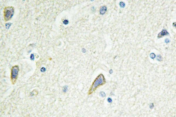
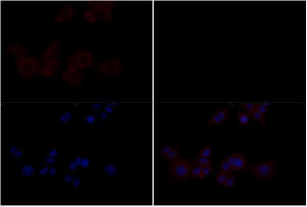
A00690 and A00690-2 have paraffin-section IHC images (catalog IHC captions); both list human and mouse reactivity (catalog reactivity), while A00690-2 also lists rat reactivity and has an IF image (catalog reactivity/IF caption).
A00690 shows IHC on paraffin-embedded human tonsil and lists human and mouse reactivity (A00690 IHC caption/catalog reactivity). A00690-2 shows IHC on paraffin-embedded human brain and IF in MCF-7 cells, and lists human, mouse and rat reactivity (A00690-2 IHC and IF captions/catalog reactivity).
Which to pick: For tissue IHC, choose A00690 if its human tonsil example with 1:200 primary dilution and Tris-EDTA retrieval at pH 9.0 fits your plan; A00690-2 has a paraffin-embedded human brain example (respective IHC captions). For IF, A00690-2 has an MCF-7 image at 1:200; for ICC, A00690-3 lists ICC as an application but provides no ICC image (A00690-2 IF caption; A00690-3 catalog applications/image captions). For rat samples, A00690-2 and A00690-3 list rat reactivity; the rendered antibodies have no clone reported, and neither IHC caption reports a fixative (catalog reactivity/clone fields; IHC captions).